Plasticity of Temporal Information Processing in the Primary Auditory ...
Plasticity of Temporal Information Processing in the Primary Auditory ...
Plasticity of Temporal Information Processing in the Primary Auditory ...

Create successful ePaper yourself
Turn your PDF publications into a flip-book with our unique Google optimized e-Paper software.
© 1998 Nature America Inc. • http://neurosci.nature.com<br />
articles<br />
<strong>Plasticity</strong> <strong>of</strong> temporal <strong>in</strong>formation<br />
process<strong>in</strong>g <strong>in</strong> <strong>the</strong> primary auditory<br />
cortex<br />
Michael P. Kilgard 1 and Michael M. Merzenich 1,2<br />
1 Coleman Laboratory, Departments <strong>of</strong> Otolaryngology and Physiology, Keck Center for Integrative Neuroscience,<br />
University <strong>of</strong> California at San Francisco, San Francisco, California 94143-0444, USA<br />
2 Scientific Learn<strong>in</strong>g Corporation, 1995 University Avenue, Berkeley, California 94104-1075, USA<br />
Correspondence should be addressed to M.P.K. (kilgard@phy.ucsf.edu)<br />
© 1998 Nature America Inc. • http://neurosci.nature.com<br />
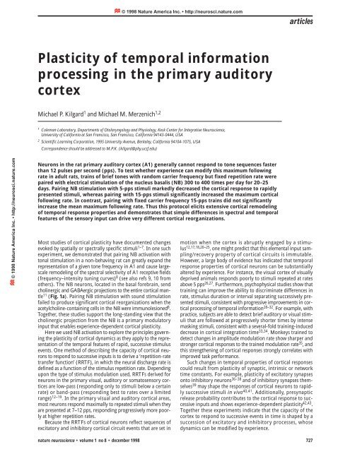
Neurons <strong>in</strong> <strong>the</strong> rat primary auditory cortex (A1) generally cannot respond to tone sequences faster<br />
than 12 pulses per second (pps). To test whe<strong>the</strong>r experience can modify this maximum follow<strong>in</strong>g<br />
rate <strong>in</strong> adult rats, tra<strong>in</strong>s <strong>of</strong> brief tones with random carrier frequency but fixed repetition rate were<br />
paired with electrical stimulation <strong>of</strong> <strong>the</strong> nucleus basalis (NB) 300 to 400 times per day for 20–25<br />
days. Pair<strong>in</strong>g NB stimulation with 5-pps stimuli markedly decreased <strong>the</strong> cortical response to rapidly<br />
presented stimuli, whereas pair<strong>in</strong>g with 15-pps stimuli significantly <strong>in</strong>creased <strong>the</strong> maximum cortical<br />
follow<strong>in</strong>g rate. In contrast, pair<strong>in</strong>g with fixed carrier frequency 15-pps tra<strong>in</strong>s did not significantly<br />
<strong>in</strong>crease <strong>the</strong> mean maximum follow<strong>in</strong>g rate. Thus this protocol elicits extensive cortical remodel<strong>in</strong>g<br />
<strong>of</strong> temporal response properties and demonstrates that simple differences <strong>in</strong> spectral and temporal<br />
features <strong>of</strong> <strong>the</strong> sensory <strong>in</strong>put can drive very different cortical reorganizations.<br />
Most studies <strong>of</strong> cortical plasticity have documented changes<br />
evoked by spatially or spectrally specific stimuli 1–7 . In one such<br />
experiment, we demonstrated that pair<strong>in</strong>g NB activation with<br />
tonal stimulation <strong>in</strong> a non-behav<strong>in</strong>g rat can greatly expand <strong>the</strong><br />
representation <strong>of</strong> a given tone frequency <strong>in</strong> A1 and cause largescale<br />
remodel<strong>in</strong>g <strong>of</strong> <strong>the</strong> spectral selectivity <strong>of</strong> A1 receptive fields<br />
(frequency–<strong>in</strong>tensity tun<strong>in</strong>g curves) 8 (see also refs 9, 10 from<br />
o<strong>the</strong>rs). The NB neurons, located <strong>in</strong> <strong>the</strong> basal forebra<strong>in</strong>, send<br />
chol<strong>in</strong>ergic and GABAergic projections to <strong>the</strong> entire cortical mantle<br />
11 (Fig. 1a). Pair<strong>in</strong>g NB stimulation with sound stimulation<br />
failed to produce significant cortical reorganizations when <strong>the</strong><br />
acetylchol<strong>in</strong>e-conta<strong>in</strong><strong>in</strong>g cells <strong>in</strong> <strong>the</strong> NB were immunolesioned 8 .<br />
Toge<strong>the</strong>r, <strong>the</strong>se studies support <strong>the</strong> long-stand<strong>in</strong>g view that <strong>the</strong><br />
chol<strong>in</strong>ergic projection from <strong>the</strong> NB is a primary modulatory<br />
<strong>in</strong>put that enables experience-dependent cortical plasticity.<br />
Here we used NB activation to explore <strong>the</strong> pr<strong>in</strong>ciples govern<strong>in</strong>g<br />
<strong>the</strong> plasticity <strong>of</strong> cortical dynamics as <strong>the</strong>y apply to <strong>the</strong> representation<br />
<strong>of</strong> <strong>the</strong> temporal features <strong>of</strong> rapid, successive stimulus<br />
events. One method <strong>of</strong> describ<strong>in</strong>g <strong>the</strong> capacity <strong>of</strong> cortical neurons<br />
to respond to successive <strong>in</strong>puts is to derive a ‘repetition-rate<br />
transfer function’ (RRTF), <strong>in</strong> which <strong>the</strong> neural discharge rate is<br />
def<strong>in</strong>ed as a function <strong>of</strong> <strong>the</strong> stimulus repetition rate. Depend<strong>in</strong>g<br />
upon <strong>the</strong> type <strong>of</strong> stimulus modulation used, RRTFs derived for<br />
neurons <strong>in</strong> <strong>the</strong> primary visual, auditory or somatosensory cortices<br />
are low-pass (respond<strong>in</strong>g only to stimuli below a certa<strong>in</strong><br />
rate) or band-pass (respond<strong>in</strong>g best to rates over a limited<br />
range) 12–19 . In <strong>the</strong> primary visual and auditory cortical areas,<br />
most neurons respond maximally to repeated stimuli when <strong>the</strong>y<br />
are presented at 7–12 pps, respond<strong>in</strong>g progressively more poorly<br />
at higher repetition rates.<br />
Because <strong>the</strong> RRTFs <strong>of</strong> cortical neurons reflect sequences <strong>of</strong><br />
excitatory and <strong>in</strong>hibitory cortical circuit events that are set <strong>in</strong><br />
motion when <strong>the</strong> cortex is abruptly engaged by a stimulus<br />
12,17,18,20–25 , one might predict that this elemental <strong>in</strong>put sampl<strong>in</strong>g/recovery<br />
property <strong>of</strong> cortical circuits is immutable.<br />
However, a large body <strong>of</strong> evidence has <strong>in</strong>dicated that temporal<br />
response properties <strong>of</strong> cortical neurons can be substantially<br />
altered by experience. For <strong>in</strong>stance, <strong>the</strong> visual cortex <strong>of</strong> visually<br />
deprived animals responds poorly to stimuli repeated at rates<br />
above 5 pps 26,27 . Fur<strong>the</strong>rmore, psychophysical studies show that<br />
tra<strong>in</strong><strong>in</strong>g can improve <strong>the</strong> ability to discrim<strong>in</strong>ate differences <strong>in</strong><br />
rate, stimulus duration or <strong>in</strong>terval separat<strong>in</strong>g successively presented<br />
stimuli, consistent with progressive improvements <strong>in</strong> cortical<br />
process<strong>in</strong>g <strong>of</strong> temporal <strong>in</strong>formation 28–32 . For example, with<br />
practice, subjects are able to detect brief auditory or visual stimuli<br />
that are followed at progressively shorter times by <strong>in</strong>tense<br />
mask<strong>in</strong>g stimuli, consistent with a several-fold tra<strong>in</strong><strong>in</strong>g-<strong>in</strong>duced<br />
decrease <strong>in</strong> cortical <strong>in</strong>tegration time 33,34 . Monkeys tra<strong>in</strong>ed to<br />
detect changes <strong>in</strong> amplitude modulation rate show sharper and<br />
stronger cortical responses to <strong>the</strong> tra<strong>in</strong>ed modulation rate 35 , and<br />
this streng<strong>the</strong>n<strong>in</strong>g <strong>of</strong> cortical responses strongly correlates with<br />
improved task performance.<br />
Such changes <strong>in</strong> temporal properties <strong>of</strong> cortical responses<br />
could result from plasticity <strong>of</strong> synaptic, <strong>in</strong>tr<strong>in</strong>sic or network<br />
time constants. For example, plasticity <strong>of</strong> excitatory synapses<br />
onto <strong>in</strong>hibitory neurons 36–38 and <strong>of</strong> <strong>in</strong>hibitory synapses <strong>the</strong>mselves<br />
39 may shape <strong>the</strong> responses <strong>of</strong> cortical neurons to rapidly<br />
successive stimuli <strong>in</strong> vivo 40,41 . Additionally, presynaptic<br />
release probability contributes to <strong>the</strong> cortical response to successive<br />
<strong>in</strong>puts and shows experience-dependent plasticity 42,43 .<br />
Toge<strong>the</strong>r <strong>the</strong>se experiments <strong>in</strong>dicate that <strong>the</strong> capacity <strong>of</strong> <strong>the</strong><br />
cortex to respond to successive events <strong>in</strong> time is shaped by a<br />
succession <strong>of</strong> excitatory and <strong>in</strong>hibitory processes, whose<br />
dynamics can be modified by experience.<br />
nature neuroscience • volume 1 no 8 • december 1998 727
articles<br />
© 1998 Nature America Inc. • http://neurosci.nature.com<br />
a<br />
a<br />
Cortex<br />
b<br />
CPu<br />
GP<br />
Thal<br />
Nucleus<br />
Basalis<br />
© 1998 Nature America Inc. • http://neurosci.nature.com<br />
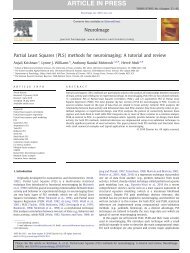
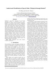
Fig. 1. Response <strong>of</strong> rat C<br />
auditory cortex neurons<br />
to repeated stimuli.<br />
(a) Schematic diagram <strong>of</strong><br />
<strong>the</strong> projection <strong>of</strong> nucleus<br />
basalis to <strong>the</strong> neocortex.<br />
(b) Number <strong>of</strong> rats and<br />
penetrations for each<br />
experimental condition.<br />
(c) Dot rasters and repetition-rate<br />
transfer function<br />
(RRTF) <strong>of</strong> primary auditory<br />
cortex (A1) neurons from<br />
a naïve (control) rat. Each dot represents<br />
a s<strong>in</strong>gle action potential. The<br />
short horizontal l<strong>in</strong>es <strong>in</strong>dicate <strong>the</strong><br />
spike-collection w<strong>in</strong>dows that were<br />
used to generate <strong>the</strong> RRTF. The RRTF<br />
quantifies <strong>the</strong> generally low-pass nature<br />
<strong>of</strong> <strong>the</strong> responses <strong>of</strong> A1 cortical neurons<br />
to <strong>the</strong>se pulsed stimuli <strong>in</strong> <strong>the</strong> rat. The<br />
solid vertical l<strong>in</strong>e <strong>in</strong> <strong>the</strong> RRTF shows <strong>the</strong><br />
average number <strong>of</strong> spikes evoked by <strong>the</strong><br />
first tone <strong>in</strong> <strong>the</strong> tra<strong>in</strong>. (d) Mean normalized<br />
RRTF for all penetrations recorded<br />
from normal (control) rats. Error bars<br />
<strong>in</strong>dicate standard errors.<br />
Repetition rate (pps)<br />
d<br />
Normalized spike rate<br />
ms<br />
Repetition rate (pps)<br />
Repetition rate (pps)<br />
Spikes/tone<br />
Results<br />
In this study, NB activation was paired with temporally modulated<br />
acoustic stimuli to <strong>in</strong>vestigate plasticity <strong>of</strong> <strong>the</strong> cortical representation<br />
<strong>of</strong> time-vary<strong>in</strong>g <strong>in</strong>formation. Stimulat<strong>in</strong>g electrodes<br />
were chronically implanted <strong>in</strong> <strong>the</strong> right NB <strong>of</strong> 15 adult rats. After<br />
recovery, animals were placed <strong>in</strong> a sound-attenuation chamber,<br />
and tra<strong>in</strong>s <strong>of</strong> six tone bursts were paired with NB stimulation<br />
dur<strong>in</strong>g daily sessions. Tone tra<strong>in</strong>s and NB stimulation occurred<br />
randomly every 8–40 seconds, repeated three- to four-hundred<br />
times per day for 20–25 days. Rats were unanes<strong>the</strong>tized and unrestra<strong>in</strong>ed<br />
throughout this procedure. The tra<strong>in</strong> repetition rate for<br />
each rat was fixed at 5, 7.5 or 15 pps. The tonal (carrier) frequency<br />
was varied randomly trial by trial. The seven carrier frequencies<br />
that were used extended across most <strong>of</strong> <strong>the</strong> frequency range represented<br />
<strong>in</strong> <strong>the</strong> primary auditory cortex (A1) <strong>of</strong> <strong>the</strong> rat. Twentyfour<br />
hours after <strong>the</strong> last pair<strong>in</strong>g session, each animal was<br />
anes<strong>the</strong>tized, and <strong>the</strong> responses <strong>of</strong> A1 neurons were recorded<br />
from 30–60 microelectrode penetrations distributed evenly across<br />
A1. Frequency–<strong>in</strong>tensity tun<strong>in</strong>g curves and RRTFs were derived<br />
to characterize <strong>the</strong> spectral and temporal response properties <strong>of</strong><br />
neurons <strong>in</strong> every penetration.<br />
In naïve animals, at repetition rates up to about 9 pps, each<br />
brief tone generally evoked <strong>the</strong> same number <strong>of</strong> spikes from<br />
A1 neurons as did <strong>the</strong> first tone <strong>in</strong> <strong>the</strong> tra<strong>in</strong> (Fig. 1). At repetition<br />
rates from 9 to 14 pps, <strong>the</strong> number <strong>of</strong> spikes per tone fell<br />
<strong>of</strong>f rapidly, and only neurons at rare sites responded at all to<br />
rates above 15 pps. In experimental rats, exposure to 15-pps<br />
stimuli at variable carrier frequencies paired with NB stimulation<br />
markedly altered <strong>the</strong> temporal responses <strong>of</strong> cortical neurons<br />
recorded all across A1. Although no specific response peak<br />
emerged at 15 pps, <strong>the</strong> cut-<strong>of</strong>f rate <strong>of</strong> low-pass RRTFs <strong>in</strong> A1<br />
rose significantly <strong>in</strong> most sampled neurons. In strik<strong>in</strong>g contrast<br />
to control rats, <strong>the</strong> average neuron <strong>in</strong> <strong>the</strong>se samples<br />
responded strongly to repetition rates between 10 and 20 pps<br />
(Fig. 2a). This <strong>in</strong>crease <strong>in</strong> <strong>the</strong> neural response to 15-pps tra<strong>in</strong>s<br />
after pair<strong>in</strong>g was highly significant (p < 0.0001; Fig. 2c). The<br />
high-rate slope <strong>of</strong> <strong>the</strong> average RRTF shifted up, reflect<strong>in</strong>g a<br />
strong response improvement across a broad range <strong>of</strong> higher<br />
modulation frequencies (Fig. 2a and c).<br />
To determ<strong>in</strong>e whe<strong>the</strong>r or not NB-<strong>in</strong>duced temporal plasticity<br />
is specific to <strong>the</strong> repetition rate <strong>of</strong> <strong>the</strong> paired acoustic stimulus,<br />
5-pps tra<strong>in</strong>s were paired with NB activation. The normalized<br />
response evoked by stimuli repeated at 5 pps was <strong>in</strong>creased significantly<br />
(p < 0.01), result<strong>in</strong>g <strong>in</strong> RRTFs with bandpass characteristics<br />
(response maxima near 5 pps). The 5-pps pair<strong>in</strong>g resulted<br />
<strong>in</strong> two dist<strong>in</strong>ct classes <strong>of</strong> cortical responsiveness to faster rates.<br />
728 nature neuroscience • volume 1 no 8 • december 1998
© 1998 Nature America Inc. • http://neurosci.nature.com<br />
articles<br />
© 1998 Nature America Inc. • http://neurosci.nature.com<br />
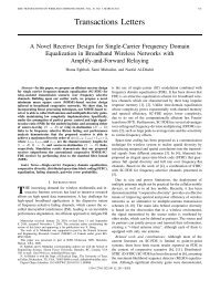
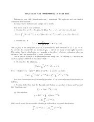
Fig. 2. <strong>Temporal</strong> response<br />
plasticity <strong>in</strong>duced by<br />
nucleus basalis stimulation.<br />
a 3.2<br />
6.8<br />
(a) Response to repeated<br />
8.4<br />
tones after pair<strong>in</strong>g NB stimulation<br />
with 15-pps tra<strong>in</strong>s 9.8<br />
applied at 7 different carrier<br />
frequencies. The maximum<br />
follow<strong>in</strong>g rate and <strong>the</strong> range<br />
over which strong stimulusby-stimulus<br />
11.1<br />
13.9<br />
16.9<br />
responses were<br />
evoked was <strong>in</strong>creased all -100 0 100 200 300 400 500 600 700<br />
across A1 <strong>in</strong> this sample,<br />
compared to controls. b<br />
3.2<br />
(b) Response to repeated<br />
tones after pair<strong>in</strong>g NB stimulation<br />
6.8<br />
with 5-pps tra<strong>in</strong>s <strong>of</strong><br />
tones at a randomly varied<br />
carrier frequency. The maximum<br />
follow<strong>in</strong>g rate was<br />
8.4<br />
9.8<br />
11.1<br />
significantly decreased compared<br />
to controls. (c) Mean<br />
13.9<br />
normalized RRTFs for penetrations<br />
16.9<br />
recorded from ani-<br />
mals that received NB -100 0 100 200 300 400 500 600 700<br />
stimulation paired with 5-,<br />
Milliseconds ms<br />
7.5- or 15-pps tra<strong>in</strong>s <strong>of</strong> tone<br />
(carrier) frequencies that varied from c<br />
1.2<br />
tra<strong>in</strong> to tra<strong>in</strong>, compared to control<br />
RRTFs. The RRTF <strong>of</strong> each site was normalized<br />
us<strong>in</strong>g <strong>the</strong> number <strong>of</strong> spikes<br />
1<br />
evoked by <strong>the</strong> first tone <strong>in</strong> each tra<strong>in</strong>.<br />
Error bars <strong>in</strong>dicate standard error. The<br />
rates that were significantly different from<br />
0.8<br />
controls are marked with dots (one-way<br />
Control<br />
ANOVA, Fisher’s PLSD, p < 0.05).<br />
0.6<br />
5 pps<br />
Repetition rate Rate (pps) (pps)<br />
Repetition rate Rate (pps)<br />
Normalized Spike spike Rate rate<br />
0.4<br />
7.5 pps<br />
15 pps<br />
Repetition Rate rate (pps)<br />
Repetition Rate rate (pps)<br />
5<br />
10<br />
15<br />
20<br />
0 2 4<br />
5<br />
10<br />
15<br />
20<br />
0 2<br />
Spikes/Tone<br />
Spikes/tone<br />
0.2<br />
3 5 7 9 11 13 15 17 19<br />
Repetition Rate rate (pps)<br />
In three <strong>of</strong> four animals, <strong>the</strong> entire RRTF was shifted leftward,<br />
caus<strong>in</strong>g significantly lower maximum follow<strong>in</strong>g rates (Fig. 2b<br />
and c). In <strong>the</strong> rema<strong>in</strong><strong>in</strong>g animal, 75% <strong>of</strong> sites showed strong facilitation<br />
to both 5- and 10-pps stimuli, but poor responses to stimuli<br />
repeated at 7.5 pps (data not shown). Thus NB-<strong>in</strong>duced<br />
plasticity reliably <strong>in</strong>creased <strong>the</strong> strength <strong>of</strong> <strong>the</strong> cortical response to<br />
stimuli presented at <strong>the</strong> paired rate, even though <strong>the</strong> RRTF plasticity<br />
took two different forms.<br />
Pair<strong>in</strong>g 7.5-pps stimuli at randomly varied carrier frequency<br />
with NB stimulation selectively streng<strong>the</strong>ned <strong>the</strong> cortical response<br />
to stimuli repeated at rates near 7.5 pps. The mean RRTF aga<strong>in</strong><br />
took a bandpass form, with a substantially stronger-than-normal<br />
response to modulated stimuli emerg<strong>in</strong>g at <strong>the</strong> paired stimulus<br />
rate (Fig. 2c). The normalized response <strong>of</strong> 1.2 <strong>in</strong>dicates that<br />
20% more spikes were evoked on average by each tone <strong>in</strong> <strong>the</strong> context<br />
<strong>of</strong> a 7-pps tra<strong>in</strong> compared to <strong>the</strong> same tone <strong>in</strong> isolation.<br />
As reported previously, pair<strong>in</strong>g tones at 15 pps with a constant<br />
carrier frequency <strong>of</strong> 9 kHz markedly enlarged <strong>the</strong> region <strong>of</strong> A1<br />
represent<strong>in</strong>g 9 kHz <strong>in</strong> every animal 8 . Surpris<strong>in</strong>gly, pair<strong>in</strong>g NB<br />
stimulation with tra<strong>in</strong>s <strong>of</strong> 9-kHz tones repeated at 15 pps did not<br />
cause <strong>the</strong> rightward shift <strong>in</strong> <strong>the</strong> mean RRTF that resulted from<br />
pair<strong>in</strong>g NB stimulation with multiple-frequency tra<strong>in</strong>s repeated at<br />
15 pps. No significant alteration <strong>in</strong> <strong>the</strong> mean response to <strong>the</strong><br />
paired repetition rate (15 pps) was detected <strong>in</strong> <strong>the</strong> population<br />
RRTF analysis (0.27 versus 0.28, p > 0.5; one-way ANOVA).<br />
Although it is unclear how topographic map reorganization and<br />
temporal plasticity are related, it is <strong>in</strong>terest<strong>in</strong>g to note that pair<strong>in</strong>g<br />
random-frequency 15-pps tra<strong>in</strong>s with NB stimulation significantly<br />
<strong>in</strong>creased <strong>the</strong> maximum stimulus follow<strong>in</strong>g rate <strong>of</strong> cortical<br />
neurons, but did not systematically alter <strong>the</strong> A1 frequency map.<br />
Discussion<br />
Paired NB and sensory stimulation provides a simple model system<br />
for study<strong>in</strong>g <strong>the</strong> rules that operate <strong>in</strong> <strong>the</strong> cortex to transform<br />
sensory <strong>in</strong>put structure and schedules <strong>in</strong>to distributed cortical<br />
response patterns. Previous studies focus<strong>in</strong>g on <strong>the</strong> plasticity <strong>of</strong><br />
<strong>the</strong> cortical representation <strong>of</strong> spectral <strong>in</strong>formation have demonstrated<br />
that chol<strong>in</strong>ergic modulation is sufficient to shift A1 tun<strong>in</strong>g<br />
curves toward <strong>the</strong> frequency paired with NB<br />
stimulation 8–10,44,45 . In this study, we used NB activation to<br />
nature neuroscience • volume 1 no 8 • december 1998 729
articles<br />
© 1998 Nature America Inc. • http://neurosci.nature.com<br />
© 1998 Nature America Inc. • http://neurosci.nature.com<br />
explore temporal <strong>in</strong>formation process<strong>in</strong>g, and we showed that<br />
<strong>the</strong> temporal response properties <strong>of</strong> A1 neurons can be altered<br />
markedly to ref<strong>in</strong>e or degrade <strong>the</strong> capacity <strong>of</strong> <strong>the</strong> cortex to<br />
respond to rapid successive <strong>in</strong>put events. We also showed that<br />
this plasticity has a large capacity to exaggerate <strong>the</strong> representation<br />
<strong>of</strong> specific, heavily presented sensory <strong>in</strong>put rates. F<strong>in</strong>ally, we<br />
demonstrated that A1 neuronal networks can generate spectrally<br />
and temporally selective responses and that <strong>the</strong>se networks can<br />
reorganize topographic representations <strong>of</strong> tone frequency with<br />
no evident change <strong>in</strong> <strong>the</strong> representation <strong>of</strong> temporal <strong>in</strong>formation,<br />
or vice versa, all as an apparent function <strong>of</strong> <strong>the</strong> spectrotemporal<br />
structures and schedules <strong>of</strong> sensory <strong>in</strong>puts.<br />
This strik<strong>in</strong>g capacity for learn<strong>in</strong>g-based revision <strong>of</strong> <strong>the</strong> basic<br />
<strong>in</strong>tegration/segmentation times <strong>of</strong> <strong>the</strong> cortical process<strong>in</strong>g mach<strong>in</strong>ery<br />
could result from plasticity <strong>of</strong> synaptic time constants, <strong>of</strong><br />
<strong>in</strong>tr<strong>in</strong>sic temporal characteristics and/or <strong>of</strong> network dynamics<br />
12,17,21,42,46 . Paired-pulse facilitation and slow <strong>in</strong>hibitory potentials,<br />
for example, almost certa<strong>in</strong>ly are important <strong>in</strong> <strong>the</strong> cortical<br />
recovery <strong>of</strong> responsiveness follow<strong>in</strong>g any brief stimulation<br />
12,23,24,40–42,47 . NB-<strong>in</strong>duced plasticity will provide a powerful<br />
experimental approach for relat<strong>in</strong>g <strong>the</strong> cortical representation<br />
<strong>of</strong> temporal <strong>in</strong>formation to changes <strong>in</strong> basic cortical dynamics<br />
that shape <strong>the</strong> cortical responses to time-vary<strong>in</strong>g stimuli.<br />
Methods<br />
PREPARATION. Plat<strong>in</strong>um bipolar stimulat<strong>in</strong>g electrodes were lowered<br />
7 mm below <strong>the</strong> cortical surface, 3.3 mm lateral and 2.3 mm posterior<br />
to bregma <strong>in</strong> barbiturate-anes<strong>the</strong>tized female rats (~300 g) and cemented<br />
<strong>in</strong>to place us<strong>in</strong>g sterile techniques approved under UCSF Animal Care<br />
Facility protocols. After two weeks <strong>of</strong> recovery, tra<strong>in</strong>s <strong>of</strong> six 25-ms tones<br />
were paired with 200 ms <strong>of</strong> NB electrical stimulation <strong>in</strong> a sound-shielded,<br />
calibrated test chamber (five days per week). The frequency <strong>of</strong> <strong>the</strong><br />
tone was ei<strong>the</strong>r one <strong>of</strong> seven frequencies (1.3, 2, 3, 5, 9, 14 or 19 kHz) or<br />
was fixed (9 kHz). Tone amplitude was 20–30 dB above <strong>the</strong> m<strong>in</strong>imum<br />
rat hear<strong>in</strong>g threshold 48 . In experiments us<strong>in</strong>g multiple carrier frequencies,<br />
<strong>the</strong> frequency <strong>of</strong> <strong>the</strong> tones with<strong>in</strong> each tra<strong>in</strong> was constant, whereas <strong>the</strong><br />
frequencies used from tra<strong>in</strong> to tra<strong>in</strong> were randomly varied. The tone pips<br />
<strong>in</strong> stimulus tra<strong>in</strong>s were presented <strong>in</strong> a given rat at 5, 7.5 or 15 pps. Electrical<br />
stimulation began with <strong>the</strong> onset <strong>of</strong> <strong>the</strong> fourth tone. The stimulat<strong>in</strong>g<br />
current level (70–150 µA) was <strong>the</strong> m<strong>in</strong>imum necessary to<br />
desynchronize <strong>the</strong> EEG dur<strong>in</strong>g slow-wave sleep for 1–2 seconds. Stimulation<br />
consisted <strong>of</strong> 100-pps capacitatively coupled biphasic pulses <strong>of</strong> 0.1-<br />
ms duration. Several microdialysis studies have shown that this<br />
stimulation protocol results <strong>in</strong> <strong>the</strong> release <strong>of</strong> cortical acetylchol<strong>in</strong>e 49,50 .<br />
Ei<strong>the</strong>r chol<strong>in</strong>ergic antagonists or lesions <strong>of</strong> <strong>the</strong> chol<strong>in</strong>ergic cells <strong>in</strong> <strong>the</strong><br />
NB with 192 immunoglobul<strong>in</strong> G-sapor<strong>in</strong> are sufficient to block this plasticity<br />
generated by NB stimulation 8,9 . Tonal and electrical stimuli did not<br />
evoke any observable behavioral responses (that is, <strong>the</strong>y did not cause<br />
rats to stop groom<strong>in</strong>g, or if sleep<strong>in</strong>g, to awaken).<br />
ELECTROPHYSIOLOGY. Twenty-four hours after <strong>the</strong> last pair<strong>in</strong>g, animals<br />
were anes<strong>the</strong>tized with sodium pentobarbital, <strong>the</strong> right auditory cortex<br />
was surgically exposed, and neural responses were recorded with parylene-coated<br />
tungsten microelectrodes (FHC #070-02-01, 2 MΩ). Because<br />
we used barbiturate anes<strong>the</strong>sia, <strong>the</strong> modulation <strong>of</strong> responses recorded <strong>in</strong><br />
this study may not be identical to <strong>the</strong> responses <strong>of</strong> awake animals. Penetration<br />
sites were chosen to evenly sample <strong>the</strong> cortical surface while avoid<strong>in</strong>g<br />
blood vessels. To m<strong>in</strong>imize <strong>the</strong> possibility <strong>of</strong> experimenter bias or<br />
response variability due to variable record<strong>in</strong>g depth, at every penetration<br />
site <strong>the</strong> electrode was lowered to ~550 µm below <strong>the</strong> pial surface<br />
(layers 4/5), which yielded vigorous driven responses. Frequency/<strong>in</strong>tensity<br />
response areas were reconstructed <strong>in</strong> detail by present<strong>in</strong>g 45 pure<br />
tone frequencies (50-ms duration, 3-ms ramps) at each <strong>of</strong> 15 sound<br />
<strong>in</strong>tensities to <strong>the</strong> contralateral ear at a rate <strong>of</strong> 2 stimuli per s. The evoked<br />
spikes <strong>of</strong> a neuron or a small cluster <strong>of</strong> 2–5 neurons were collected at<br />
each site. <strong>Primary</strong> auditory cortex was def<strong>in</strong>ed on <strong>the</strong> basis <strong>of</strong> <strong>the</strong> short<br />
latency (8–20 ms) <strong>of</strong> its evoked neuronal spike responses and its cont<strong>in</strong>uous<br />
tonotopy. (Best frequency <strong>in</strong>creases from posterior to anterior.)<br />
Responsive sites that had clearly discont<strong>in</strong>uous best frequencies, along<br />
with long-latency responses, high thresholds or very broad tun<strong>in</strong>g were<br />
considered to be non-A1 sample sites and were not <strong>in</strong>cluded <strong>in</strong> <strong>the</strong>se<br />
sample data.<br />
To determ<strong>in</strong>e <strong>the</strong> RRTF for each site, six tones (25 ms with 5-ms<br />
ramps, 70 dB SPL) were presented twelve times at each <strong>of</strong> sixteen repetition<br />
rates. To m<strong>in</strong>imize adaptation effects, repetition rates were randomly<br />
<strong>in</strong>terleaved, and two seconds <strong>of</strong> silence separated each tra<strong>in</strong>. The twosecond<br />
<strong>in</strong>terval between tra<strong>in</strong>s allowed <strong>the</strong> response strength to 0.5-pps<br />
tra<strong>in</strong>s to be approximated. RRTFs were def<strong>in</strong>ed us<strong>in</strong>g <strong>the</strong> tone frequency<br />
<strong>of</strong> <strong>the</strong> seven presented dur<strong>in</strong>g <strong>the</strong> pair<strong>in</strong>g period that was closest to<br />
<strong>the</strong> best frequency <strong>of</strong> each site. To reduce <strong>the</strong> variability result<strong>in</strong>g from<br />
different numbers <strong>of</strong> neurons <strong>in</strong>cluded <strong>in</strong> different ‘multi-unit’ responses<br />
recorded <strong>in</strong> this study, response amplitude was normalized us<strong>in</strong>g <strong>the</strong><br />
number <strong>of</strong> spikes evoked at each site to an isolated tone. The normalized<br />
RRTF was def<strong>in</strong>ed as <strong>the</strong> average number <strong>of</strong> spikes evoked for each <strong>of</strong><br />
<strong>the</strong> last five tones <strong>in</strong> <strong>the</strong> tra<strong>in</strong> divided by <strong>the</strong> number <strong>of</strong> spikes evoked by<br />
<strong>the</strong> first tone <strong>in</strong> <strong>the</strong> tra<strong>in</strong>. Thus, a normalized spike rate <strong>of</strong> one <strong>in</strong>dicates<br />
that, at <strong>the</strong> given repetition rate, each <strong>of</strong> <strong>the</strong> tones <strong>in</strong> <strong>the</strong> tra<strong>in</strong>, on average,<br />
evoked <strong>the</strong> same number <strong>of</strong> spikes as <strong>the</strong> first tone. Values greater than<br />
one <strong>in</strong>dicate facilitation; values less than one <strong>in</strong>dicate response adaptation.<br />
Only spikes occurr<strong>in</strong>g from 5–40 ms after each tone onset were used to<br />
calculate <strong>the</strong> RRTF. The RRTF data could not be viewed on-l<strong>in</strong>e and were<br />
analyzed only after each experiment was completed. All analyses were<br />
automatized and <strong>the</strong>refore were not subject to experimenter bias or error.<br />
The effect <strong>of</strong> NB pair<strong>in</strong>g on mean RRTF across all conditions was determ<strong>in</strong>ed<br />
with analysis <strong>of</strong> variance; pairwise comparisons were analyzed by<br />
Fisher’s PLSD (protected least significant differences) test.<br />
Acknowledgements<br />
This work was supported by NIH grant NS-10414, ONR grant N00014-96-102,<br />
Hear<strong>in</strong>g Research Inc. and an NSF predoctoral fellowship. We thank<br />
H.W. Mahncke, R.C. deCharms and C.E. Schre<strong>in</strong>er for technical advice, and<br />
S.S. Nagarajan, W.J. Mart<strong>in</strong>, D.V. Buonomano, P. Bedenbaugh, A.I. Basbaum,<br />
K. Miller, C.E. Schre<strong>in</strong>er and E. Knudsen for comments on <strong>the</strong> manuscript.<br />
RECEIVED 18 JUNE: ACCEPTED 30 SEPTEMBER 1998<br />
1. Bak<strong>in</strong>, J. S. & We<strong>in</strong>berger, N. M. Classical condition<strong>in</strong>g <strong>in</strong>duces CS-specific<br />
receptive field plasticity <strong>in</strong> <strong>the</strong> auditory cortex <strong>of</strong> <strong>the</strong> gu<strong>in</strong>ea pig. Bra<strong>in</strong> Res.<br />
536, 271–286 (1990).<br />
2. Bak<strong>in</strong>, J. S., South, D. A. & We<strong>in</strong>berger, N. M. Induction <strong>of</strong> receptive field<br />
plasticity <strong>in</strong> <strong>the</strong> auditory cortex <strong>of</strong> <strong>the</strong> gu<strong>in</strong>ea pig dur<strong>in</strong>g <strong>in</strong>strumental<br />
avoidance condition<strong>in</strong>g. Behav. Neurosci. 110, 905–913 (1996).<br />
3. Recanzone, G. H., Merzenich, M. M. & Jenk<strong>in</strong>s, W. M. Frequency<br />
discrim<strong>in</strong>ation tra<strong>in</strong><strong>in</strong>g engag<strong>in</strong>g a restricted sk<strong>in</strong> surface results <strong>in</strong> an<br />
emergence <strong>of</strong> a cutaneous response zone <strong>in</strong> cortical area 3a. J. Neurophysiol.<br />
67, 1057–1070 (1992).<br />
4. Recanzone, G. H., Merzenich, M. M., Jenk<strong>in</strong>s, W. M., Grajski, K. A. & D<strong>in</strong>se,<br />
H. R. Topographic reorganization <strong>of</strong> <strong>the</strong> hand representation <strong>in</strong> cortical area<br />
3b owl monkeys tra<strong>in</strong>ed <strong>in</strong> a frequency-discrim<strong>in</strong>ation task. J. Neurophysiol.<br />
67, 1031–1056 (1992).<br />
5. Recanzone, G. H., Schre<strong>in</strong>er, C. E. & Merzenich, M. M. <strong>Plasticity</strong> <strong>in</strong> <strong>the</strong><br />
frequency representation <strong>of</strong> primary auditory cortex follow<strong>in</strong>g<br />
discrim<strong>in</strong>ation tra<strong>in</strong><strong>in</strong>g <strong>in</strong> adult owl monkeys. J. Neurosci. 13, 87–103 (1993).<br />
6. We<strong>in</strong>berger, N. M. Learn<strong>in</strong>g-<strong>in</strong>duced changes <strong>of</strong> auditory receptive fields.<br />
Curr. Op<strong>in</strong>. Neurobiol. 3, 570–577 (1993).<br />
7. Xerri, C., Coq, J. O., Merzenich, M. M. & Jenk<strong>in</strong>s, W. M. Experience-<strong>in</strong>duced<br />
plasticity <strong>of</strong> cutaneous maps <strong>in</strong> <strong>the</strong> primary somatosensory cortex <strong>of</strong> adult<br />
monkeys and rats. J. Physiol. (Paris) 90, 277–287 (1996).<br />
8. Kilgard, M. P. & Merzenich, M. M. Cortical map reorganization enabled by<br />
nucleus basalis activity [see comments]. Science 279, 1714–1718 (1998).<br />
9. Bak<strong>in</strong>, J. S. & We<strong>in</strong>berger, N. M. Induction <strong>of</strong> a physiological memory <strong>in</strong> <strong>the</strong><br />
cerebral cortex by stimulation <strong>of</strong> <strong>the</strong> nucleus basalis [see comments]. Proc.<br />
Natl. Acad. Sci. USA 93, 11219–11224 (1996).<br />
10. Bjordahl, T. S., Dimyan, M. A. & We<strong>in</strong>berger, N. M. Induction <strong>of</strong> long-term<br />
receptive field plasticity <strong>in</strong> <strong>the</strong> auditory cortex <strong>of</strong> <strong>the</strong> wak<strong>in</strong>g gu<strong>in</strong>ea pig by<br />
stimulation <strong>of</strong> <strong>the</strong> nucleus basalis. Behav. Neurosci. 112, 467–479 (1998).<br />
11. Mesulam, M. M., Mufson, E. J., Wa<strong>in</strong>er, B. H. & Levey, A. I. Central<br />
chol<strong>in</strong>ergic pathways <strong>in</strong> <strong>the</strong> rat: an overview based on an alternative<br />
nomenclature (Ch1-Ch6). Neuroscience 10, 1185–1201 (1983).<br />
730 nature neuroscience • volume 1 no 8 • december 1998
© 1998 Nature America Inc. • http://neurosci.nature.com<br />
articles<br />
© 1998 Nature America Inc. • http://neurosci.nature.com<br />
12. De Ribaupierre, F., Goldste<strong>in</strong>, M. H. Jr & Yeni-Komshian, G. Cortical cod<strong>in</strong>g<br />
<strong>of</strong> repetitive acoustic pulses. Bra<strong>in</strong> Res. 48, 205–225 (1972).<br />
13. Tolhurst, D. J. & Movshon, J. A. Spatial and temporal contrast sensitivity <strong>of</strong><br />
striate cortical neurones. Nature 257, 674–675 (1975).<br />
14. Movshon, J. A., Thompson, I. D. & Tolhurst, D. J. Spatial and temporal<br />
contrast sensitivity <strong>of</strong> neurones <strong>in</strong> areas 17 and 18 <strong>of</strong> <strong>the</strong> cat’s visual cortex. J.<br />
Physiol. (Lond.) 283, 101–120 (1978).<br />
15. Schre<strong>in</strong>er, C. E. & Urbas, J. V. Representation <strong>of</strong> amplitude modulation <strong>in</strong> <strong>the</strong><br />
auditory cortex <strong>of</strong> <strong>the</strong> cat. II. Comparison between cortical fields. Hear. Res.<br />
32, 49–63 (1988).<br />
16. Schre<strong>in</strong>er, C. E. & Langer, G. <strong>in</strong> <strong>Auditory</strong> Function (eds Edelman, G., Gall, E.<br />
& Cowan, M.) 337–362 (John Wiley, New York, 1986).<br />
17. Eggermont, J. J. & Smith, G. M. Synchrony between s<strong>in</strong>gle-unit activity and<br />
local field potentials <strong>in</strong> relation to periodicity cod<strong>in</strong>g <strong>in</strong> primary auditory<br />
cortex. J. Neurophysiol. 73, 227–245 (1995).<br />
18. Gaese, B. H. & Ostwald, J. <strong>Temporal</strong> cod<strong>in</strong>g <strong>of</strong> amplitude and frequency<br />
modulation <strong>in</strong> <strong>the</strong> rat auditory cortex. Eur. J. Neurosci. 7, 438–450 (1995).<br />
19. Hawken, M. J., Shapley, R. M. & Gros<strong>of</strong>, D. H. <strong>Temporal</strong>-frequency selectivity<br />
<strong>in</strong> monkey visual cortex. Vis. Neurosci. 13, 477–492 (1996).<br />
20. De Ribaupierre, F., Goldste<strong>in</strong>, M. H. J. & Yeni-Komshian, G. Intracellular<br />
study <strong>of</strong> <strong>the</strong> cat’s primary auditory cortex. Bra<strong>in</strong> Res. 48, 185–204 (1972).<br />
21. Kenmochi, M. & Eggermont, J. J. Autonomous cortical rhythms affect<br />
temporal modulation transfer functions. Neuroreport 8, 1589–1593 (1997).<br />
22. Chance, F. S., Nelson, S. B. & Abbott, L. F. Synaptic depression and <strong>the</strong><br />
temporal response characteristics <strong>of</strong> V1 cells. J. Neurosci. 18, 4785–4799<br />
(1998).<br />
23. Brosch, M. & Schre<strong>in</strong>er, C. E. Time course <strong>of</strong> forward mask<strong>in</strong>g tun<strong>in</strong>g curves<br />
<strong>in</strong> cat primary auditory cortex. J. Neurophysiol. 77, 923–943 (1997).<br />
24. Schre<strong>in</strong>er, C. E., Mendelson, J., Raggio, M. W., Brosch, M. & Krueger, K.<br />
<strong>Temporal</strong> process<strong>in</strong>g <strong>in</strong> cat primary auditory cortex. Acta Otolaryngol. Suppl.<br />
(Stockh.) 532, 54–60 (1997).<br />
25. Cartl<strong>in</strong>g, B. Control <strong>of</strong> computational dynamics <strong>of</strong> coupled <strong>in</strong>tegrate-andfire<br />
neurons. Biol. Cybern. 76, 383–395 (1997).<br />
26. Beaulieu, C. & Cynader, M. Effect <strong>of</strong> <strong>the</strong> richness <strong>of</strong> <strong>the</strong> environment on<br />
neurons <strong>in</strong> cat visual cortex. II. Spatial and temporal frequency<br />
characteristics. Dev. Bra<strong>in</strong> Res. 53, 82–88 (1990).<br />
27. Pizzorusso, T., Fagiol<strong>in</strong>i, M., Porciatti, V. & Maffei, L. <strong>Temporal</strong> aspects <strong>of</strong><br />
contrast visual evoked potentials <strong>in</strong> <strong>the</strong> pigmented rat: effect <strong>of</strong> dark rear<strong>in</strong>g.<br />
Vision Res. 37, 389–395 (1997).<br />
28. Woodrow, H. The effect <strong>of</strong> practice upon time-order errors <strong>in</strong> <strong>the</strong><br />
comparison <strong>of</strong> temporal <strong>in</strong>tervals. Psychol. Rev. 42, 127–152 (1935).<br />
29. Neisser, U. & Hirst, W. Effect <strong>of</strong> practice on <strong>the</strong> identification <strong>of</strong> auditory<br />
sequences. Percept. Psychophys. 15, 391–398 (1974).<br />
30. Recanzone, G. H., Jenk<strong>in</strong>s, W. M., Hradek, G. T. & Merzenich, M. M.<br />
Progressive improvement <strong>in</strong> discrim<strong>in</strong>ative abilities <strong>in</strong> adult owl monkeys<br />
perform<strong>in</strong>g a tactile frequency discrim<strong>in</strong>ation task. J. Neurophysiol. 67,<br />
1015–1030 (1992).<br />
31. Nagarajan, S. S., Blake, D. T., Wright, B. A., Byl, N. & Merzenich, M. M.<br />
Practice-related improvements <strong>in</strong> somatosensory <strong>in</strong>terval discrim<strong>in</strong>ation are<br />
temporally specific but generalize across sk<strong>in</strong> location, hemisphere, and<br />
modality. J. Neurosci. 18, 1559–1570 (1998).<br />
32. Wright, B. A., Buonomano, D. V., Mahncke, H. W. & Merzenich, M. M.<br />
Learn<strong>in</strong>g and generalization <strong>of</strong> auditory temporal-<strong>in</strong>terval discrim<strong>in</strong>ation <strong>in</strong><br />
humans. J. Neurosci. 17, 3956–3963 (1997).<br />
33. Ahissar, M. & Hochste<strong>in</strong>, S. Attentional control <strong>of</strong> early perceptual learn<strong>in</strong>g.<br />
Proc. Natl. Acad. Sci. USA 90, 5718–5722 (1993).<br />
34. Merzenich, M. M. et al. <strong>Temporal</strong> process<strong>in</strong>g deficits <strong>of</strong> language-learn<strong>in</strong>g<br />
impaired children ameliorated by tra<strong>in</strong><strong>in</strong>g [see comments]. Science 271,<br />
77–81 (1996).<br />
35. Recanzone, G. H., Merzenich, M. M. & Schre<strong>in</strong>er, C. E. Changes <strong>in</strong> <strong>the</strong><br />
distributed temporal response properties <strong>of</strong> SI cortical neurons reflect<br />
improvements <strong>in</strong> performance on a temporally based tactile discrim<strong>in</strong>ation<br />
task. J. Neurophysiol. 67, 1071–1091 (1992).<br />
36. Charpier, S., Behrends, J. C., Triller, A., Faber, D. S. & Korn, H. “Latent”<br />
<strong>in</strong>hibitory connections become functional dur<strong>in</strong>g activity-dependent<br />
plasticity. Proc. Natl. Acad. Sci. USA 92, 117–120 (1995).<br />
37. Grabauskas, G. & Bradley, R. M. Tetanic stimulation <strong>in</strong>duces short-term<br />
potentiation <strong>of</strong> <strong>in</strong>hibitory synaptic activity <strong>in</strong> <strong>the</strong> rostral nucleus <strong>of</strong> <strong>the</strong><br />
solitary tract. J. Neurophysiol. 79, 595–604 (1998).<br />
38. Hollrigel, G. S., Morris, R. J. & Soltesz, I. Enhanced bursts <strong>of</strong> IPSCs <strong>in</strong> dentate<br />
granule cells <strong>in</strong> mice with regionally <strong>in</strong>hibited long-term potentiation. Proc.<br />
R. Soc. Lond. B Biol. Sci. 265, 63–69 (1998).<br />
39. Fischer, T. M., Blazis, D. E., Priver, N. A. & Carew, T. J. Metaplasticity at<br />
identified <strong>in</strong>hibitory synapses <strong>in</strong> Aplysia [see comments]. Nature 389,<br />
860–865 (1997).<br />
40. Buonomano, D. V., Hickmott, P. W. & Merzenich, M. M. Context-sensitive<br />
synaptic plasticity and temporal-to-spatial transformations <strong>in</strong> hippocampal<br />
slices. Proc. Natl. Acad. Sci. USA 94, 10403–10408 (1997).<br />
41. Buonomano, D. V. & Merzenich, M. M. <strong>Temporal</strong> <strong>in</strong>formation transformed<br />
<strong>in</strong>to a spatial code by a neural network with realistic properties. Science 267,<br />
1028–1030 (1995).<br />
42. Markram, H. & Tsodyks, M. Redistribution <strong>of</strong> synaptic efficacy between<br />
neocortical pyramidal neurons [see comments]. Nature 382, 807–810 (1996).<br />
43. Tsodyks, M. V. & Markram, H. The neural code between neocortical<br />
pyramidal neurons depends on neurotransmitter release probability<br />
[published erratum appears <strong>in</strong> Proc. Natl. Acad. Sci. USA 94, 5495, 1997].<br />
Proc. Natl. Acad. Sci. USA 94, 719–723 (1997).<br />
44. Me<strong>the</strong>rate, R. & We<strong>in</strong>berger, N. M. Acetylchol<strong>in</strong>e produces stimulus-specific<br />
receptive field alterations <strong>in</strong> cat auditory cortex. Bra<strong>in</strong> Res. 480, 372–377<br />
(1989).<br />
45. McKenna, T. M., Ashe, J. H. & We<strong>in</strong>berger, N. M. Chol<strong>in</strong>ergic modulation <strong>of</strong><br />
frequency receptive fields <strong>in</strong> auditory cortex: I. Frequency-specific effects <strong>of</strong><br />
muscar<strong>in</strong>ic agonists. Synapse 4, 30–43 (1989).<br />
46. D<strong>in</strong>se, H. R. et al. Low-frequency oscillations <strong>of</strong> visual, auditory and<br />
somatosensory cortical neurons evoked by sensory stimulation. Int. J.<br />
Psychophysiol. 26, 205–227 (1997).<br />
47. Abbott, L. F., Varela, J. A., Sen, K. & Nelson, S. B. Synaptic depression and<br />
cortical ga<strong>in</strong> control [see comments]. Science 275, 220–224 (1997).<br />
48. Kelly, J. B. & Masterton, B. <strong>Auditory</strong> sensitivity <strong>of</strong> <strong>the</strong> alb<strong>in</strong>o rat. J. Comp.<br />
Physiol. Psychol. 91, 930–936 (1977).<br />
49. Jimenez-Capdeville, M. E., Dykes, R. W. & Myasnikov, A. A. Differential<br />
control <strong>of</strong> cortical activity by <strong>the</strong> basal forebra<strong>in</strong> <strong>in</strong> rats: a role for both<br />
chol<strong>in</strong>ergic and <strong>in</strong>hibitory <strong>in</strong>fluences. J. Comp. Neurol. 381, 53–67 (1997).<br />
50. Rasmusson, D. D., Clow, K. & Szerb, J. C. Frequency-dependent <strong>in</strong>crease <strong>in</strong><br />
cortical acetylchol<strong>in</strong>e release evoked by stimulation <strong>of</strong> <strong>the</strong> nucleus basalis<br />
magnocellularis <strong>in</strong> <strong>the</strong> rat. Bra<strong>in</strong> Res. 594, 150–154 (1992).<br />
nature neuroscience • volume 1 no 8 • december 1998 731