Proefschrift - anjob Nathalie de Gast
Proefschrift - anjob Nathalie de Gast
Proefschrift - anjob Nathalie de Gast

Create successful ePaper yourself
Turn your PDF publications into a flip-book with our unique Google optimized e-Paper software.
Anjob <strong>Nathalie</strong> <strong>de</strong> <strong>Gast</strong>
NEW DEVELOPMENTS<br />
IN IMAGING AND TREATMENT OF<br />
INTRACRANIAL ANEURYSMS<br />
ACADEMISCH PROEFSCHRIFT<br />
ter verkrijging van <strong>de</strong> graad van doctor<br />
aan <strong>de</strong> Universiteit van Amsterdam<br />
op gezag van <strong>de</strong> Rector Magnificus<br />
prof.dr. D.C. van <strong>de</strong>n Boom<br />
ten overstaan van een door het college voor promoties ingestel<strong>de</strong><br />
commissie, in het openbaar te ver<strong>de</strong>digen in <strong>de</strong> Agnietenkapel<br />
op dinsdag 8 april 2008, te 14.00 uur<br />
door Anjob <strong>Nathalie</strong> <strong>de</strong> <strong>Gast</strong><br />
geboren te Groningen
Promotiecommissie:<br />
Promotor:<br />
Co-promotores:<br />
Prof.dr. W.J.J. van Rooij<br />
Dr. M. Sluzewski<br />
Dr. C.B.L.M. Majoie<br />
Overige le<strong>de</strong>n:<br />
Prof.dr. F. Barkhof<br />
Prof.dr. W.P. Van<strong>de</strong>rtop<br />
Prof.dr. G.J.E. Rinkel<br />
Dr. Y.B. Roos<br />
Dr. R. van <strong>de</strong>n Berg<br />
faculteit <strong>de</strong>r Geneeskun<strong>de</strong><br />
COLOFON<br />
Anjob N. <strong>de</strong> <strong>Gast</strong> 2008<br />
All rights reserved. No part of this book may be reproduced in any form, by print,<br />
photoprint, microfilm or any other means, without written permission from the<br />
hol<strong>de</strong>r of the copyright.<br />
ISBN: 978-90-9022902-7<br />
Cover photo:<br />
CIRKEL, stalen kunstwerk op roton<strong>de</strong> Leigraaf/Schoenaeker te Beuningen door<br />
Anneke van Bergen 1983<br />
Layout and cover <strong>de</strong>sign:<br />
Denk • strategieconceptvormgeving, Almelo, www.<strong>de</strong>nkendoen.nl<br />
Printed by:<br />
Lulof druktechniek, Almelo.<br />
Financial support for publication was partly provi<strong>de</strong>d by C&E Bankiers and<br />
VieCuri Medisch Centrum voor Noord-Limburg
New <strong>de</strong>velopments in imaging and treatment of intracranial aneurysms<br />
Contents<br />
Chapter 1 5<br />
General introduction.<br />
Chapter 2 9<br />
Transforming growth factor ß-coated platinum coils for endovascular<br />
treatment of aneurysms: an animal study.<br />
Neurosurgery 2001; 49: 690-694<br />
Chapter 3 21<br />
Results of 101 aneurysms treated with PGLA microfilament Nexus<br />
coils compared with historical controls treated with standard coils.<br />
AJNR Am J Neuroradiol 2008, In Press<br />
Chapter 4 37<br />
Coiling of truly inci<strong>de</strong>ntal intracranial aneurysms.<br />
AJNR Am J Neuroradiol 2006; 27: 293-296<br />
Chapter 5 47<br />
Midterm clinical and magnetic resonance imaging follow-up of large<br />
and giant carotid artery aneurysms after therapeutic carotid artery<br />
occlusion.<br />
Neurosurgery 2007; 60: 1025-1029<br />
Chapter 6 61<br />
Long term 3T-MRA follow-up after therapeutic occlusion of the internal<br />
carotid artery to <strong>de</strong>tect possible <strong>de</strong> novo aneurysm formation.<br />
AJNR Am J Neuroradiol 2007; 28: 508-510<br />
Chapter 7 71<br />
Fenestrations of the anterior communicating artery: inci<strong>de</strong>nce on 3D<br />
angiography and relationship to aneurysms.<br />
AJNR Am J Neuroradiol, 2008; 29: 296-8
New <strong>de</strong>velopments in imaging and treatment of intracranial aneurysms<br />
Chapter 8 79<br />
How long does it take to coil an intracranial aneurysm?<br />
Neuroradiology 2008; 50: 53-6<br />
Chapter 9 89<br />
Summary<br />
Chapter 10 95<br />
Samenvatting (Dutch summary)<br />
Acknowledgements in Dutch<br />
Curriculum Vitae
Introduction<br />
Chapter 1<br />
Introduction<br />
5
New <strong>de</strong>velopments in imaging and treatment of intracranial aneurysms<br />
In the past two <strong>de</strong>ca<strong>de</strong>s, new techniques in imaging and treatment of intracranial<br />
aneurysms have been introduced that greatly influenced clinical practise.<br />
Since the introduction of <strong>de</strong>tachable coils in the early nineties of the last century,<br />
the endovascular treatment of intracranial aneurysms has largely overtaken<br />
the surgical approach. Coiling has several advantages over surgery: it is less<br />
invasive, has better outcome, and can be performed in patients with acutely<br />
ruptured aneurysms in poor clinical condition. The only disadvantage of coiling<br />
is the possibility of reopening of the aneurysm with time due to coil compaction<br />
necessitating additional coil treatment. This occurs in about 10% of coiled<br />
intracranial aneurysms and imaging follow up of all coiled aneurysms is mandatory.<br />
Follow up angiography is invasive and is associated with patient discomfort and<br />
relatively high costs. Non-invasive imaging with sufficient resolution to replace<br />
follow up angiography would be welcomed by patients and institutions.<br />
In an attempt to reduce the reopening rate of aneurysms after coiling, several<br />
new types of coils, coated with “biologically active” substances, have been<br />
<strong>de</strong>veloped. These substances were inten<strong>de</strong>d to accelerate the natural biological<br />
processes involved in cellular proliferation and healing of the aneurysm orifice on<br />
the parent artery thereby reducing the risk of reopening.<br />
Parallel to this dramatic change in aneurysm therapy, improvements in imaging<br />
of intracranial aneurysms has facilitated diagnosis and follow up. Digital<br />
subtraction catheter angiography (DSA) has moved to a higher level owing to<br />
the introduction of 3D Rotational Angiography (3DRA). With this technique, high<br />
resolution 3D images of the cerebral vessels can be acquired that can be freely<br />
rotated. With 3DRA, small aneurysms are more easily <strong>de</strong>picted and evaluation<br />
of local aneurysm anatomy is more reliable. With use of 3DRA, time-consuming<br />
evaluation of aneurysm anatomy with multiple 2D projections is redundant.<br />
With 3DRA new angiographic insight in anatomic variations such as arterial<br />
fenestrations is offered. Another major step forwards is the <strong>de</strong>velopment of noninvasive<br />
angiographic imaging techniques with CT and MR. CT is performed<br />
with increasing multi-slice techniques and MR with increasing field strength.<br />
With CT Angiography (CTA) and MR Angiography (MRA), imaging of cerebral<br />
vessels can be performed without the need for catheterization and with less<br />
patient discomfort. Image resolution of CTA and MRA is not yet sufficient for<br />
reliable aneurysm <strong>de</strong>piction and evaluation of anatomy in the acute setting of<br />
aneurysm rupture but can play a major role in follow up imaging after treatment<br />
and in screening. The increased use of CT and MRI in general has resulted in<br />
<strong>de</strong>tection of more asymptomatic intracranial aneurysms. In the <strong>de</strong>cision whether<br />
or not to treat these inci<strong>de</strong>ntally found aneurysms, risk of rupture has to be<br />
balanced against the risk of treatment.<br />
6
Introduction<br />
The aim of this thesis is to <strong>de</strong>scribe consequences for diagnosis, treatment and<br />
imaging follow-up in some aspects of these new <strong>de</strong>velopments in imaging and<br />
treatment.<br />
In chapter 1, we compared a newly <strong>de</strong>veloped coil coated with transforming<br />
growth factor-ß (TGF-ß) with a standard platinum coil in a rabbit mo<strong>de</strong>l for cellular<br />
proliferation on the transition of aneurysm and parent artery.<br />
In chapter 2, we clinically evaluated another new type of coil with interwoven<br />
polyglycolic/polyactid (PGLA) microfilament threads in 101 aneurysms and<br />
results were compared to historical results of aneurysms treated with standard<br />
coils.<br />
In chapter 3, we evaluated the clinical results of coiling of 58 asymptomatic<br />
aneurysms in 48 patients, discovered on CT or MRI performed for reasons<br />
unrelated to the aneurysm. Treatment complications and results of follow-up<br />
were compared with natural history of patients with inci<strong>de</strong>ntal aneurysms that<br />
were not treated. In addition, clinical <strong>de</strong>cision making in indication for treatment<br />
or non treatment was observed.<br />
In chapter 5, we used a newly installed MRI at 3.0 Tesla as mid term imaging<br />
follow up in 39 patients with unruptured large and giant carotid artery aneurysms<br />
that presented with symptoms of mass effect and were treated with therapeutic<br />
carotid artery occlusion. In particular, we assessed whether aneurysm size<br />
<strong>de</strong>creased with time and sought to find a relation with evolution of clinical<br />
symptoms.<br />
In chapter 6, we used a newly <strong>de</strong>veloped high resolution 3.0 Tesla MRA protocol<br />
to assess the inci<strong>de</strong>nce of <strong>de</strong> novo aneurysm formation and growth of existing<br />
untreated aneurysms in 39 patients mid-term after therapeutic carotid artery<br />
occlusion for carotid artery aneurysms.<br />
In chapter 7, we used 3D Rotational Angiography to assess the inci<strong>de</strong>nce of<br />
visible fenestrations of the anterior communicating artery and evaluated the<br />
relationship with aneurysms on this location.<br />
In chapter 8, we evaluated whether introduction of 3D Rotational Angiography,<br />
among other factors, reduced procedural time of coiling. Procedural time of<br />
coiling of 642 aneurysms was assessed and risk factors for long procedural time<br />
were sought for.<br />
In chapter 9, a summary of this thesis is provi<strong>de</strong>d.<br />
7
New <strong>de</strong>velopments in imaging and treatment of intracranial aneurysms<br />
8
Transforming growth factor ß-coated<br />
platinum coils for endovascular treatment<br />
of aneurysms: an animal study<br />
Chapter 2<br />
Transforming growth factor ß-coated<br />
platinum coils for endovascular treatment<br />
of aneurysms: an animal study<br />
A.N. <strong>de</strong> <strong>Gast</strong> 1) , M.D., T.A. Altes M.D. 2) , W.F. Marx M.D. 2) , H.M. Do M.D. 3) ,<br />
G.A. Helm M.D. 2) , Ph.D. 2) , D. Kallmes M.D. 2)<br />
1)<br />
Department of Radiology, University Medical Centre, Utrecht, The Netherlands<br />
2)<br />
Department of Radiology and Neurosurgery, University of Virginia Health Services,<br />
Charlottesville, Virginia, USA<br />
3)<br />
Department of Radiology, Stanford University Medical Center, Stanford, California, USA<br />
Neurosurgery 2001;49:690-694<br />
9
New <strong>de</strong>velopments in imaging and treatment of intracranial aneurysms<br />
ABSTRACT<br />
OBJECTIVE<br />
To test the hypothesis that coating platinum coils with transforming growth factor<br />
ß (TGFß) would improve the cellular proliferation within experimental aneurysms<br />
relative to uncoated coils.<br />
MATERIALS AND METHODS<br />
Elastase-induced saccular aneurysms were created in 12 New Zealand White<br />
rabbits. These aneurysms were embolized with platinum coils, either “control”<br />
(unmodified) coils or “test” (coated with TGFß) coils. Subjects were killed either<br />
2 weeks (n= 3, control; n= 3, test) or 6 weeks (n= 3, control; n= 3, test) after<br />
embolization. Aneurysm tissue was embed<strong>de</strong>d in plastic, sectioned, and stained<br />
with hematoxylin and eosin. The thickness of tissue covering the coils at the<br />
coil-lumen interface was measured by use of a digital microscope, and was<br />
compared between groups by use of the Stu<strong>de</strong>nt’s t test.<br />
RESULTS<br />
Two-week implantation samples <strong>de</strong>monstrated mean thickness of tissue<br />
overlying TGFß -coated coils of 36 ± 15μm and mean thickness of overlying<br />
control coils of 3 ± 5 μm, indicating significantly thicker tissue growth covering<br />
test versus control coils (P = 0.02). Six-week implantation samples <strong>de</strong>monstrated<br />
mean thickness of tissue overlying TGFß -coated coils of 86 ± 74 μm versus<br />
mean thickness overlying control coils of 37 ± 6 μm this difference did not reach<br />
statistical significance (P = 0.30). Thickness of tissue covering TGFß -coated<br />
coils did not change significantly from 2 to 6 weeks (P = 0.31). Tissue thickness<br />
over control coils increased significantly between 2 and 6 weeks (P = 0.002).<br />
CONCLUSION<br />
TGFß -coated platinum coils un<strong>de</strong>rgo earlier cellular coverage than standard<br />
platinum coils, but differences in coverage between coated and control coils are<br />
no longer present at later time points. These data suggest that improvements in<br />
intra-aneurysmal cellular proliferation resulting from coil modifications, although<br />
significant in the early postembolization phase, may dissipate over time.<br />
KEY WORDS<br />
Aneurysm, Animal mo<strong>de</strong>l, Embolization, Growth factors<br />
10
Transforming growth factor ß-coated<br />
platinum coils for endovascular treatment<br />
of aneurysms: an animal study<br />
INTRODUCTION<br />
Guglielmi <strong>de</strong>tachable coils (GDCs) (Target Therapeutics, Fremont, CA) represent<br />
an important advance in minimally invasive therapy of cerebral aneurysms.<br />
Numerous reports have <strong>de</strong>monstrated the clinical efficacy of GDCs in diminishing<br />
rates of rehemorrhage in ruptured aneurysms (9,11,16) . The relative efficacy of GDCs<br />
is influenced by aneurysm size; small aneurysms <strong>de</strong>monstrate excellent longterm<br />
occlusion rates, but large and giant aneurysms <strong>de</strong>monstrate disappointing<br />
rates of stable, aneurysm occlusion (5,8,10,11,16,21) .<br />
Because of relatively disappointing results in the treatment of large and giant<br />
aneurysms with GDCs, several investigators have proposed modifications to<br />
the coil to improve occlusion rates in these aneurysms (1,3,6,7,12,14,15,17,19,22,23) . These<br />
coil modifications are aimed primarily at increasing the “biological activity” of<br />
the GDC, because standard GDCs arc relatively biologically inert and fail to<br />
induce scar formation in large aneurysms (8,10,11,16) . Three classes of biological<br />
modification have been proposed previously. First, the addition of a collagen<br />
filament to the core of the GDC has been tested in a swine mo<strong>de</strong>l (6,7,23) . Second,<br />
tissue allografts, including fibroblasts and smooth muscle cells, have been ad<strong>de</strong>d<br />
to embolic agents to improve cell growth in experimental aneurysms (15,19,22) .<br />
And last, a number of coil coatings have been proposed to increase biological<br />
activity. These coatings inclu<strong>de</strong> basement membrane and extracellular matrix<br />
proteins, proteins involved in the fibrinolytic pathway, and pepti<strong>de</strong> growth factors<br />
(1,3,12,14,17)<br />
. Several of these coatings have <strong>de</strong>monstrated promise in a swine mo<strong>de</strong>l<br />
of saccular aneurysms, but a recent report in a rabbit mo<strong>de</strong>l with modulators<br />
of the fibrinolytic system <strong>de</strong>monstrated no improved efficacy in increasing intraaneurysmal<br />
fibrosis as compared with standard coils (3) .<br />
Transforming growth factor ß (TGFß) is an ubiquitous growth factor that has<br />
been <strong>de</strong>monstrated to increase collagen synthesis, and endothelialization in vivo.<br />
These biological functions would be consi<strong>de</strong>red beneficial in the setting of coil<br />
embolization of aneurysms. TGFß has been <strong>de</strong>monstrated previously to adhere<br />
to the surface of platinum coils in vitro (13) . In the current study, we explore the<br />
histological differences, by use of a rabbit aneurysm mo<strong>de</strong>l, between standard<br />
platinum coils and platinum coils coated with TGFß, specifically regarding the<br />
rate and <strong>de</strong>gree of cell growth over intra-aneurysmal coils.<br />
MATERIALS AND METHODS<br />
Aneurysm creation<br />
All animal experimentation was approved by the animal research committee at<br />
our institution. Details of aneurysm creation are available in published reports<br />
(2,4)<br />
. Twelve New Zealand White Rabbits (3-4 kg) were used for this study.<br />
Each animal was anesthetized with 60 mg/kg of intramuscularly administered<br />
ketamine (Ketaved; Vedco, St. Joseph, MO) and 6 mg/kg xylazine (Traquived;<br />
Vedco). Heparin, 100 U/kg, was administered intravenously. By use of sterile<br />
technique, a 3-cm longitudinal incision was ma<strong>de</strong> in the midline of the neck.<br />
11
New <strong>de</strong>velopments in imaging and treatment of intracranial aneurysms<br />
The right common carotid artery (CCA) was exposed for approximately 1.5 cm.<br />
Three 3-0 silk sutures were passed around the artery. The distal aspect of the<br />
artery was legated with one of the silk sutures. An arteriotomy was performed<br />
and a 5-French vascular sheath was passed retrogra<strong>de</strong> into the CCA, with<br />
its tip resting approximately 3 cm cephalad to the origin of the CCA. Un<strong>de</strong>r<br />
fluoroscopic guidance, a 3-French Fogarty balloon catheter (Baxter Healthcare<br />
Corp., Deerfield, IL) and a Tracker 10 microcatheter (Target Therapeutics) were<br />
passed through the diaphragm of the sheath. The balloon was inflated at the<br />
bifurcation of the right CCA and the right subclavian artery. Porcine elastase,<br />
200 U, (Sigma Chemical Co., St. Louis, MO, and Worthington Biochemical,<br />
Lakewood, NJ) was mixed with Omnipaque 300 iodinated contrast (Nycomed,<br />
Princeton, NJ) and saline. This elastase-contrast-saline mixture was injected<br />
through the microcatheter and incubated within the CCA lumen for 20 minutes.<br />
The Fogarty balloon was <strong>de</strong>flated and removed. After the procedure, the animaIs<br />
were allowed to recover for 2 weeks before embolization.<br />
Coating of platinum coils with TGFß<br />
Previous studies have <strong>de</strong>monstrated prolonged adherence of TGFß to the surface<br />
of platinum coils by simple co-incubation of the coils with the growth factor (13, 20) .<br />
TGFß (Sigma) reconstituted in bovine serum albumin to a concentration 1 μg/ml<br />
was prepared. Coil samples were incubated in this solution for 2 hours, followed<br />
by immediate implantation into aneurysms.<br />
Aneurysm embolization procedure<br />
With the elastase-induced aneurysm technique as used in New Zealand White<br />
Rabbits, it is impossible to precisely predict the size and shape of resultant<br />
aneurysm. For the current study, we specifically chose aneurysms with relatively<br />
narrow necks (dome-to-neck ratio 6 1,5:1) to facilitate coil embolizations. Aneurysm<br />
sizes were approximately 4 mm diameter for all cases. Before embolization, the<br />
animals each were premedicated as indicated previously. Heparin (100 U/kg)<br />
was given intravenously to prevent thromboembolic complications. A 4-French<br />
vascular sheath was placed in the right common femoral artery. A 4-French<br />
catheter was advanced to the origin of the brachiocephalic artery. An external<br />
sizing <strong>de</strong>vice was placed on the chest to aid in choice of appropriate coils. Digital<br />
subtraction angiography was performed. By use of coaxial technique, a Tracker<br />
10 microcatheter was advanced into the aneurysm cavity. GDC embolization<br />
of the aneurysms was performed by use of either control (untreated) GDCs<br />
(n=6) or TGFß-coated T10 coils (n=6), sized appropriately to the diameter of the<br />
aneurysm cavity. A single T10 coil, 4 mm x 10 cm, was used for embolization<br />
in each case. Because we were highly focused on the interface between the<br />
coil and flowing blood within an aneurysm, we chose to use a single coil for<br />
embolization to ensure some ongoing intra-aneurysmal flow after coil insertion.<br />
After embolization, the microcatheters and guiding catheters were removed, the<br />
femoral sheath was removed, and the femoral artery was legated. The skin over<br />
the femoral artery was closed with running suture.<br />
12
Transforming growth factor ß-coated<br />
platinum coils for endovascular treatment<br />
of aneurysms: an animal study<br />
Histological evaluation<br />
Animals survived for either 2 weeks (n=3, control animals; n=3, TGFß-coated coil<br />
animals) or 6 weeks (n=3, control animals; n=3, TGFß-coated coil animals). The<br />
animals were killed with a lethal dose of pentobarbital. The aortic arc and the<br />
brachiocephalic vessels were removed en bloc and placed in formalin for at least<br />
24 hours. The samples were embed<strong>de</strong>d in methylmethacrylate, sectioned at 30<br />
μm increments, and stained with hematoxylin and eosin. Qualitative histological<br />
evaluation was performed by two experienced observers. These observers<br />
characterized the cellular infiltration within the aneurysm lumen globally, with<br />
particular attention to the type of cellular infiltration (nucleated cells, red blood<br />
cells, or thrombus). Detailed characterization of the cells immediately adjacent<br />
to the coils also was performed.<br />
Quantitative histopathology<br />
Stained tissue sections were viewed by two experienced observers, blin<strong>de</strong>d to<br />
the treatment group, by use of an Olympus BH2 microscope (Olympus Optical<br />
Co., Tokyo, Japan) connected to an MTI (Irvine, CA) digital camera. A calibrated<br />
sli<strong>de</strong> with 10-m minor units and 100-m major units was used to set the calibration<br />
scale. The thickness of tissue overlying two coil loops was measured for each<br />
sample. The thickness of tissue was compared between control versus test coils<br />
at 2 weeks and at 6 weeks, between control coils at 2 weeks versus control coils<br />
at 6 weeks, and between test coils at 2 weeks versus 6 weeks by use of the<br />
Stu<strong>de</strong>nt’s t test.<br />
13
New <strong>de</strong>velopments in imaging and treatment of intracranial aneurysms<br />
RESULTS<br />
Qualitative histopathology<br />
Samples of the aneurysms with standard GDCs collected 2 weeks after embolization<br />
<strong>de</strong>monstrated unorganized thrombus involving the majority of the aneurysm cavity.<br />
Red blood cells were noted within the central lumina of the coil winds. There was<br />
no evi<strong>de</strong>nce of cellular proliferation on the implanted coils, within the central lumen<br />
of the coils, or across the necks of the aneurysms (Fig. 1).<br />
<br />
Fig 1. Control coil, 2 weeks after implantation. Coil winds have been displaced slightly from<br />
their original position, leaving serrated clear spaces that indicate the initial position of the<br />
coil. AII tissue surrounding all coil loops consists of red blood cells, without evi<strong>de</strong>nce of<br />
infiltration by nucleated cells (hematoxylin and eosin; original magnification, x 100).<br />
Conversely, TGFß-coated coil samples at 2 weeks <strong>de</strong>monstrated multiple<br />
nucleated cells proliferating over the surface of the coils and within the central<br />
lumen of the coils. Measurable tissue was present over the surface of the coils<br />
at their point of contact with the aneurysm neck (Fig. 2).<br />
14
Transforming growth factor ß-coated<br />
platinum coils for endovascular treatment<br />
of aneurysms: an animal study<br />
Fig 2. TGFß-coated coil, 2 weeks after implantation. Coil winds have been displaced slightly<br />
from their original position, leaving serrated clear spaces that indicate the initial position of<br />
the coil. There is a measurable thickness of cell growth over the coil loop (arrows). Nucleated<br />
cells also are present within the central lumen of the coil (hematoxylin and eosin; original<br />
magnification, x 100).<br />
Fig 3. Control coil, 6 weeks after implantation. There is a measurable thickness of cell growth<br />
over the coil loop (arrows). Nucleated cells also are present within the central lumen of the<br />
coil (hematoxylin and eosin; original magnification, x 100).<br />
15
New <strong>de</strong>velopments in imaging and treatment of intracranial aneurysms<br />
Among the samples of the aneurysms with standard GDCs collected 6 weeks<br />
after embolization, coil winds were found primarily along the periphery of the<br />
aneurysms. As such, it was straight forward to compare similar portions of the<br />
aneurysm in different cases.<br />
Although most of the aneurysm lumens were filled with involuting thrombus,<br />
measurable areas of cellular proliferation were present over the surface of the<br />
coils in these 6-week control samples (Fig. 3). Qualitative histopathology findings<br />
for the 6-week TGFß-coated GDCs were nearly i<strong>de</strong>ntical to those observed in<br />
the 2-week TGFß-coated samples.<br />
<br />
Fig 4. TGFß-coated coil, 6 weeks after implantation. Cell growth is observed over the surface<br />
of the coil (arrows) (hematoxylin and eosin: original magnification, x 100).<br />
Specifically, there were multiple nucleated cells proliferating over the surface of the coils and<br />
within the central lumen of the coils. Measurable tissue was present over the surface of the<br />
coils at their point of contact with the aneurysm neck (Fig. 4).<br />
Quantitative histopathology<br />
We noted a statistically significant difference at 2 weeks in the thickness of<br />
the cellular layer covering the TGFß-coated coils (mean thickness, 36 ± 15<br />
μm) versus that of covering control coils (mean thickness, 3 ± 5 μm) (P = 0.02).<br />
Cellular coverage was nearly completely absent over all coil loops in the control<br />
samples at 2 weeks, and a measurable thickness of tissue was noted over all<br />
TGFß-coated coils at 2 weeks.<br />
At 6 weeks after implantation, the control coils <strong>de</strong>monstrated a significant increase<br />
in thickness covering the coils as compared with control coils at 2 weeks (37 ±<br />
6 μm at 6 wk versus 3 ± 4 μm for control coils at 6 and 2 wk, respectively; p =<br />
0.002). TGFß-coated coils <strong>de</strong>monstrated a trend toward increased thickness from<br />
16
Transforming growth factor ß-coated<br />
platinum coils for endovascular treatment<br />
of aneurysms: an animal study<br />
2 to 6 weeks, but this difference did not reach statistical significance. Notably,<br />
there was no statistically significant difference at 6 weeks between the thickness<br />
covering control coils versus that covering TGFß-coated coils (37 ± 6 μm versus<br />
86 ± 74 μm for control and TGFß-coated coils, respectively; P=0.30).<br />
DISCUSSION<br />
In this study, we compared the “biological activity,” <strong>de</strong>fined as the <strong>de</strong>gree of<br />
cellular proliferation around and within coils, of standard platinum coils with that<br />
of platinum coils coated with TGFß in a rabbit mo<strong>de</strong>l of secular aneurysms. TGFß<br />
was chosen for this study because it is a growth factor that plays a prominent<br />
role in wound healing and because it binds nonselectively to various inorganic<br />
surfaces, including platinum coils (13,20) . We <strong>de</strong>monstrated that coating with<br />
TGFß improves cellular coverage early after coil implantation as compared with<br />
standard coils. However, the difference in <strong>de</strong>gree of cellular proliferation over<br />
coils dissipated over time; in our mo<strong>de</strong>l, no difference was observed between<br />
TGFß coated coils and control coils at 6 weeks.<br />
Numerous authors have proposed methods for increasing the “biological<br />
activity” of platinum coils to improve occlusion rates in cerebral aneurysms.<br />
Collagen and other basement membrane coatings, collagen filaments, growth<br />
factors and other proteins, polymers, and cells have been ad<strong>de</strong>d to platinum<br />
coils to improve fibrotic reaction to implanted coils (6,7,17,23) . Although some of<br />
these techniques <strong>de</strong>monstrated promise in preclinical testing, none has reached<br />
clinical application. Most of these coil modification techniques were tested in<br />
a swine mo<strong>de</strong>l of aneurysms; this mo<strong>de</strong>l evi<strong>de</strong>ntly un<strong>de</strong>rgoes spontaneous<br />
thrombosis and fibrosis even without treatment. To our knowledge, none of the<br />
preclinical testing in swine of modified platinum coils has inclu<strong>de</strong>d an untreated<br />
control arm, ren<strong>de</strong>ring it impossible to interpret histological findings in treated<br />
aneurysms. A recent report used a surgically created aneurysm in rabbits to test<br />
the efficacy of plasminogen activation inhibitor in improvement of intra-aneurysm<br />
healing (3) . These latter authors failed to <strong>de</strong>monstrate improvement in cellular<br />
proliferation over the coils by use of plasminogen activation inhibitor coatings.<br />
We hypothesize that thin coatings of bioactive substances on platinum coils<br />
are unable to effect metabolic changes at any significant distance from the<br />
coil surface. Because only a small minority of the volume of a coil-embolized<br />
aneurysm contains coil, and the majority of the volume represents blood, it is<br />
reasonable to assume that improvements in healing must reach long distances<br />
relative to coating thickness. Our TGFß coating was able to improve cellular<br />
coverage at early time points after embolization, but these differences did not<br />
persist. We hypothesize that more durable improvements may occur with the<br />
use of bioactive substances that either permeate further into the thrombus<br />
surrounding the coil loops or are produced during a prolonged period of time.<br />
Although our study failed to <strong>de</strong>monstrate prolonged “bioactivity” within<br />
experimental aneurysms by use of a TGFß coating, we remain optimistic that<br />
TGFß, when <strong>de</strong>livered in larger amounts or in a more prolonged fashion than in<br />
17
New <strong>de</strong>velopments in imaging and treatment of intracranial aneurysms<br />
this study, may improve intra-aneurismal fibrosis. As stated above, we chose to<br />
focus on TGFß among many available growth factors, not only because it plays<br />
a prominent role in wound healing but also because it has been <strong>de</strong>monstrated<br />
to bind nonselectively to various inorganic surfaces (18,20) . TGFß is released<br />
from platelet a-granules and influences many of the cells involved in wound<br />
healing. TGFß enhances epithelial cell coverage (12) and stimulates fibroblasts<br />
to produce Type I collagen and fibronectin (17) . All of these biological actions are<br />
consi<strong>de</strong>red by us to be <strong>de</strong>sirable within coil-embolized aneurysms. Prolonged<br />
<strong>de</strong>livery of growth factors may be achieved either by binding the proteins to<br />
various nonmetallic carriers or by implantation of tissue allografts that have been<br />
biologically modified to produce and secrete the protein (12, 14) .<br />
Our study had several important limitations. As observed with many protocols<br />
that use metallic implants, <strong>de</strong>tailed immunohistochemical processing is difficult<br />
or impossible. Because of these severe limitations in tissue processing, we<br />
have not attempted to i<strong>de</strong>ntify the cell types observed proliferating adjacent to<br />
implanted coils. We have simply i<strong>de</strong>ntified nucleated versus no nucleated cells.<br />
The former probably represent fibroblasts, smooth muscle cells, or inflammatory<br />
cells, and the latter almost certainly represent red blood cells in various stages<br />
of breakdown. Even within this limitation, we consi<strong>de</strong>r it useful to i<strong>de</strong>ntify any<br />
type of nucleated cell adjacent to implanted coils, if the control coils fail to<br />
<strong>de</strong>monstrate such findings. Another limitation of this study was the small sample<br />
size. Even with the small sample size, however, we were able to <strong>de</strong>monstrate<br />
statistically significant differences between test and control coils at an early<br />
time point. Furthermore, our study was performed by use of a rabbit mo<strong>de</strong>l<br />
of aneurysms that may not perfectly simulate human intracranial aneurysms.<br />
Among the many animal mo<strong>de</strong>ls available, however, the mo<strong>de</strong>l used herein<br />
has numerous advantages. It shares the morphological features observed in<br />
humans, with parent arteries of approximately 3 to 4 mm and aneurysm cavities<br />
of approximately 6 mm, and it arises at a prominent vessel curve. The wall is<br />
arterial, rather than venous as observed in other animal mo<strong>de</strong>ls of aneurysms,<br />
and there is no local surgery around the aneurysm cavity. Last, we did not inclu<strong>de</strong><br />
angiographic follow-up as a part of this study. Our primary interest was to analyze<br />
the histological reaction to the coated coil rather than coil morphology.<br />
A further limitation of our study was that we could not match packing <strong>de</strong>nsity<br />
exactly in each aneurysm, and the <strong>de</strong>gree of packing <strong>de</strong>nsity probably is an<br />
important <strong>de</strong>terminant in the biological response to implanted coils. We remain<br />
highly focused on the interaction between the coil surface and the intraaneurysmal<br />
tissue, and our mo<strong>de</strong>l of a single coil allows clear i<strong>de</strong>ntification of<br />
this interaction. Numerous coil winds were readily assessed regarding thickness<br />
of overlying tissue in each sample. Another reason to purposely “un<strong>de</strong>r pack”<br />
the aneurysm cavities is that biological modifications probably will have most<br />
impact in large and giant aneurysms, in which <strong>de</strong>nse packing is impossible in<br />
most cases. Because our animal mo<strong>de</strong>l usually results in small aneurysms, we<br />
used the “un<strong>de</strong>rpacked” small aneurysm as a surrogate for mo<strong>de</strong>rately packed<br />
large and giant aneurysms.<br />
18
Transforming growth factor ß-coated<br />
platinum coils for endovascular treatment<br />
of aneurysms: an animal study<br />
REFERENCES<br />
1. Ahuja AA, Hergenrother RW, Strother C, Rappe AA, Cooper SL, Graves VB: Platinum coil<br />
coatings to increase thrombogenicity: A preliminary study in rabbits. AJNR Am J NeuroradioI<br />
1993;14:794-798.<br />
2. Altes AT, Cloft HJ, Short JG, <strong>de</strong> <strong>Gast</strong> A, Do HM, Helm GA, Kallmes DF: 1999 ARRS Executive Council<br />
Award: Creation of saccular aneurysms in the rabbit-A mo<strong>de</strong>l suitable for testing endovascular<br />
<strong>de</strong>viccs: American Roentgen Ray Society. AJR Am J Roentgenol 2000:174:349-354.<br />
3. Bavinzski G, Richling B, Bin<strong>de</strong>r BR, Gruber A, Talazoglu V, Dietrich W, Schwendtenwein I, Plenk<br />
H Jr: Histopathological findings in experimental aneurysms embolized with conventional and<br />
thrombogenic/antithrombolytic Guglielmi coils. Minim Invasive Neurosurg 1999;42:167-174.<br />
4. Cloft HJ, Altes TA, Marx WF, Raibe RJ, Hudson SB, Helm GA, Man<strong>de</strong>ll JW, Jensen ME, Dion<br />
JE, Kallmes DF: Endovascular creation of an in vivo bifurcation aneurysm mo<strong>de</strong>l in rabbits.<br />
Radiology 1999;213:223-228.<br />
5. Cognard C, Pierot L, Boulin A, Weill A, Toevi M, Castaings L, Rey A, Moret J: lntracranial<br />
aneurysms: Endovascular treatment with mechanical <strong>de</strong>tachable spiraIs in 60 aneurysms.<br />
Radiology 1997;202:783-792.<br />
6. Dawson RC, Krisht AF, Barrow DL, Joseph GJ, Shengelaia GC, Bonner G: Treatment of<br />
experimental aneurysms using collagen-coated microcoils. Neurosurgery 1995;36:133-140.<br />
7. Dawson RCM, Shengelaia GG, Krisht AF, Bonner GD: Histologic effects of collagen-filled<br />
interlocking <strong>de</strong>tachable coils in the ablation of experimental aneurysms in swine. ANJR Am J<br />
Neuroradiol 1996;17:853-858.<br />
8. Gruber A, Killer M, Bavinzski G, Richling B: Clinical and angiographic results of endosaccular<br />
coiling treatment of giant and very large intracranial aneurysms: A 7-year, single-center<br />
experience. Neurosurgery 1999;45:793-803.<br />
9. Guglielmi G, Viñuela F, Dion J, Duckwiler G: Electrothrombosis of saccular aneurysms via<br />
endovascular approach: Part II-Preliminary clinical experience. J Neurosurg 1991;75:8-14.<br />
10. Guglielmi G, Viñuela F, Duckwiler G, Dion J, Lylyk P, Berenstein A, Strother C, Graves V,<br />
Halbach V, Nichols D, Hopkins N, Ferguson R, Sepetka I: Endovascular treatment of posterior<br />
circulation aneurysms by electrothrombosis using electrically <strong>de</strong>tachable coils. J Neurosurg<br />
1992;77:515-524.<br />
11. Guglielmi G, Viñuela F, Sepetka I, Macellari V: Electrothrombosis of saccular aneurysms via<br />
endovascular approach: Part I-Electrochemical basis, technique, and experimental results. J<br />
Neurosurg 1991;75:1-7.<br />
12. Kallmes DF, Borland MK, Cloft HJ, Altes TA, Dion JE, Jensen ME, Hankins GR, Helm GA: In vitro<br />
proliferation and adhesion of basic fibroblast growth factor-producing fibroblasts on platinum<br />
coils. Radiology 1998;206:237-243.<br />
13. Kallmes DF, Hankins GR, Borland MK, Cloft HJ, Jensen ME, Dion JE, Helm GA: Transforming<br />
growth factor-ß binds reversibly in vitro to Guglielmi <strong>de</strong>tachabIe coils. Intervent NeuroradioI<br />
1998;4:21-26.<br />
14. Kallmes DF, Williams AD, Cloft HJ, Lopez MB, Hankins GR, Helm GA: Platinum coil-mediated<br />
implantation of growth factor-secreting endovascular tissue grafts: An in vivo study. Radiology<br />
1998;207:519-523.<br />
15. Koh GY, Kim SJ, Klug MG, Park K, Soonpaa MH, Field LJ: Targeted expression of transforming<br />
growth factor-ß 1 in intra-cardiac grafts promotes vascular endothelial cell DNA synthesis. J Clin<br />
Invest 1995;95:114-121.<br />
19
New <strong>de</strong>velopments in imaging and treatment of intracranial aneurysms<br />
16. Malisch TW, Guglielmi G, Vinuela F, Duckwiler G, Gobin YP, Martin NA, Frazee JG: Intracranial<br />
aneurysms treated with the Guglielmi <strong>de</strong>tachabIe coil: Midterm clinical results in a consecutive<br />
series of 100 patients. J Neurosurg 1997;87:176-183.<br />
17. Murayama Y, Viñuela F, Suzuki Y, Do HM, Massoud TF, Guglielmi G, Ji C, Iwaki M, Kusakabe<br />
M, Kamio M, Abe T: Ion implantation and protein coating of <strong>de</strong>tachable coils for endovascular<br />
treatment of cerebral aneurysms: Concepts and preliminary results in swine mo<strong>de</strong>ls. Neurosurgery<br />
1997;40:1233-1244.<br />
18. Paralkar VM, Vukicevic S, Reddi AH: Transforming growth factor–ß type 1 binds to collagen IV of<br />
basement membrane matrix: Implications for <strong>de</strong>velopment. Dev Biol 1991;143:303-308.<br />
19. Raymond J, Desfaits AC, Roy D: Fibrinogen and vascular smooth muscle cell grafts promote<br />
healing of experimental aneurysms treated by embolization. Stroke 1999;30:1657-1664.<br />
20. Reisenbichler H, Jirtle RL: BSA treatment of plasticware reduces TGF β binding. Biotechniques<br />
1994;17:675-676.<br />
21. Reul J, Weis J, Spetzger U, Konert T, Fricke C, Thron A: Long-term angiographic and<br />
histopathologic findings in experimental aneurysms of the carotid bifurcation embolized with<br />
platinum and tungsten coils. A]NR Am J NeuroradioI 1997;18:35-42.<br />
22. Scott NA, Candal FJ, Robinson KA, A<strong>de</strong>s EW: Seeding of intracoronary stents with immortalized<br />
human microvascular endothelial cells. Am Heart J 1995;129:860-866.<br />
23. Szikora I, Wakhloo AK, Guterman LR, Chavis TD, Dawson RC III, Hergenrother RW, Twyford RH,<br />
Hopkins LN: Initial experience with collagen-filled Guglielmi <strong>de</strong>tachable coils for endovascular<br />
treatment of experimental aneurysms. AJNR Am J NeuroradioI 1997;18:667-672.<br />
20
Results of 101 aneurysms treated with pgla microfilament nexus coils<br />
compared with historical controls treated with standard coils<br />
Chapter 3<br />
Results of 101 aneurysms treated with<br />
pgla microfilament nexus coils<br />
compared with historical controls<br />
treated with standard coils<br />
A.N. <strong>de</strong> <strong>Gast</strong> M.D., W.J. van Rooij M.D., Ph.D., M. Sluzewski M.D., Ph.D.<br />
Department of Radiology, St. Elisabeth Ziekenhuis, Tilburg, The Netherlands<br />
AJNr Am J Neuroradiol 2008, In Press<br />
21
New <strong>de</strong>velopments in imaging and treatment of intracranial aneurysms<br />
ABSTRACT<br />
BACKGROUND AND PURPOSE<br />
Polyglycolic/polylactic acid (PGLA) addition to bare platinum coils is inten<strong>de</strong>d to<br />
reduce reopening rate of coiled intracranial aneurysms. Nexus coils are standard<br />
complex platinum coils with interwoven PGLA microfilament threads. We present<br />
the clinical results of coiling of 101 intracranial aneurysms with Nexus coils.<br />
PATIENTS AND METHODS<br />
Results of coiling of 101 aneurysms treated with Nexus coils were compared<br />
with historical results of coiling of 120 aneurysms with GDC 10 coils and 115<br />
with Cordis TruFill coils using i<strong>de</strong>ntical methodology. Complication rate, mean<br />
aneurysm volume, packing <strong>de</strong>nsity, incomplete aneurysm occlusion at 6 months<br />
and retreatment rates were compared.<br />
RESULTS<br />
Initial occlusion in aneurysms treated with Nexus coils was (near) complete in 97<br />
aneurysms and incomplete in 4 aneurysms. There were no permanent procedural<br />
complications (0 in 95 patients, 0%, 97.5% CI 0.0-3.3%). Mean aneurysm<br />
volume was 180.2 mm 3 (median 71, range 5-1624 mm 3 ). Mean packing was<br />
19.4% (median 18.3%, range 7.5-38.9%). Six months angiographic follow up<br />
in 87 of 101 aneurysms showed incomplete occlusion in 14 (16%) and 12 of<br />
those (14%) were additionally coiled. Mean packing of 19.4% of Nexus coils was<br />
significantly lower than 22.9% for GDC 10 and 29.7% for Cordis TruFill coils.<br />
Other clinical results were not statistically different.<br />
CONCLUSION<br />
PGLA microfilament Nexus coils are safe to use with clinical results comparable<br />
to those of standard platinum coils. Handling properties are not optimal. This<br />
study gives further evi<strong>de</strong>nce of lack of beneficial effect of PGLA addition to<br />
reduce recurrence rate.<br />
22
Results of 101 aneurysms treated with pgla microfilament nexus coils<br />
compared with historical controls treated with standard coils<br />
INTRODUCTION<br />
Endovascular treatment of intracranial aneurysms with bare platinum coils has<br />
become an accepted alternative to surgery (1) . The most significant drawback<br />
of this technique is the possibility of aneurysm reopening over time occurring<br />
at an inci<strong>de</strong>nce of around 10%. In particular, large and giant aneurysms and<br />
initially incompletely occlu<strong>de</strong>d aneurysms are at risk for reopening (2-4) . Animal<br />
mo<strong>de</strong>ls and human studies have shown that the biologic response of intracranial<br />
aneurysms to bare coils is complex, requiring stability of the initial occlusion for a<br />
consi<strong>de</strong>rable period of time for a stable and durable treatment result. As for now,<br />
high packing <strong>de</strong>nsity is the only proven factor predictive of stable occlusion (2) .<br />
Recently, coating the surface of bare platinum coils with a co-polymer consisting<br />
of polyglycolic/polylactic acid (PGLA) was proposed in an attempt to accelerate<br />
the biologic response to coils with inten<strong>de</strong>d reduction of reopening rate. After<br />
an experimental study in 26 swine and a registry of 100 patients (5,6) , Boston<br />
Scientific (Fremont, CA) introduced the first PGLA coated coil (Matrix) on the<br />
market in 2003, later followed by other manufacturers. Matrix coils have a thick<br />
PGLA coating over a platinum core with different physical properties (softness,<br />
surface smoothness and other) from standard bare platinum coils resulting in<br />
different handling. The Nexus PGLA microfilament coil was introduced by EV3<br />
(Irvine, CA) in 2005. Nexus coils are standard bare platinum coils with additional<br />
PGLA microfilament threads interwoven in the primary coil. The concept of<br />
microfilament threads was chosen by the manufacturer in or<strong>de</strong>r to maintain the<br />
physical properties of the standard bare platinum coil. In this study, we present<br />
our results with coiling of 101 intracranial aneurysms with Nexus microfilament<br />
coils. In addition, we compare the results of Nexus coils with historical data from<br />
our institution of two types of bare platinum coils (7) .<br />
PATIENTS AND METHODS<br />
Description of coils<br />
Nexus coils have a thickness of 0.010 inch and are available in helical and two different<br />
complex shapes (Morpheus and Tetris). Nexus coils have PGLA microfilament<br />
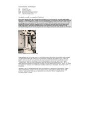
threads interwoven in the primary coil (fig 1). In addition, the coils have a nitinol<br />
inner core inten<strong>de</strong>d to make the coil resistant to stretch and compaction.<br />
23
New <strong>de</strong>velopments in imaging and treatment of intracranial aneurysms<br />
Fig1 Schematic representation of complex shaped Nexus coil with interwoven PGLA<br />
microfilament threads.<br />
Patients<br />
Between May 15, 2006 and May 5, 2007, 101 aneurysms in 95 patients were<br />
treated with Nexus coils. Patients were not consecutive; in the same period<br />
another 23 aneurysms were treated with other types of coils for the following<br />
reasons: in the beginning and ending of the study period stock of Nexus coils<br />
was incomplete (12 aneurysms), operator preference for (thicker) GDC 18 coils<br />
in aneurysms of 10-20 mm (7 aneurysms), and conversion from Nexus coils to<br />
other types of coils (4 aneurysms). There were 39 men and 56 women with a<br />
mean age of 53.6 years (median 53, range 14-89 years). Of 101 aneurysms, 77<br />
24
Results of 101 aneurysms treated with pgla microfilament nexus coils<br />
compared with historical controls treated with standard coils<br />
had ruptured and 24 had not. Of 24 unruptured aneurysms, 12 were inci<strong>de</strong>ntally<br />
discovered, 8 were additional to another ruptured aneurysm and 4 presented<br />
with symptoms of mass effect. Of 77 patients with a ruptured aneurysm, Hunt<br />
and Hess (HH) grading at the time of treatment was HH I-II in 41, HH III in 16, HH<br />
IV-V in 20 patients. Timing of treatment after SAH was 0-3 days in 55 patients,<br />
4-7 days in 13 patients and >7 days in 9 patients.<br />
Location of 101 aneurysms was anterior communicating artery in 34, middle<br />
cerebral artery in 20, posterior communicating artery in 15, ophthalmic artery in<br />
10, basilar tip in 7, posterior inferior cerebellar artery in 5, pericallosal artery in 4,<br />
anterior choreoi<strong>de</strong>al artery in 2, superior cerebellar artery in 2, posterior cerebral<br />
artery in 1 and vertebral artery in 1. Mean aneurysm size was 6.6 mm (median 6,<br />
range 2-16 mm).<br />
Coiling procedure and complication registration<br />
Coiling of aneurysms was performed on a biplane angiographic unit (Integris BN<br />
3000, Philips Medical Systems, Best, The Netherlands) with the patient un<strong>de</strong>r<br />
general anaesthesia. The aim of coiling was to obtain a <strong>de</strong>nse packing of the<br />
aneurysm, until not one coil could be placed. After location of the aneurysm<br />
on 2D angiography, 3D Rotational Angiography was performed of the vessel<br />
harbouring the aneurysm. From 3D images, coil projection was assessed and<br />
measurements of aneurysm diameter and aneurysm volume were performed.<br />
Used types of coils, number and lengths of coils and total volume of inserted<br />
coils was assessed for every aneurysm and packing, <strong>de</strong>fined as coil volume /<br />
aneurysm volume x 100% was calculated. Coil volume was calculated with<br />
a spreadsheet provi<strong>de</strong>d by the manufacturer containing volumes per cm of<br />
every type of coil. Angiographic occlusion was dichotomized in (near) complete<br />
occlusion (90-100%) or incomplete occlusion (
New <strong>de</strong>velopments in imaging and treatment of intracranial aneurysms<br />
Patients with unruptured aneurysms treated with stent assisted coiling were<br />
preloa<strong>de</strong>d with Clopidogrel 75 mg and Aspirin 80 mg and this was continued<br />
for 3-6 months after the procedure. Patients with acutely ruptured aneurysms<br />
treated with stent assisted coiling received intravenous Aspirin 500 mg before<br />
placement of the stent. In all patients treated with stents, response to antiplatelet<br />
medication was tested with VerifyNow P2Y12 Assay (Accumetrics, San Diego,<br />
CA) before stent placement.<br />
When thrombus formation on the coil mesh was angiographically visible or when<br />
coil loops protru<strong>de</strong>d outsi<strong>de</strong> the aneurysm in the parent artery, intravenous<br />
infusion of a glycoprotein IIb/IIIa antagonist (tirofiban, Aggrastat, Merck & Co.,<br />
Inc., Whitehouse Station, NJ), was started for 24-48 hours duration.<br />
Clinical and angiographic follow up<br />
Patients who survived the hospital admission period were scheduled for a<br />
follow up visit at 6 weeks and for angiographic follow up at 6 months. Results<br />
of angiographic follow up were classified in the same way as initial angiographic<br />
occlusion. Need for additional treatment was assessed in a weekly meeting with<br />
neuroradiologists, neurologists and neurosurgeons. Outcome according to the<br />
Glasgow Outcome Scale (GOS) was assessed at 6 months.<br />
Comparison with historical data of bare platinum coils<br />
Results of coiling of 101 aneurysms with Nexus coils were compared with historical<br />
results of coiling of 120 aneurysms with GDC 10 coils and 115 aneurysms with<br />
Cordis TruFill coils using i<strong>de</strong>ntical methodology (7) .<br />
Mean aneurysm volume, packing, incomplete aneurysm occlusion at 6 months<br />
follow up angiography and retreatment rates for Nexus coils were compared<br />
with both GDC 10 coils and Cordis TruFill coils. Differences were statistically<br />
analyzed using the unpaired t test for comparison of means and Chi-square test<br />
for comparison of proportions. P values < 0.05 were consi<strong>de</strong>red significant.<br />
26
Results of 101 aneurysms treated with pgla microfilament nexus coils<br />
compared with historical controls treated with standard coils<br />
RESULTS<br />
Initial angiographic results and packing<br />
Initial occlusion was (near) complete in 97 aneurysms and incomplete in 4<br />
aneurysms. Mean aneurysm volume was 180.2 mm 3 (median 71, range 5-1624<br />
mm 3 ). Mean packing was 19.4% (median 18.3%, range 7.5-38.9%). Of 95<br />
patients, 58 (61%) were prescribed anticoagulation after the procedure.<br />
In 101 aneurysms, 426 coils were used (mean 4.2, median 3, range 1-14). The<br />
426 coils had a total length of 5,776 cm and total coil volume was 3,088 mm3. Of<br />
5,776 cm total length of coils, 2,775 cm (48%) was from helical coils, 2,372 cm<br />
(41%) was from complex Morpheus coils and 629 cm (11%) was from complex<br />
Tetris coils. Inserted coil length per mm 3 aneurysm volume was 0.32 cm (total<br />
coil length 5,776 / total aneurysm volume 18,201).<br />
Clinical complications<br />
There were no complications leading to transient or permanent morbidity or<br />
mortality (0 in 95 patients, 0%, 97.5% CI 0.0-3.3%).<br />
Technical complications and conversions<br />
Coil loops protruding from the aneurysm in the parent vessel occurred in 11<br />
patients, in two patients with angiographic visible thrombus formation on the<br />
coils. Aggrastat infusion was administered in 7 of 11 patients. In all cases,<br />
malposition of coil loops occurred after correct placement of the first coil and<br />
was caused by insertion of additional coils that displaced loops of previously<br />
inserted coils (figs 2 and 3).<br />
27
New <strong>de</strong>velopments in imaging and treatment of intracranial aneurysms<br />
Fig 2 50-year-old woman with an inci<strong>de</strong>ntally discovered unruptured ophthalmic aneurysm.<br />
A: Lateral internal carotid angiogram <strong>de</strong>monstrates 9 mm ophthalmic aneurysm.<br />
B: After insertion of three 8x30 Nexus Morpheus coils an a<strong>de</strong>quate basket is formed.<br />
C: After failed attempt to <strong>de</strong>liver and withdrawal of a 7x30 Nexus Morpheus coil: coil loops<br />
of previous coils protruding in the parent artery (arrow).<br />
D: Final result with coil loops in carotid artery. The patient received Aggrastat infusion for 48<br />
hours. Good clinical outcome.<br />
28
Results of 101 aneurysms treated with pgla microfilament nexus coils<br />
compared with historical controls treated with standard coils<br />
Fig 3 62-year-old man with gra<strong>de</strong> III SAH from anterior communicating artery aneurysm.<br />
A: Internal carotid angiogram shows bilobated 5 mm anterior communicating artery<br />
aneurysm.<br />
B: First coil (5x15 Morpheus) forms a<strong>de</strong>quate basket.<br />
C: during insertion of the second coil (2x8mm helical), sud<strong>de</strong>nly a coil loop protru<strong>de</strong>d in the<br />
parent artery (arrow). After withdrawal of this coil in the micro catheter, the protruding<br />
loop persisted proving this was a displaced loop of the first coil.<br />
D: final result with persisting protrusion of the coil loop (arrow). Aggrastat infusion was<br />
started. Good clinical outcome.<br />
29
New <strong>de</strong>velopments in imaging and treatment of intracranial aneurysms<br />
In four aneurysms inten<strong>de</strong>d to be treated with Nexus coils, this proved impossible<br />
and these aneurysms were treated with other types of coils. In two wi<strong>de</strong> necked<br />
aneurysms (one 7 mm middle cerebral artery aneurysm and one 10 mm anterior<br />
communicating aneurysm) treated with balloon assisted coiling, the first inserted<br />
Nexus coil did not retain its shape after insertion with balloon assistance but<br />
retook its original shape after <strong>de</strong>flating the balloon resulting in protrusion of<br />
loops in the parent artery (fig. 4). Both aneurysms were treated with GDC 18<br />
coils. In one 2 mm anterior communicating artery aneurysm, a 2 mm helical<br />
Nexus coil could not be <strong>de</strong>livered and this aneurysm was treated with a 2 mm<br />
GDC 10 Ultrasoft coil. Finally, first inserted 10 mm Nexus Morpheus coil in a 12<br />
mm middle cerebral artery aneurysm could not be placed satisfactory and this<br />
aneurysm was treated with GDC 18 coils.<br />
30
Results of 101 aneurysms treated with pgla microfilament nexus coils<br />
compared with historical controls treated with standard coils<br />
Fig 4 57-year-old woman with gra<strong>de</strong> II SAH from a middle cerebral artery aneurysm.<br />
A: internal carotid angiogram shows wi<strong>de</strong> necked 7 mm middle cerebral artery aneurysm.<br />
B: A<strong>de</strong>quate placement of the first coil (7x21 Morpheus) with assistance of Hyperform 7 mm<br />
balloon (EV3, Irvine, CA).<br />
C: after <strong>de</strong>flation of the balloon expansion of the coil with protrusion into the parent artery.<br />
This coil was withdrawn.<br />
D: Final result after balloon assisted treatment with GDC 18 coils (Boston Scientific, Fremont,<br />
CA).<br />
Clinical follow up<br />
Clinical follow up at 6 months was available for all patients. Of 77 patients with ruptured<br />
aneurysms, 7 died in the hospital from initial impact of SAH or vasospasm (GOS 1).<br />
Two patients were in a nursing home (GOS 3), 4 patients had non disabling neurological<br />
<strong>de</strong>ficits (GOS 4) as a result of vasospasm and 64 patients had good outcomes (GOS<br />
5). All 18 patients with unruptured aneurysms were neurologically intact.<br />
31
New <strong>de</strong>velopments in imaging and treatment of intracranial aneurysms<br />
Angiographic follow up<br />
Angiographic follow up at 6 months was available for 82 patients with 87 aneurysms<br />
(86%). Thirteen patients (with 14 aneurysms) had no follow up angiography for<br />
the following reasons: <strong>de</strong>ath after SAH in 7, refusal in 5 and advanced age (78<br />
years) in 1 patient. Occlusion status for 87 aneurysms at 6 months was (near)<br />
complete in 73 (84%) and incomplete in 14 (16%) aneurysms. Of 14 incompletely<br />
occlu<strong>de</strong>d aneurysms, 12 were additionally coiled. In 2 aneurysms, both for 80%<br />
occlu<strong>de</strong>d, further follow up is scheduled. Overall retreatment rate was 12% (12<br />
of 101) and retreatment rate for aneurysms with angiographic follow up was<br />
14% (12 of 87). Additional coilings were without complications.<br />
Comparison with historical data of bare platinum coils<br />
Results of comparison of Nexus coated coils with GDC 10 coils and with Cordis<br />
TruFill coils are displayed in Table. Mean packing of 19.2% of Nexus coils was<br />
significantly lower than 22.9% of GDC 10 coils (P
Results of 101 aneurysms treated with pgla microfilament nexus coils<br />
compared with historical controls treated with standard coils<br />
DISCUSSION<br />
In this study, we found that results of treating aneurysms with PGLA microfilament<br />
Nexus coils are similar to those of two types of standard bare platinum coils.<br />
In other words, a beneficial effect of PGLA addition on stability at follow up<br />
could not be <strong>de</strong>monstrated. Nexus coils were safe to use: no neurological<br />
complications occurred in 95 patients with 101 aneurysms. The significantly<br />
lower packing of Nexus coils compared to both GDC 10 coils and Cordis TruFill<br />
coils had no significant effect on reopening and retreatment rates in this relatively<br />
small aneurysm groups, although there was a trend to lower retreatment rate for<br />
Cordis Trufill coils. Technical complications occurred rather frequently, in our<br />
opinion predominantly related to the inner nitinol core of the coil, inten<strong>de</strong>d to<br />
make the coil resistant to compaction and stretch. Nitinol has the propensity<br />
to regain its original shape after being forced into a different shape. In clinical<br />
practise, this physical property of the coil resulted in displacement of loops of<br />
already inserted coils during placement of additional coils resulting in coil loops<br />
protruding in the parent artery (figs 2 and 3). In our experience, we have not<br />
encountered this phenomenon with the use of other types of (bare platinum) coils.<br />
In patients with coil loops protruding outsi<strong>de</strong> the aneurysm in the parent artery,<br />
we administered an intravenous infusion of a glycoprotein IIb/IIIa antagonist<br />
for 24-48 hours, since Nexus coils have additional PGLA microfilaments with<br />
possible increased thrombogenicity. With this protocol, no permanent thromboembolic<br />
complications occurred.<br />
Another disadvantage of the physical property of the nitinol core is the dire<br />
performance in balloon assisted treatment. The first inserted Nexus coil usually<br />
did not retain its shape after insertion with balloon assistance but retook its<br />
original shape after <strong>de</strong>flating the balloon resulting in protrusion of loops in the<br />
parent artery, thereby neutralizing the inten<strong>de</strong>d effect of balloon assistance<br />
(fig.4). In these cases, treatment was continued with other types of coils with<br />
good results.<br />
The coil manufacturer (EV3, Irvine, CA) has recently introduced a new range<br />
of coils of several thicknesses, from 0.0115-0.0145 inch (Axium) <strong>de</strong>pendant<br />
on the loop diameter. This new coil is inten<strong>de</strong>d to replace the current available<br />
Nexus range. The new Axium coil has no nitinol core and will be available as bare<br />
platinum coil and as microfilament coil with either PGLA or nylon.<br />
In general, the results of this study are comparable to studies with other types<br />
coils with PGLA addition such as Matrix and Cerecyte coils (8-18) . As in our study,<br />
a beneficial effect of PGLA in terms of better stability at follow up was absent or<br />
not significant in previous studies (8-18) .<br />
Should we continue to use these PGLA coils to treat intracranial aneurysms?<br />
In our opinion (19-21) , supported by others (22-24) , the answer to this question is a<br />
clearly no for the following reasons. First and most important, clinical results<br />
of coils with PGLA addition are not better than those of bare platinum coils.<br />
Second, in some PGLA coils (Matrix), coating modifies physical coil properties,<br />
negatively affecting ease of handling. Third, PGLA coils are more expensive.<br />
33
New <strong>de</strong>velopments in imaging and treatment of intracranial aneurysms<br />
Large prospective randomized trials, instigated by coil manufacturers (24) ,<br />
comparing performance of PGLA coils and standard coils are not warranted,<br />
based on current available data.<br />
CONCLUSION<br />
PGLA microfilament Nexus coils are safe to use with results comparable to<br />
those of standard platinum coils. Handling properties are not optimal. This study<br />
gives further evi<strong>de</strong>nce of lack of beneficial effect of PGLA addition to reduce<br />
recurrence rate.<br />
34
Results of 101 aneurysms treated with pgla microfilament nexus coils<br />
compared with historical controls treated with standard coils<br />
REFERENCES<br />
1. Molyneux A, Kerr R, Stratton I, San<strong>de</strong>rcock P, Clarke M, Shrimpton J, Holman R; International<br />
Subarachnoid Aneurysm Trial (ISAT) Collaborative Group: International Subarachnoid Aneurysm<br />
Trial (ISAT) of neurosurgical clipping versus endovascular coiling in 2143 patients with ruptured<br />
intracranial aneurysms: A randomised trial. Lancet 2002;360:1267–1274<br />
2. Slob MJ, van Rooij WJ, Sluzewski M: The relation between packing and reopening in coiled<br />
intracranial aneurysms: a prospective study. Neuroradiology 2005;47:942-5<br />
3. Sluzewski M, van Rooij WJ, Slob MJ, Bescos JO, Slump CH, Wijnalda D: Relation between<br />
aneurysm volume, packing, and compaction in 145 cerebral aneurysms treated with coils.<br />
Radiology 2004;231:653-8<br />
4. van Rooij WJ, Sprengers ME, Sluzewski M, Beute GN: Intracranial aneurysms that repeatedly<br />
reopen over time after coiling: imaging characteristics and treatment outcome. Neuroradiology<br />
2007;49:343-9<br />
5. Murayama Y, Tateshima S, Gonzalez NR, et al.: Matrix and bioabsorbable polymeric coils accelerate<br />
healing of intracranial aneurysms: long-term experimental study. Stroke 2003;34:2031–37<br />
6. Matrix Newsletter 2004, Boston Scientific, Fremont, CA<br />
7. Slob MJ, van Rooij WJ, Sluzewski M: Influence of coil thickness on packing, re-opening and<br />
retreatment of intracranial aneurysms: a comparative study between two types of coils. Neurol<br />
Res 2005;27 Suppl 1:S116-9<br />
8. Taschner CA, Leclerc X, Rachdi H, et al.: Matrix <strong>de</strong>tachable coils for the endovascular treatment<br />
of intracranial aneurysms: analysis of early angiographic and clinical outcomes. Stroke<br />
2005;36:2176–80<br />
9. Linfante I, Akkawi NM, Perlow A, et al.: Polyglycoli<strong>de</strong>/polylacti<strong>de</strong>-coated platinum coils for<br />
patients with ruptured and unruptured cerebral aneurysms: a single-center experience. Stroke<br />
2005;36:1948–53<br />
10. Katsaridis V, Papagiannaki C, Violaris C: Guglielmi Detachable Coils versus Matrix coils:<br />
A comparison of the immediate posttreatment results of the embolization of 364 cerebral<br />
aneurysms in 307 patients: A single-center, single-surgeon experience. AJNR Am J Neuroradiol<br />
2006;27:1841-1848<br />
11. Rivet DJ, Moran CJ, Mazumdar A, Pilgram TK, Der<strong>de</strong>yn CP, Cross DT: Single-institution<br />
experience with Matrix coils in the treatment of intracranial aneurysms: comparison with samecenter<br />
outcomes with the use of platinum coils. AJNR Am J Neuroradiol 2007;28:1736-1742<br />
12. Fiorella D, Albuquerque FC, McDougall CG: Durability of aneurysm embolization with Matrix<br />
<strong>de</strong>tachable coils. Neurosurgery 2006;58:51–9<br />
13. Niimi Y, Song J, Madrid M, et al.: Endosaccular treatment of intracranial aneurysms using Matrix<br />
coils: early experience and midterm follow-up. Stroke 2006;37:1028–32<br />
14. Kang HS, Han MH, Kwon BJ, et al.: Short-term outcome of intracranial aneurysms treated with<br />
polyglycolicacid/lacti<strong>de</strong> copolymer-coated coils compared to historical controls treated with<br />
bare platinum coils: a single-center experience. AJNR Am J Neuroradiol 2005;26:1921–8<br />
15. Pierot L, Bonafé A, Bracard S, Leclerc X for the French Matrix Registry Investigators: Endovascular<br />
treatment of intracranial aneurysms with Matrix Detachable Coils: Immediate posttreatment<br />
results from a prospective multicenter registry. AJNR Am J Neuroradiol 2006;27:1693-1699<br />
35
New <strong>de</strong>velopments in imaging and treatment of intracranial aneurysms<br />
16. Pierot L, Bonafé A, Bracard S, Leclerc X for the French Matrix Registry Investigators: Endovascular<br />
Treatment of Intracranial Aneurysms with Matrix Detachable Coils: Midterm Anatomic Follow-Up<br />
from a Prospective Multicenter Registry. AJNR Am J Neuroradiol 2007 Oct 5; [Epub ahead of<br />
print]<br />
17. Bendszus M, Solymosi L: Cerecyte coils in the treatment of intracranial aneurysms: A preliminary<br />
clinical study. AJNR Am J Neuroradiol 2006;27:2053-2057<br />
18. Butteriss D, Gholkar A, Mitra D, Birchall D, Jayakrishnan V: Single-center experience of Cerecyte<br />
coils in the treatment of intracranial aneurysms: Initial experience and early follow-up results.<br />
AJNR Am J Neuroradiol 2007 Oct 5.<br />
19. Sluzewski M, van Rooij WJ: Questionable interpretation of results of ACTIVE study on Matrix<br />
coils by Boston Scientific [letter]. AJNR Am J Neuroradiol 2005;26:1882–3<br />
20. Sluzewski M, van Rooij WJ: Reply to letter regarding interpretation of results of ACTIVE Study.<br />
AJNR Am J Neuroradiol 2005;26:2436-7<br />
21. van Rooij WJ, Sluzewski M: Registry on Matrix coils: bias in inclusion, exclusion, and publication.<br />
AJNR Am J Neuroradiol 2007;28:398-9<br />
22. Cloft HJ: Have you been smoking something that is biologically active? AJNR Am J Neuroradiol<br />
2006;27:240-2<br />
23. Cloft HJ: Matrix reloa<strong>de</strong>d. AJNR Am J Neuroradiol 2007 Oct 9.<br />
24. Kallmes DF, Cloft HJ: Ready or not, here they come: randomized trials evaluating new<br />
endovascular aneurysm therapies. AJNR Am J Neuroradiol 2007;28:799-803<br />
36
Coiling of truly inci<strong>de</strong>ntal<br />
intracranial aneurysms<br />
Chapter 4<br />
Coiling of truly inci<strong>de</strong>ntal<br />
intracranial aneurysms<br />
A.N. <strong>de</strong> <strong>Gast</strong> M.D. 1) , W.J. van Rooij M.D., Ph.D. 1) , M. Sluzewski M.D., Ph.D. 1) ,<br />
P.C. Nijssen M.D. 2) , G.N. Beute M.D. 3)<br />
1)<br />
Department of Radiology, St. Elisabeth Ziekenhuis, Tilburg, The Netherlands<br />
2)<br />
Department of Neurology, St. Elisabeth Ziekenhuis, Tilburg, The Netherlands<br />
3)<br />
Department of Neurosurgery, St. Elisabeth Ziekenhuis, Tilburg, The Netherlands<br />
AJNR Am J Neuroradiol 2006; 27: 293-296<br />
37
New <strong>de</strong>velopments in imaging and treatment of intracranial aneurysms<br />
ABSTRACT<br />
BACKGROUND AND PURPOSE<br />
The purpose of this study is to report the morbidity, mortality, and angiographic<br />
results of coiling of asymptomatic inci<strong>de</strong>ntal aneurysms and compare the<br />
characteristics of these aneurysms with other asymptomatic inci<strong>de</strong>ntal<br />
aneurysms that were not treated.<br />
PATIENTS AND METHODS<br />
During a 10-year period, 97 patients without previous subarachnoid hemorrhage,<br />
presented with inci<strong>de</strong>ntally found intracranial aneurysms. In 48 patients, 58<br />
aneurysms were coiled. The mean size of the 58 coiled inci<strong>de</strong>ntal aneurysms was<br />
10.9 mm (median, 9 mm; range, 3–40 mm). Twenty-six of 58 coiled aneurysms<br />
(44.8%) were 6 10 mm.<br />
RESULTS<br />
Permanent morbidity of coiling was 2.1% (1 of 48), mortality was 0%. Compared<br />
with untreated patients with inci<strong>de</strong>ntal aneurysms, coiled patients were younger<br />
and more often had multiple aneurysms. Aneurysms of coiled patients more often<br />
had a small neck, were more often located on the carotid artery, and were less<br />
often located on the middle cerebral artery. Of 46 aneurysms with angiographic<br />
follow up, 45 were completely or near completely occlu<strong>de</strong>d. To obtain these<br />
results, 3 aneurysms were coiled more than once. Coiled inci<strong>de</strong>ntal aneurysms<br />
did not rupture during a median follow-up period of 28.5 months. Mean hospital<br />
stay per patient was 2.5 days.<br />
CONCLUSION<br />
Coiling of inci<strong>de</strong>ntal intracranial aneurysms has a low complication rate in<br />
selected aneurysms and patients. Coiling should be the first treatment option in<br />
inci<strong>de</strong>ntal aneurysms suitable for this technique.<br />
38
Coiling of truly inci<strong>de</strong>ntal<br />
intracranial aneurysms<br />
INTRODUCTION<br />
The wi<strong>de</strong>spread use of imaging of the brain with CT scanning and MR imaging<br />
enhaces the chance of discovering an intracranial aneurysm unrelated to the<br />
presenting symptoms of the patient. Bleeding from an intracranial aneurysm may<br />
have <strong>de</strong>vastating consequences, but the chance of rupture of an inci<strong>de</strong>ntally<br />
discovered aneurysm is generaly low and is mainly <strong>de</strong>pen<strong>de</strong>nt on size and<br />
location. In making the <strong>de</strong>cision whether to treat an unruptured aneurysm, many<br />
factors have to be consi<strong>de</strong>red: location, size, and morphology of the aneurysm,<br />
patient age and life expectancy, and the skills and experience of the treating<br />
physician.<br />
Unruptured aneurysms can be classified as symptomatic (by mass effect) or<br />
asymptomatic. Most asymptomatic unruptured aneurysms are additional<br />
aneurysms discovered in the diagnostic work-up of another ruptured aneurysm.<br />
Truly inci<strong>de</strong>ntal aneurysms form a specific subset of unruptured aneurysms with<br />
a lower chance of rupture than additional or symptomatic unruptured aneurysms<br />
(1-3)<br />
. Most reports about endovascular treatment of unruptured aneurysms contain<br />
data about all 3 types of unruptured aneurysms (4-7) . In this study, we report our<br />
experience during 10 years with coiling of 58 inci<strong>de</strong>ntal aneurysms in 48 patients<br />
and compare patient and aneurysm characteristics with 53 inci<strong>de</strong>ntal aneurysms<br />
in 41 patients who were not treated during the same period.<br />
PATIENTS AND METHODS<br />
General<br />
Between January 1995 and April 2005, 97 patients presented with inci<strong>de</strong>ntally<br />
found aneurysms. All patients were discussed in a multidisciplinary working<br />
group consisting of 2 neurosurgeons, 2 neurologists, and 2 neuroradiologists.<br />
Asymptomatic aneurysms, diagnosed on imaging studies performed for<br />
symptoms unrelated to the presence of the aneurysm in patients without a<br />
history of subarachnoid hemorrhage, were classified as inci<strong>de</strong>ntal. Imaging<br />
studies inclu<strong>de</strong>d CT scanning, MR imaging, and angiography performed for<br />
a variety of symptoms: headache, transient ischemic attacks, carotid artery<br />
stenosis, seizures, multiple sclerosis, tinnitus, dizziness, collapse, meningeoma,<br />
memory disturbances, suspected metastases, etc. In patients with an inci<strong>de</strong>ntal<br />
aneurysm diagnosed on CT or MR imaging and for whom treatment was<br />
consi<strong>de</strong>red, a complete angiogram was performed; in the past 4 years, this<br />
inclu<strong>de</strong>d a 3D rotational angiogram. After completion of the angiographic imaging,<br />
the possibility of and need for treatment were discussed in the working group<br />
and the consensus opinion was discussed with the patient in the outpatient<br />
clinic. In general, coiling was the first treatment option for all aneurysms in the<br />
posterior circulation and for aneurysms with a well-<strong>de</strong>fined neck in the anterior<br />
circulation. Surgery was consi<strong>de</strong>red in aneurysms not suitable for coiling or<br />
endovascular parent vessel occlusion, generally wi<strong>de</strong>-necked aneurysms in the<br />
anterior circulation. The working group tried to balance the benefit and risk<br />
39
New <strong>de</strong>velopments in imaging and treatment of intracranial aneurysms<br />
of treatment against the presumed annual risk of bleeding from the aneurysm.<br />
Multiple patient and aneurysm factors were taken into consi<strong>de</strong>ration: patient age,<br />
general medical condition, comorbidity and wish for treatment, aneurysm size,<br />
location, anatomy, and presence of intraluminal thrombus or calcifications.<br />
Patients<br />
Of the 97 patients presenting with an inci<strong>de</strong>ntal aneurysm, 56 (57.1%) were<br />
treated, 48 with coil embolization, 7 with surgical clipping, and one with parent<br />
vessel balloon occlusion. The 48 patients with inci<strong>de</strong>ntal aneurysms that were<br />
coiled are the subject of the present study. There were 38 women and 10 men<br />
with a mean age of 53.6 years (median age, 52 years; range, 26–75 years). Of<br />
the 48 patients, 19 (39.6%) had multiple inci<strong>de</strong>ntal aneurysms for a total of 85<br />
aneurysms: 8 patients had 2 aneurysms, 8 patients had 3 aneurysms, 2 patients<br />
had 4 aneurysms, and one patient had 8 aneurysms. Of these 85 aneurysms<br />
in 48 patients, 66 were treated, 58 with coil embolization and 8 with surgical<br />
clipping: 8 patients had 2 treated inci<strong>de</strong>ntal aneurysms, 2 patients had 3 treated<br />
aneurysms, and one patient had 7 treated aneurysms. In 6 patients, 2 inci<strong>de</strong>ntal<br />
aneurysms were coiled in the same session, and in one patient 4 aneurysms<br />
were coiled in the same session. The mean size of the 58 coiled inci<strong>de</strong>ntal<br />
aneurysms was 10.9 mm (median, 9 mm; range, 3–40 mm). Twenty-six of 58<br />
coiled aneurysms (44.8%) were 6 10 mm. Locations were the ophthalmic artery<br />
(15)<br />
, basilar tip (14) , posterior communicating artery (8), anterior communicating<br />
artery (6), middle cerebral artery (5), carotid tip (4), posterior cerebral artery (2),<br />
posterior inferior cerebellar artery (2), superior cerebellar artery (one), and basilar<br />
trunk (one). During the same period, 184 other unruptured aneurysms were<br />
treated by endovascular means in our hospital: 97 were additional to another<br />
ruptured aneurysm, and 87 presented with symptoms of mass effect. Thus,<br />
the proportion of inci<strong>de</strong>ntal aneurysms was 24% (58 of 242) of endovascularly<br />
treated unruptured aneurysms.<br />
Coiling Procedure<br />
Coiling of aneurysms was performed with the patient un<strong>de</strong>r general anesthesia<br />
and systemic heparinization. Heparin was continued intravenously or<br />
subcutaneously for 48 hours after the procedure, followed by low-dose aspirin for<br />
3 months orally. Coiling was performed with Guglielmi <strong>de</strong>tachable coils (Boston<br />
Scientific, Fremont, Calif) or TruFill DCS coils (Cordis, Miami, Fla). Some large<br />
aneurysms were coiled with very long mechanically <strong>de</strong>tachable coils (Detach 18;<br />
Cook Inc., Copenhagen, Denmark). The aim of coiling was to pack the aneurysm<br />
as <strong>de</strong>nsely as possible until not a single additional coil could be placed. Wi<strong>de</strong>necked<br />
aneurysms were coiled with a temporary supporting balloon (Sentry;<br />
Boston Scientific) or after placement of a permanent supporting <strong>de</strong>vice (TriSpan<br />
or Neuroform stent; Boston Scientific). Complications of coiling were recor<strong>de</strong>d.<br />
Initial angiographic results of coiling were classified as complete occlusion<br />
(98%–100%), near-complete occlusion (90%–98%), or incomplete occlusion<br />
(< 90%).<br />
40
Coiling of truly inci<strong>de</strong>ntal<br />
intracranial aneurysms<br />
Duration of Hospital Stay<br />
Patients with inci<strong>de</strong>ntal aneurysms scheduled for coiling or additional coiling were<br />
admitted in the neurosurgical ward in the morning, coiling was performed in the<br />
afternoon, and discharge was the next afternoon in uncomplicated procedures.<br />
Duration of hospital stay was recor<strong>de</strong>d in all patients.<br />
Follow-Up<br />
Patients with coiled inci<strong>de</strong>ntal aneurysms were scheduled for an outpatient clinic<br />
visit at 6 weeks and a follow-up angiogram after 6 months. Results of angiographic<br />
follow-up were classified in the same way as the initial postembolization occlusion.<br />
Incomplete occlusion at any point in time was consi<strong>de</strong>red an indication for further<br />
therapy, unless clinical or anatomic factors dictated otherwise. Results and<br />
consequences of angiographic follow-up were discussed in the working group.<br />
During the meeting, <strong>de</strong>cisions were ma<strong>de</strong> for the need for additional treatment<br />
or exten<strong>de</strong>d angiographic follow-up. Patients with aneurysms with complete<br />
or near-complete occlusion at 6-month follow-up angiography were generally<br />
discar<strong>de</strong>d from further angiographic follow-up. When additional treatment was<br />
performed, the result was evaluated in the weekly meeting and 6-month followup<br />
angiogram after additional treatment was scheduled.<br />
In April 2005, a telephone survey was done by one of us (A.d.G.) for all treated<br />
patients with an outpatient clinic visit more than 6 months earlier. Patients or their<br />
next of kin were asked about general health, hospital admissions, and episodic<br />
headaches that might be contributing to subarachnoid hemorrhage.<br />
Untreated Patients<br />
Of the 97 patients with inci<strong>de</strong>ntal aneurysms, 41 were not treated. The main<br />
reasons for not treating the 41 patients inclu<strong>de</strong>d unfavorable aneurysm anatomy<br />
(wi<strong>de</strong> neck) in 29, patient refusal in 3, near- complete thrombosis in 2, small<br />
aneurysm size (5 mm) in 2, advanced patient age in 2, failed surgical clipping<br />
in one, location in the cavernous sinus in one, and one 65-year-old man with a<br />
30-mm basilar trunk dissecting aneurysm discovered on MR imaging performed<br />
for transient ischemic attacks in the right carotid artery territory who died of<br />
subarachnoid hemorrhage shortly before scheduled treatment. There were 23<br />
women and 18 men, with a mean age of 60.6 years (median age, 63 years;<br />
range, 46–74 years). Of the 41 patients, 7 (17%) had multiple aneurysms, for<br />
a total of 53 aneurysms: 4 patients had 2 aneurysms and 3 patients had 3<br />
aneurysms. The mean size of the aneurysms was 9.2 mm (median, 8 mm; range,<br />
3–30 mm). The locations of 53 aneurysms were the middle cerebral artery (15),<br />
anterior communicating artery (9), basilar tip (9), cavernous sinus (5), posterior<br />
communicating artery (4), ophthalmic artery (3), superior cerebellar artery (2),<br />
carotid tip (2), posterior cerebral artery (one), basilar trunk (one), pericallosal<br />
artery (one), and vertebral artery (one).<br />
41
New <strong>de</strong>velopments in imaging and treatment of intracranial aneurysms<br />
Comparison of Treated and Untreated Patients<br />
For patients with coiled versus untreated inci<strong>de</strong>ntal aneurysms, the following<br />
patient and aneurysm characteristics were compared: mean patient age, mean<br />
aneurysm size, proportion of wi<strong>de</strong>-necked aneurysms, proportion of patients<br />
with multiple inci<strong>de</strong>ntal aneurysms, and proportion of aneurysms located on<br />
anterior cerebral artery, middle cerebral artery, carotid artery, and vertebrobasilar<br />
artery. Statistical analysis was performed by using a t test for comparison of<br />
means and a x 2 test for comparison of proportions (MedCalc statistical software,<br />
Mariakerke, Belgium).<br />
RESULTS<br />
Comparison of Treated and Untreated Patients<br />
In the Table, the characteristics of 48 patients with 58 inci<strong>de</strong>ntal aneurysms that<br />
were coiled and of 41 patients with 53 inci<strong>de</strong>ntal aneurysms that were not treated<br />
are summarized. Mean age of 53.7 years of 48 coiled patients was significantly<br />
lower than mean age of 60.6 years in 41 untreated patients. Mean aneurysm<br />
size did not differ for both groups. Eight of 58 coiled aneurysms (13.8%) had a<br />
wi<strong>de</strong> neck versus 29 of 53 (54.7%) untreated aneurysms. This difference was<br />
statistically significant. Of the 48 coiled patients, 19 had multiple inci<strong>de</strong>ntal<br />
aneurysms (39.6%) versus 7 of 41 untreated patients (17.1%). This difference<br />
was also statistically significant. Aneurysms located in the carotid artery were<br />
significantly more often coiled, whereas aneurysms located in the middle cerebral<br />
artery were—to a statistically significant <strong>de</strong>gree—more often left untreated.<br />
Characteristics of 48 patients with 58 inci<strong>de</strong>ntal aneurysms that were coiled and of 41 patients with 53<br />
inci<strong>de</strong>ntal aneurysms that were not treated.<br />
48 Patients with 58 41 Patients with 53<br />
Coiled Aneurysms Untreated Aneurysms P Value<br />
Mean age 53.6 y 60.6 y .0006<br />
(median 52, range 26-75) (median 63, range 46-74)<br />
Multiple aneurysms 19/48 (39.6%) 7/41 (17,1%) .035<br />
Mean aneurysms size 10.9 mm 9.2 mm .461<br />
(median 9, range 3-40) (median 8, range 3-30)<br />
Aneurysms with wi<strong>de</strong> neck 8/58 (13,8%) 29/53 (54,7%)
Coiling of truly inci<strong>de</strong>ntal<br />
intracranial aneurysms<br />
Duration of Hospital Stay<br />
Hospitalization was 2 days for 57 coiling procedures in 47 patients and 5 days<br />
for the one patient who suffered a thromboembolic complication. Mean hospital<br />
stay per patient, including additional coil treatments, was 2.5 days.<br />
Clinical Follow-Up<br />
All 48 coiled patients had clinical follow-up of a median 28.5 months (mean,<br />
35.1 months; range, 2-112 months), totaling 1688 months (140.6 patient-years).<br />
A 68-year-old man died of pneumonia 6 months after coiling of a basilar tip<br />
aneurysm. A 70-year-old woman had mild aphasia after clipping of an additional<br />
inci<strong>de</strong>ntal 15 mm left middle cerebral artery aneurysm. All other patients were<br />
alive and neurologically intact at the time of follow-up, and there were no hospital<br />
admissions or episo<strong>de</strong>s of headache that could be attributed to subarachnoid<br />
hemorrhage.<br />
Angiographic Results<br />
Of the 58 aneurysms treated in 48 patients, 6 wi<strong>de</strong>-necked aneurysms were<br />
coiled with the aid of a temporary supporting balloon and one with the aid of a<br />
stent. One 14 mm basilar tip aneurysm was initially coiled with the aid of a TriSpan<br />
supporting <strong>de</strong>vice and 6 months later was additionally coiled after placement of<br />
a stent. Initial postembolization occlusion in 58 coiled aneurysms was complete<br />
occlusion in 49, near-complete occlusion in 6, and incomplete occlusion in 3<br />
aneurysms. Two of the 3 incompletely occlu<strong>de</strong>d aneurysms were additionally<br />
coiled, one 16 mm basilar tip aneurysm was additionally coiled with the aid of a<br />
stent, and one 26-mm partially thrombosed basilar tip aneurysm was additionally<br />
coiled 5 times during a 3-year period. One 10-mm ophthalmic aneurysm that was<br />
80% occlu<strong>de</strong>d remained stable on follow-up and was not additionally treated.<br />
Follow-up angiography at 6 months was performed in 42 patients with 46 coiled<br />
aneurysms (6 patients with 12 coiled aneurysms are scheduled for follow-up).<br />
One initial completely occlu<strong>de</strong>d 30 mm basilar tip aneurysm showed reopening<br />
and was additionally coiled for 4 times during a 2-year period to obtain a stable,<br />
almost-complete occlusion.<br />
DISCUSSION<br />
In this study we found that coiling of inci<strong>de</strong>ntal aneurysms has a low complication<br />
rate in selected patients and aneurysms. Selection was biased toward younger<br />
patients and aneurysms with well-<strong>de</strong>fined necks. Aneurysms located on<br />
the carotid artery were treated significantly more often, and aneurysms on<br />
the middle cerebral artery were more often not treated. This may reflect the<br />
difference in morphology of aneurysms located at these sites: carotid artery<br />
aneurysms are usually si<strong>de</strong>wall aneurysms, whereas most middle cerebral artery<br />
aneurysms are located at bi-or trifurcations of arteries. Moreover, if carotid artery<br />
aneurysms are wi<strong>de</strong>-necked, coiling with balloon protection of the carotid artery<br />
is relatively straight forward, whereas balloon protection in the middle cerebral<br />
43
New <strong>de</strong>velopments in imaging and treatment of intracranial aneurysms<br />
artery aneurysms is hazardous. In the present study, none of coiled aneurysms<br />
ruptured during a median follow-up period of 28.5 months, but longer followup<br />
is necessary to establish the efficacy of coiling of inci<strong>de</strong>ntal aneurysms.<br />
Although selection was biased toward suitability of aneurysms for coiling, almost<br />
half of the coiled inci<strong>de</strong>ntal aneurysms were 6 10 mm and in 9 procedures<br />
coiling was possible only with assistance of balloon protection of the parent<br />
artery or placement of a stent or TriSpan <strong>de</strong>vice. Despite these more challenging<br />
treatments, the complication rate was low. The natural history of unruptured<br />
intracranial aneurysms is still unclear and is influenced by many factors, such as<br />
previous subarachnoid hemorrhage from another aneurysm, history of cigarette<br />
smoking, coexisting medical conditions, and aneurysm characteristics such as<br />
size, location, and morphology (1-3,8-11) . In the International Study of Unruptured<br />
Intracranial Aneurysms (ISUIA) (1) , 5-year cumulative rupture rates for patients<br />
who did not have a history of subarachnoid hemorrhage with aneurysms<br />
located in the internal carotid artery, anterior communicating artery, anterior<br />
cerebral artery, and middle cerebral artery were 0%, 2.6%, 14.5%, and 40% for<br />
aneurysms
Coiling of truly inci<strong>de</strong>ntal<br />
intracranial aneurysms<br />
REFERENCES<br />
1. Wiebers DO, Whisnant JP, Huston J 3rd, et al.: Unruptured intracranial aneurysms: natural history,<br />
clinical outcome, and risks of surgical and endovascular treatment. Lancet 2003;362:103–10<br />
2. The International Study of Unruptured Intracranial Aneurysms Investigators. Unruptured<br />
intracranial aneurysms: risk of rupture and risks of surgical intervention. N Engl J Med<br />
1998;339:1725–33<br />
3. Rinkel GJ, Djibuti M, van Gijn J: Prevalence and risk of rupture of intracranial aneurysms: a<br />
systematic review. Stroke 1998;29:251–56<br />
4. Wanke I, Doerfler A, Dietrich U, et al.: Endovascular treatment of unruptured intracranial<br />
aneurysms. AJNR Am J Neuroradiol 2002;23:756–61<br />
5. Johnston SC, Dudley RA, Gress DR, et al.: Surgical and endovascular treatment of unruptured<br />
cerebral aneurysms at university hospitals. Neurology1999;52:1799–805<br />
6. Johnston SC, Wilson C, Halbach V, et al.: Endovascular and surgical treatment of unruptured<br />
cerebral aneurysms: comparison of risks. Ann Neurol 2000;4:11–19<br />
7. Lanterna LA, Tredici G, Dimitrov BD, et al.: Treatment of unruptured cerebral aneurysms by<br />
embolization with Guglielmi <strong>de</strong>tachable coils: case-fatality, morbidity, and effectiveness in<br />
preventing bleeding: a systematic review of the literature. Neurosurgery 2004;55:767–75<br />
8. Juvela S: Recommendations for the management of patients with unruptured intracranial<br />
aneurysms. Stroke 2001;32:815<br />
9. Vindlacheruvu RR, Men<strong>de</strong>low AD, Mitchell P: Risk-benefit analysis of the treatment of unruptured<br />
intracranial aneurysms. J Neurol Neurosurg Psychiatry 2005;76:234–39<br />
10. Tsutsumi K, Ueki K, Morita A, et al.: Risk of rupture from inci<strong>de</strong>ntal cerebral aneurysms. J<br />
Neurosurg 2000;93:550–53<br />
11. Be<strong>de</strong>rson J, Awad I, Wiebers D, et al.: Recommendations forthe management of patients with<br />
unruptured intracranial aneurysms. Stroke 2000;31:2742–50<br />
12. Raaymakers T, Rinkel GJ, Limburg M, et al.: Mortality and morbidity of surgery for unruptured<br />
intracranial aneurysms. Stroke 1998;29:1531–38<br />
13. King JT, Berlin JA, Flamm ES: Morbidity and mortality from elective surgery for asymptomatic,<br />
unruptured, intracranial aneurysms: a meta-analysis. J Neurosurg 1994;81:837–42<br />
45
New <strong>de</strong>velopments in imaging and treatment of intracranial aneurysms<br />
46
Midterm clinical and magnetic resonance imaging follow-up of large<br />
and giant carotid artery aneurysms after therapeutic carotid artery occlusion<br />
Chapter 5<br />
Midterm clinical and magnetic<br />
resonance imaging follow-up of large<br />
and giant carotid artery aneurysms after<br />
therapeutic carotid artery occlusion<br />
A.N. <strong>de</strong> <strong>Gast</strong> M.D. 1) , M.E. Sprengers M.D. 2) , W.J. van Rooij M.D., Ph.D. 1) ,<br />
C. Lavini M.Sc., M. Phil. 2) , M. Sluzewski M.D., Ph.D. 1) , C.B. Majoie M.D., Ph.D. 2)<br />
1)<br />
Department of Radiology, St. Elisabeth Ziekenhuis, Tilburg, The Netherlands<br />
2)<br />
Department of Radiology, Aca<strong>de</strong>misch Medisch Centrum Amsterdam, The Netherlands<br />
Neurosurgery 2007; 60: 1025-1029<br />
47
New <strong>de</strong>velopments in imaging and treatment of intracranial aneurysms<br />
ABSTRACT<br />
OBJECTIVE<br />
The purpose of this study was to evaluate aneurysm size and clinical symptoms<br />
midterm after therapeutic carotid artery occlusion in 39 patients with large or<br />
giant carotid artery aneurysms.<br />
METHODS<br />
Between January 1996 and August 2004, 39 patients with large or giant carotid<br />
artery aneurysms were treated with therapeutic carotid artery occlusion and<br />
had clinical and magnetic resonance imaging follow-up of at least 3 months<br />
(mean, 35.9 mo; median, 29 mo; range, 3–107 mo; 117 patient-yr). Initial clinical<br />
presentation was mass effect caused by the aneurysm in 32 (82%) of the 39<br />
patients. Three patients presented<br />
with subarachnoid hemorrhage (SAH) and one presented with epistaxis; two<br />
aneurysms were an inci<strong>de</strong>ntal finding and one was additional to another ruptured<br />
aneurysm.<br />
RESULTS<br />
There were no early or late complications of therapeutic carotid artery occlusion.<br />
All aneurysms seemed to have thrombosed completely after carotid artery<br />
occlusion as observed on early and late magnetic resonance imaging (MRI) and<br />
magnetic resonance angiographic (MRA) follow-up studies. At the time of the<br />
most recent MRI follow-up study, 29 (74%) of the 39 aneurysms involuted totally,<br />
two aneurysms <strong>de</strong>creased to 25% of the original diameter, two aneurysms<br />
<strong>de</strong>creased to 50%, and five aneurysms <strong>de</strong>creased to 75%. Two aneurysms<br />
remained unchanged in size after 49 and 58 months, respectively. At the most<br />
recent clinical follow-up evaluation, symptoms of mass effect were cured in<br />
19 (60%), improved in 10 (31%), and remained unchanged in 3 (9%) of the 32<br />
patients.<br />
CONCLUSION<br />
Therapeutic carotid artery occlusion was a simple, safe, and effective treatment<br />
for large and giant carotid artery aneurysms. Almost all aneurysms involute<br />
completely or substantially <strong>de</strong>crease in size. Alleviation of symptoms of mass<br />
effect was achieved in most patients.<br />
KEY WORDS<br />
Aneurysm, Follow-up, Magnetic resonance imaging, Therapeutic carotid artery<br />
occlusion<br />
48
Midterm clinical and magnetic resonance imaging follow-up of large<br />
and giant carotid artery aneurysms after therapeutic carotid artery occlusion<br />
INTRODUCTION<br />
Large and giant carotid artery aneurysms are located extradurally in the cavernous<br />
sinus or intradurally in its supraclinoid segments. Clinical presentation is usually<br />
with symptoms of mass effect on one or more of the adjacent cranial nerves (CN)<br />
II–VI resulting in diplopia, ophthalmoparesis, <strong>de</strong>creased visual acuity, trigeminal<br />
neuralgia, or a combination thereof. Rupture may result in SAH, carotid cavernous<br />
fistulae, or epistaxis, <strong>de</strong>pending on aneurysm location.<br />
For large and giant aneurysms of the internal carotid artery, therapeutic carotid<br />
artery balloon occlusion is a simple, safe, and effective therapy in patients who<br />
can tolerate sacrifice of the carotid artery (6,13,16) . Tolerance to internal carotid<br />
artery occlusion can be evaluated reliably by angiographic test occlusion, even<br />
in patients un<strong>de</strong>r general anesthesia (1,8,14,15) . The aim of therapeutic carotid artery<br />
occlusion is thrombosis of the internal carotid artery, including the aneurysm.<br />
In this study, we evaluated aneurysm size and clinical symptoms midterm after<br />
therapeutic carotid artery occlusion in 39 patients with large or giant carotid<br />
artery aneurysms.<br />
PATIENTS AND METHODS<br />
Patients<br />
Between January 1996 and August 2004, 52 patients were treated with carotid<br />
artery balloon occlusion for large or giant carotid aneurysms. Technical and<br />
clinical aspects of carotid artery occlusion in these patients have been published<br />
in previous articles (14,15) . During the same period, another 26 patients with<br />
carotid artery aneurysms inten<strong>de</strong>d to be treated with carotid artery occlusion<br />
did not tolerate the test occlusion. Of these 26 patients, four with cavernous<br />
sinus aneurysms symptomatic only by abducens palsy were not treated at all.<br />
Seven patients had bypass surgery preceding carotid artery occlusion. Fifteen<br />
aneurysms were coiled, two of them after placement of a stent and six with the<br />
aid of a supporting balloon. In June 2005, a follow-up survey was completed<br />
by the 52 patients with aneurysms treated with carotid artery balloon occlusion<br />
without preceding bypass surgery. Eight patients died during the follow-up<br />
period: one patient died shortly after carotid occlusion of the initial SAH, one<br />
patient died of trauma, two el<strong>de</strong>rly patients died in the hospital from pneumonia<br />
and multiple organ failure, one patient died in the hospital as a result of chronic<br />
obstructive pulmonary disease, one patient died of lung cancer, one patient<br />
died in the hospital from cardiac infarction, and one patient died at home of<br />
old age. None of these patients died as a result of SAH. Of the 44 surviving<br />
patients, seven could not be located in June 2005 but had previous follow-up<br />
data. The remaining 37 patients were contacted and asked to complete a phone<br />
questionnaire about the evolution<br />
of their clinical symptoms after carotid artery occlusion and their current state<br />
of health. Follow-up MRI and MRA on a 3.0 Tesla system (Philips Intera RIO;<br />
Philips Medical Systems, Best, The Netherlands) were offered to all 37 patients<br />
49
New <strong>de</strong>velopments in imaging and treatment of intracranial aneurysms<br />
and were eventually performed in 26. Basic ophthalmological assessment was<br />
performed at the same time. Of the 11 patients who refused long-term MRI<br />
follow-up, the seven patients who could not be located, and the eight patients<br />
who had died, previous clinical and MRI follow-up data for 3 months or longer<br />
was available in the medical records of 13, allowing these patients to be inclu<strong>de</strong>d<br />
in the analysis. Thus, the final study group consisted of 39 patients with large or<br />
giant carotid artery aneurysms treated with therapeutic carotid artery occlusion<br />
and with clinical and MRI follow-up of at least 3 months (mean, 35.9 mo; median,<br />
29 mo; range, 3–107 mo; 117 patient yr). There were 32 women and 7 men with<br />
a mean age of 56 years (range, 26–78 yr) at the time of carotid artery occlusion.<br />
The location of the large and giant carotid artery aneurysms was the carotid<br />
cavernous sinus in 28 of the patients, the carotid hypophyseal segment in four,<br />
the carotid ophthalmic segment in three, the carotid supraclinoid segment in<br />
three, and the internal carotid bifurcation in one.<br />
The initial clinical presentation was mass effect caused by the aneurysm in 32<br />
(82%) of the 39 patients. Oculomotor dysfunction with intact visual acuity was<br />
present in 27 patients with large or giant cavernous sinus aneurysms, including<br />
isolated abducens palsy in five patients and ophthalmoparesis in 22 patients (in<br />
two patients with additional trigeminal neuralgia). Decreased visual acuity was<br />
the presenting symptom in four patients with carotid ophthalmic or hypophyseal<br />
aneurysms; one patient with an internal carotid bifurcation aneurysm presented<br />
with hemiparesis. Retro-orbital pain accompanied symptoms of mass effect in<br />
18 of the 32 patients. Three patients presented with SAH and were treated with<br />
carotid artery occlusion more than 3 weeks after SAH, two of them after initial<br />
coiling in the acute phase of SAH. One patient presented with epistaxis. Two<br />
aneurysms were an inci<strong>de</strong>ntal finding and one aneurysm was additional to another<br />
ruptured aneurysm. Seven (18%) of the 39 aneurysms contained intraluminal<br />
thrombus at the time of presentation. The presence of intraluminal thrombus<br />
was assessed from both cross-sectional imaging and angiography. This study<br />
was approved by the ethical committees of both participating hospitals.<br />
Initial and Follow-up MRI Protocol<br />
In all 39 patients, MRI was performed before carotid artery occlusion. MRI was<br />
repeated 2 to 5 days after carotid artery occlusion and again 3 months later.<br />
Exten<strong>de</strong>d MRI follow-up (>3 mo) was performed in 35 of the 39 patients. MRI was<br />
performed on a 0.5-, 1.0-, or 1.5-Tesla system (Philips T5, T10, or T15; Philips<br />
Medical Systems) and consisted of transverse and coronal T1 weighted images<br />
and transverse T2 weighted images. In 26 patients, midterm MRI scanning was<br />
performed on a 3.0-Tesla system.<br />
Initial and follow-up MRI studies were compared by two experienced<br />
neuroradiologists (WJvR, MS) in consensus. Thrombosis of the aneurysm<br />
after carotid artery occlusion was assessed by typical signal changes of the<br />
aneurysmal lumen on MRI scans over time, including loss of flow void and the<br />
appearance of T1-weighted hyperintensities (Fig. 1). Aneurysm diameter on<br />
follow-up MRI scans was categorized as 100, 75, 50, 25, or 0% of the initial<br />
50
Midterm clinical and magnetic resonance imaging follow-up of large<br />
and giant carotid artery aneurysms after therapeutic carotid artery occlusion<br />
aneurysm diameter. Differences in aneurysm size reduction, dichotomized as<br />
50% or more and less than 50%, were assessed for supraclinoidal aneurysms<br />
and cavernous sinus aneurysms. The evolution of clinical symptoms<br />
was categorized as worsened, unchanged, improved, or cured. All patients and<br />
their family physicians were informed regarding the imaging findings.<br />
Statistical Analysis<br />
Aneurysm size reduction for aneurysms with initial intraluminal thrombus was<br />
compared with size reduction of aneurysms without initial intraluminal thrombus<br />
using Fisher’s exact test. Differences in aneurysm size reduction, dichotomized<br />
as 50% or more and less than 50%, were compared for supraclinoidal aneurysms<br />
and cavernous sinus aneurysms using the x 2 test. In 32 patients, the relationship<br />
between aneurysm size reduction at the most recent follow-up examination and<br />
clinical symptoms of mass effect was assessed as follows: the proportion of<br />
aneurysms with almost complete obliteration (0 or 25% of the initial aneurysm<br />
diameter) was assessed for patients with clinical cure, for patients with improved<br />
symptoms, and for patients with unchanged symptoms. The x 2 test was used to<br />
i<strong>de</strong>ntify statistical relations.<br />
51
New <strong>de</strong>velopments in imaging and treatment of intracranial aneurysms<br />
Fig 1. This 51-year-old woman presented with <strong>de</strong>creased visual acuity. A, bilateral internal<br />
carotid artery angiogram <strong>de</strong>monstrating a giant carotid ophthalmic aneurysm. B, coronal<br />
T1-weighted MRI scan obtained 5 days after left internal carotid artery balloon occlusion<br />
showing complete aneurysm thrombosis. Note the mass effect on the optic chiasm. C, MRI<br />
scan obtained 2 weeks later showing unchanged aneurysm size. D, MRI scan obtained after<br />
4 months showing complete aneurysm involution. At the time of the last clinical follow-up<br />
examination (50 mo after carotid artery occlusion), vision had markedly improved.<br />
52
Midterm clinical and magnetic resonance imaging follow-up of large<br />
and giant carotid artery aneurysms after therapeutic carotid artery occlusion<br />
100<br />
75<br />
percentage of<br />
initial aneurysm<br />
diameter<br />
50<br />
25<br />
0<br />
0<br />
10 100<br />
MRI follow up in months (logarithmic scale)<br />
Fig 2. Graph showing the reduction in aneurysm size over time for 39 large and giant carotid<br />
artery aneurysms treated with carotid artery balloon occlusion. For 29 aneurysms that<br />
involuted totally, the x axis indicates the shortest available follow-up MRI scan with complete<br />
involution; for the 10 other aneurysms, the x axis indicates last follow-up MRI scan. Multiple<br />
circles with the same coordinates are piled. Solid circles, aneurysms with initial intraluminal<br />
thrombus; open circles, aneurysms without initial intraluminal thrombus.<br />
RESULTS<br />
Aneurysm Size Reduction<br />
All aneurysms seemed to have thrombosed completely after carotid artery<br />
occlusion as seen on early and late MRI scans and magnetic resonance<br />
angiography follow-up. At the time of the last MRI follow-up, 29 of the 39<br />
aneurysms (74%) were totally involuted, two <strong>de</strong>creased to 25% of the original<br />
diameter, two <strong>de</strong>creased to 50%, and five <strong>de</strong>creased to 75%. Two aneurysms<br />
remained unchanged in size after 49 and 58 months, respectively. A reduction in<br />
aneurysm size related to the MRI follow-up period is illustrated in Fig 2. Complete<br />
aneurysm involution was obvious within 3 months for nine aneurysms, within 12<br />
months for another nine aneurysms, and between 17 and 98 months for the<br />
remaining 11 aneurysms. Seven (64%) out of 11 supraclinoidal aneurysms and<br />
25 (89%) out of 28 cavernous sinus aneurysms had a reduction of 50% or more<br />
in diameter size during the follow-up period. This difference was not significant<br />
(P = 0.17). Three (43%) out of seven aneurysms with initial intraluminal thrombus<br />
and 26 (81%) out of 32 aneurysms without initial intraluminal thrombus involuted<br />
completely. This difference was not statistically significant (P = 0.11). Illustrative<br />
cases are presented in Fig 1, 3, and 4.<br />
53
New <strong>de</strong>velopments in imaging and treatment of intracranial aneurysms<br />
Fig 3. This 69-year-old woman presented with acute right-si<strong>de</strong>d ophthalmoplegia.<br />
A, transverse T2-weighted MRI scan <strong>de</strong>monstrating a giant cavernous sinus aneurysm with<br />
flow effects. B, angiography showing the giant right cavernous sinus aneurysm. C, MRI<br />
scan obtained 3 months later showing that the aneurysm has markedly <strong>de</strong>creased in size. D,<br />
MRI scan obtained 16 months after carotid artery occlusion showing that the aneurysm is<br />
involuted completely. The patient’s ophthalmoplegia was cured.<br />
54
Midterm clinical and magnetic resonance imaging follow-up of large<br />
and giant carotid artery aneurysms after therapeutic carotid artery occlusion<br />
Fig 4. This 71-year-old woman presented with acute complete left ophthalmoplegia. A,<br />
transverse T2-weighted MRI scan showing a large left cavernous sinus aneurysm with<br />
intraluminal thrombus. B, bilateral internal carotid artery angiogram showing the lumen of the<br />
cavernous sinus aneurysm. C and D, transverse and coronal MRI scans obtained 32 months<br />
after left internal carotid artery occlusion showing a <strong>de</strong>crease in aneurysm size to 50% of the<br />
original diameter. The patient’s ophthalmoplegia was cured.<br />
55
New <strong>de</strong>velopments in imaging and treatment of intracranial aneurysms<br />
Clinical Follow-up<br />
There were no early or late complications of the therapeutic artery occlusion<br />
(14, 15)<br />
. At the time of the last clinical follow-up evaluation, symptoms of mass<br />
effect were cured in 19 (60%), improved in 10 (31%), and unchanged in three<br />
(9%) out of 32 patients. Although four patients reported worsening of symptoms<br />
in the first several weeks after carotid artery occlusion, none of the patients<br />
had aggravation of existing symptoms or new clinical symptoms at the time<br />
of the last follow-up evaluation. The relationship between the initial type of<br />
clinical symptoms and evolution during the follow-up period is listed in Table 1.<br />
Accompanying retro-orbital pain was cured in all 18 patients. For most patients<br />
with clinical improvement or cure, this was obvious in the first year after carotid<br />
artery occlusion.<br />
The proportion of aneurysms with almost complete involution was 89% (17 out<br />
of 19) in patients with clinical cure, 80% (8 out of 10) in patients with improved<br />
symptoms, and 100% (3 out of 3) in patients with unchanged symptoms. These<br />
differences were not significant (P > 0.9).<br />
DISCUSSION<br />
In this study, we found that alleviation of the symptoms of mass effect is achieved<br />
in the vast majority of patients treated with therapeutic carotid artery occlusion for<br />
large and giant carotid artery aneurysms. None of the patients in this series had<br />
new clinical symptoms or aggravation of existing symptoms during the follow-up<br />
period. In addition, most aneurysms involuted totally or <strong>de</strong>creased substantially in<br />
size over time, particularly in the first year after carotid artery occlusion. Aneurysm<br />
size reduction over time was in<strong>de</strong>pen<strong>de</strong>nt of the presence of intraluminal thrombus<br />
at presentation and on location (supraclinoid versus cavernous sinus). Similar<br />
good outcomes have also been reported in previous studies (1,3,6,8,16) .<br />
Although a relationship between aneurysm size reduction and improvement of<br />
symptoms of mass effects seems likely, we were unable to i<strong>de</strong>ntify a <strong>de</strong>finite<br />
relationship: some aneurysms in cured patients were unchanged in size and some<br />
patients with totally involuted aneurysms had unchanged symptoms of mass<br />
effect. Apparently, mere thrombosis of the aneurysm may be sufficient to alleviate<br />
the symptoms of mass effect. On the other hand, when cranial nerve damage is<br />
permanent, clinical symptoms remain unchanged even after complete aneurysm<br />
shrinkage. A limitation of our retrospective study, although relatively large and<br />
with midterm follow-up data, is the wi<strong>de</strong> variation in<br />
follow-up intervals: imaging and clinical findings at various points in time could<br />
have been present at an earlier stage. Moreover, we were unable to use a validated<br />
questionnaire to <strong>de</strong>termine the clinical status of the patients, and medical<br />
records were not always complete (i.e., the duration of symptoms at the time of<br />
presentation to our hospital was not always known).<br />
Most of our patients had symptomatic aneurysms located in the cavernous sinus.<br />
There is some controversy regarding whether or not to treat these aneurysms<br />
56
Midterm clinical and magnetic resonance imaging follow-up of large<br />
and giant carotid artery aneurysms after therapeutic carotid artery occlusion<br />
because their natural history is relatively benign (3,5,7,13) . The favorable long<br />
term results obtained with therapeutic carotid artery occlusion combined with<br />
the absence of complications may justify this treatment for both symptomatic<br />
and asymptomatic large and giant cavernous sinus aneurysms in patients who<br />
can tolerate carotid artery sacrifice. We generally do not treat patients with<br />
asymptomatic cavernous sinus aneurysms who cannot tolerate carotid artery<br />
occlusion.<br />
Table 1. Evolution of clinical symptoms of mass effect at the last follow-up evaluation for 32 patients with<br />
large or giant carotid artery aneurysms treated with carotid occlusion<br />
No. of patients Cured Improved Unchanged<br />
Isolated abducens palsly 5 4 0 1<br />
Ophthalmoparesis a) 22 15 5 2<br />
Visual field <strong>de</strong>ficit 4 0 4 0<br />
Hemiparesis 1 0 1 0<br />
Total 32 19 (60%) 10 (31%) 3 (9%)<br />
TABLE 1<br />
a)<br />
Two patients presented with additional trigeminal neuralgia that was cured in both patients<br />
at the time of the last follow-up evaluation.<br />
In recent years, new endovascular techniques for occlusion of carotid aneurysms<br />
with sparing of the patency of the internal carotid artery became available in<br />
clinical practice: coiling of aneurysms with a protective balloon or stent and filling<br />
of the aneurysm with a liquid embolic agent (Onyx; Microtherapeutics, Irvine,<br />
CA). These methods are technically more challenging and complications such<br />
as thromboembolic events, instent thrombosis, stent malpositioning, carotid<br />
dissection, or migration of Onyx may occur (4,9,10,11) . Moreover, when coils are used<br />
to occlu<strong>de</strong> large and giant aneurysms, compaction is likely over time and repeat<br />
treatment is necessary (15) . In our practice, these techniques are restricted to<br />
patients with intradural aneurysms or symptomatic cavernous sinus aneurysms<br />
who cannot tolerate carotid artery occlusion.<br />
There is some concern that hemodynamic changes in the circle of Willis predispose<br />
to the formation of new aneurysms on the anterior communicating artery or on<br />
the contralateral carotid artery many years later. In our patient group, 26 patients<br />
had midterm (mean, 50 mo) follow-up magnetic resonance angiography at 3<br />
Tesla; no new aneurysms were found in these patients (2) . However, longer term<br />
follow-up data will be nee<strong>de</strong>d to draw <strong>de</strong>finitive conclusions regarding new<br />
aneurysm formation.<br />
57
New <strong>de</strong>velopments in imaging and treatment of intracranial aneurysms<br />
CONCLUSION<br />
Therapeutic carotid artery occlusion is a simple, safe, and effective treatment<br />
for large and giant carotid artery aneurysms. Almost all aneurysms involute<br />
completely or substantially <strong>de</strong>crease in size, and most aneurysms do so in<br />
the first year after carotid occlusion. Alleviation of symptoms of mass effect is<br />
achieved in most patients. Aggravation of clinical symptoms did not occur.<br />
58
Midterm clinical and magnetic resonance imaging follow-up of large<br />
and giant carotid artery aneurysms after therapeutic carotid artery occlusion<br />
REFERENCES<br />
1. Abud DG, Spelle L, Piotin M, Mounayer C, Vanzin JR, Moret J: Venous phase timing during<br />
balloon test occlusion as a criterion for permanent internal carotid artery sacrifice. AJNR Am J<br />
Neuroradiol 2005;26:2602–2609.<br />
2. <strong>de</strong> <strong>Gast</strong> AN, Sprengers ME, van Rooij WJ, Lavini C, Sluzewski M, Majoie CB: Long-term 3T MR<br />
angiography follow-up after therapeutic occlusion of the internal carotid artery to <strong>de</strong>tect possible<br />
<strong>de</strong> novo aneurysm formation. AJNR Am J Neuroradiol 2007;28:508–510.<br />
3. Gol<strong>de</strong>nberg-Cohen N, Curry C, Miller NR, Tamargo RJ, Murphey KP: Long term visual and<br />
neurological prognosis in patients with treated and untreated cavernous sinus aneurysms. J<br />
Neurol Neurosurg Psychiatry 2004;75:863–867.<br />
4. Kessler IM, Mounayer C, Piotin M, Spelle L, Vanzin JR, Moret J: The use of balloon-expandable<br />
stents in the management of intracranial arterial diseases: A 5-year single-center experience.<br />
AJNR Am J Neuroradiol 2005;26:2342–2348.<br />
5. Kupersmith MJ, Hurst R, Berenstein A, Choi IS, Jafar J, Ransohoff J: The benign course of<br />
cavernous carotid artery aneurysms. J Neurosurg 1992;77:690–693.<br />
6. Larson JJ, Tew JM Jr, Tomsick TA, van Loveren HR: Treatment of aneurysms of the internal carotid<br />
artery by intravascular balloon occlusion: Long-term follow-up of 58 patients. Neurosurgery<br />
1990;36:23–30.<br />
7. Linskey ME, Sekhar LN, Hirsch WL Jr, Yonas H, Horton JA: Aneurysms of the intracavernous<br />
carotid artery: Natural history and indications for treatment.<br />
Neurosurgery 1990;26:933–9387.<br />
8. Lubicz B, Gauvrit JY, Leclerc X, Lejeune JP, Pruvo JP: Giant aneurysms of the internal carotid<br />
artery: Endovascular treatment and long-term follow-up.<br />
Neuroradiology 45:650–655, 2003.<br />
9. Lubicz B, Piotin M, Mounayer C, Spelle L, Moret J: Selective endovascular treatment of<br />
intracranial aneurysms with a liquid embolic: A single-center experience in 39 patients with 41<br />
aneurysms. AJNR Am J Neuroradiol<br />
2005;26:885–893.<br />
10. Saatci I, Cekirge HS, Ozturk MH, Arat A, Ergungor F, Sekerci Z, Senveli E, Er U, Turkoglu S, Ozcan<br />
OE, Ozgen T: Treatment of internal carotid artery aneurysms with a covered stent: Experience in<br />
24 patients with mid-term follow-up results. AJNR Am J Neuroradiol 2004:25:1742–1749.<br />
11. Sluzewski M, van Rooij WJ, Nijssen PC, Beute GN: Balloon-assisted coil embolization of<br />
intracranial aneurysms: Inci<strong>de</strong>nce, complications, and angiography results. J Neurosurg<br />
2006;105:396–399.<br />
12. Sluzewski M, Menovsky T, van Rooij WJ, Wijnalda D: Coiling of very large or giant cerebral<br />
aneurysms: Long-term clinical and serial angiographic results. AJNR Am J Neuroradiol<br />
2003;24:257–262.<br />
13. van <strong>de</strong>r Schaaf IC, Brilstra EH, Buskens E, Rinkel GJ: Endovascular treatment of aneurysms<br />
in the cavernous sinus: A systematic review on balloon occlusion of the parent vessel and<br />
embolization with coils. Stroke 2002;33:313–318.<br />
14. van Rooij WJ, Sluzewski M, Metz NH, Nijssen PC, Wijnalda D, Rinkel GJ, Tulleken CA: Carotid<br />
balloon occlusion for large and giant aneurysms: Evaluation of a new test occlusion protocol.<br />
Neurosurgery 2000;47:116–122.<br />
59
New <strong>de</strong>velopments in imaging and treatment of intracranial aneurysms<br />
15. van Rooij WJ, Sluzewski M, Slob MJ, Rinkel GJ: Predictive value of angiographic testing<br />
for tolerance to therapeutic occlusion of the carotid artery. AJNR Am J Neuroradiol<br />
2005;1:175–178.<br />
16. Vazquez Anon V, Aymard A, Gobin YP, Casasco A, Ruffenacht D, Khayata MH, Abizanda E,<br />
Redondo A, Merland JJ: Balloon occlusion of the internal carotid artery in 40 cases of giant<br />
intracavernous aneurysm: Technical aspects, cerebral monitoring, and results. Neuroradiology<br />
1992;34:245–251.<br />
60
Long term 3T-MRA follow-up after therapeutic occlusion of the internal<br />
carotid artery to <strong>de</strong>tect possible <strong>de</strong> novo aneurysm formation<br />
Chapter 6<br />
Long term 3T-MRA follow-up after<br />
therapeutic occlusion of the internal<br />
carotid artery to <strong>de</strong>tect possible <strong>de</strong><br />
novo aneurysm formation<br />
A.N. <strong>de</strong> <strong>Gast</strong> M.D. 1) , M.E. Sprengers M.D. 2) , W.J. van Rooij M.D., Ph.D. 1) ,<br />
C. Lavini M.Sc., M. Phil. 2) , M. Sluzewski M.D., Ph.D. 1) , C.B. Majoie M.D., Ph.D. 2)<br />
1)<br />
Department of Radiology, St. Elisabeth Ziekenhuis, Tilburg, The Netherlands<br />
2)<br />
Department of Radiology, Aca<strong>de</strong>misch Medisch Centrum Amsterdam, The Netherlands<br />
AJNR Am J Neuroradiol 2007; 28: 508-510<br />
61
New <strong>de</strong>velopments in imaging and treatment of intracranial aneurysms<br />
Abstract<br />
Background and purpose<br />
The purpose of this study was to assess the inci<strong>de</strong>nce of <strong>de</strong> novo aneurysm<br />
formation, the inci<strong>de</strong>nce of subarachnoid hemorrhage (SAH) and growth of<br />
existing untreated aneurysms in 52 patients after therapeutic carotid artery<br />
balloon occlusion for carotid aneurysms.<br />
Patients and Methods<br />
Between January 1996 and August 2004, 52 patients were treated with carotid<br />
artery balloon occlusion for carotid aneurysms. In June 2005, all patients, their<br />
next of kin or family physicians were contacted and asked for episo<strong>de</strong>s of<br />
headaches or hospital admissions that could be attributed to (SAH). In addition,<br />
magnetic resonance imaging (MRI) and magnetic resonance angiographic (MRA)<br />
at 3 Tesla were performed in 26 of 44 surviving patients after a mean followup<br />
period of 50.2 months (median 43.5, range 14-107 months). MRI and MRA<br />
studies were compared with the digital subtraction angiograms at the time of<br />
carotid artery occlusion.<br />
Results<br />
During clinical follow up of mean 50.3 months (median 42.5, range 0-107 months),<br />
no episo<strong>de</strong>s of SAH were reported. In the 26 patients with follow up MR, no <strong>de</strong><br />
novo aneurysms were <strong>de</strong>tected (0%, 97.5 Confi<strong>de</strong>nce Interval 0-13.2%). Five<br />
existing untreated small aneurysms in 5 patients had not enlarged after a mean<br />
follow up of 40 months.<br />
Conclusion<br />
Therapeutic carotid artery occlusion is not associated with a higher risk for<br />
<strong>de</strong>velopment of new aneurysms or enlargement of existing untreated aneurysms<br />
over time.<br />
62
Long term 3T-MRA follow-up after therapeutic occlusion of the internal<br />
carotid artery to <strong>de</strong>tect possible <strong>de</strong> novo aneurysm formation<br />
Introduction<br />
Therapeutic carotid artery balloon occlusion is a simple, safe and effective method<br />
in the treatment of carotid aneurysms not suitable for surgical clipping or selective<br />
occlusion with coils (1-4) . After carotid occlusion, hemodynamic alterations will<br />
occur in the circle of Willis with increased flow over the contralateral carotid<br />
artery and anterior and/or posterior communicating arteries. There is some<br />
concern, expressed in anecdotal reports, that these hemodynamic changes<br />
predispose for the formation of <strong>de</strong> novo aneurysms or enlargement of existing<br />
untreated aneurysms exposing the patient to a risk of subarachnoid hemorrhage<br />
(SAH) (5-9) . In this study, we used MRI and MRA at 3.0 Tesla (10) to assess the<br />
inci<strong>de</strong>nce of <strong>de</strong> novo aneurysm formation and the occurrence of enlargement of<br />
existing untreated aneurysms long term after therapeutic carotid artery balloon<br />
occlusion. In addition we assessed the inci<strong>de</strong>nce of (SAH) during a follow up<br />
period of mean 50.3 months.<br />
Materials en Methods<br />
Patients<br />
Between January 1996 and August 2004, 52 patients were treated with carotid<br />
artery balloon occlusion for carotid aneurysms. In June 2005, a follow up survey<br />
was performed. During the follow up, 8 patients had died: one patient died shortly<br />
after carotid occlusion of initial SAH, one patient died of trauma and 6 el<strong>de</strong>rly<br />
patients died of cardiac or pulmonary disease. None of these patients had died<br />
of SAH. Of the remaining 44 surviving patients, 37 were contacted and asked<br />
for episo<strong>de</strong>s of headaches or hospital admissions that could be attributed to<br />
(SAH). Seven patients could not be traced, but previous follow up was available<br />
in medical records.<br />
Follow up MRA was offered to these 37 patients and 28 (76%) agreed to participate<br />
in this study. Between January and June 2005, MRI and MRA were performed in<br />
26 of 28 patients. In two patients, MR imaging could not be performed because<br />
of claustrophobia in one and severe cervical kyphosis in the other patient. There<br />
were 21 women and 5 men with a mean age of 60.6 years (range 28- 81 years).<br />
The mean follow-up period of 26 patients after carotid occlusion was 50.2<br />
months (median 43.5, range 14-107 months, total 1310 months). Clinical follow<br />
up of all 52 patients, including the <strong>de</strong>ceased patients and patients that could<br />
not be traced but who had a previous follow up, was 2616 months (mean 50.3<br />
months, median 42.5, range 0-107 months).<br />
This study was approved by the ethical committees of the participating<br />
hospitals.<br />
MRI protocol<br />
MRI and MRA was performed on a 3.0-Tesla system (Philips Intera R10, Philips<br />
Medical Systems, Best, The Netherlands) using the sensivity encoding (SENSE)<br />
phased array head coil (MRI Devices, Gainesville, Florida, USA).<br />
63
New <strong>de</strong>velopments in imaging and treatment of intracranial aneurysms<br />
The MR protocol inclu<strong>de</strong>d axial and coronal T2-weighted fast spin echo, coronal<br />
T1-weighted spin echo and high resolution MOTSA 3D-TOF MRA sequences.<br />
Imaging parameters for the T1-weighted spin echo sequence were 570/12 (TR/<br />
TE), 256x256 matrix (reconstructed to 512x512), 180-mm field of view, 90%<br />
rectangular field of view, 3-mm thick sections with 0.3-mm gap. Parameters for<br />
the T2-weighted fast spin echo sequence were 3394/80 (TR/TE), 400x400 matrix<br />
(reconstructed to 512x512) 230-mm field of view, 70% rectangular field of view,<br />
3-mm thick sections with 0.5-mm gap. For the MOTSA 3D-TOF MR sequence<br />
the parameters were as follows: 3D fast field echo T1-weighted sequence, 21/4<br />
(TR/TE) flip angle 20º, 512x512 matrix (reconstructed to 1024x1024), 200-mm<br />
field of view, 85% rectangular field of view, 1.0-mm thick sections, interpolated<br />
to 0.5-mm, 160 slices acquired in 8 chuncks. The measured voxel size of the<br />
MOTSA 3D-TOF MR sequence was 0.39 x 0.61 x 1 mm and the reconstructed<br />
voxel size 0.2 x 0.2 x 0.5 mm. MRI and MRA studies were interpreted by two<br />
experienced neuroradiologists in consensus and were compared with the digital<br />
subtraction angiograms at the time of carotid artery occlusion to assess the<br />
inci<strong>de</strong>nce of <strong>de</strong> novo aneurysm formation and growth of additional aneurysms.<br />
All patients and their family physicians were informed on the imaging findings.<br />
Fig 1 Follow up MRA in a 71 year old woman 28 months after right carotid artery balloon<br />
occlusion for a giant cavernous sinus aneurysm. An additional 1.7 mm anterior communicating<br />
artery aneurysm (arrow) was unchanged in size.<br />
64
Long term 3T-MRA follow-up after therapeutic occlusion of the internal<br />
carotid artery to <strong>de</strong>tect possible <strong>de</strong> novo aneurysm formation<br />
Fig 2<br />
A: left internal carotid angiogram at the time of right internal carotid artery occlusion for a<br />
giant ophthalmic aneurysm in a 43 year old woman shows additional small mirror ophthalmic<br />
aneurysm with a wi<strong>de</strong> neck.<br />
B and C: MRA MIP and source images after 27 months <strong>de</strong>monstrates unchanged size.<br />
65
New <strong>de</strong>velopments in imaging and treatment of intracranial aneurysms<br />
Results<br />
In the 26 patients, no <strong>de</strong> novo aneurysms (0%, 97.5% Confi<strong>de</strong>nce Interval<br />
0-13.2%) were i<strong>de</strong>ntified on MRA after a mean follow up of 50.2 months (median<br />
43.5, range14-107 months, 109 patient years). Five of 26 patients had one<br />
untreated additional small (2-5mm) aneurysm (fig 1 and 2) and none of these 5<br />
aneurysms had enlarged in size after a mean follow up of 40 months (range 21-60<br />
months, 16.6 patient years). During clinical follow up of all 52 patients of mean<br />
50.3 months (median 42.5, range 0-107 months, 218 patient years), no episo<strong>de</strong>s<br />
of headache or hospital admissions that could be attributed to SAH occurred.<br />
Discussion<br />
There is some concern that increased hemodynamic stress in the circle of Willis<br />
after therapeutic balloon occlusion predisposes to the formation of <strong>de</strong> novo<br />
aneurysms or enlargement of existing aneurysms. Since no systematic follow<br />
up data are available, frequency is not well established and publications are<br />
anecdotal (5-9) . In a review (6) of 19 articles published between 1962 and 2000<br />
<strong>de</strong>scribing 30 cases with angiographically proven <strong>de</strong> novo aneurysm formation<br />
after carotid occlusion, inci<strong>de</strong>nce varied from 0.7 to 3.4% with an average of<br />
2%. On the other hand, in other reviews of therapeutic carotid artery occlusion,<br />
no mention is ma<strong>de</strong> of similar cases, so cumulative inci<strong>de</strong>nce may be lower<br />
than 2% (6) , with unknown annual inci<strong>de</strong>nce rate. The interval between carotid<br />
occlusion and the onset of symptoms due to <strong>de</strong> novo aneurysms specified in<br />
28 cases varied from 3 to 25 years with an average of 9.6 years with 20 of 28<br />
(74%) between 3 and 10 years. The location of 27 <strong>de</strong> novo aneurysms was the<br />
anterior communicating artery in 11 (41%), the carotid artery in 14 (52%) and the<br />
vertebral artery in two (7%).<br />
The inci<strong>de</strong>nce of <strong>de</strong> novo aneurysm formation and recurrent SAH in patients<br />
with aneurysms treated by clipping was recently established in large prospective<br />
studies (11,12,13) . In these studies, CT angiography was used to <strong>de</strong>tect <strong>de</strong> novo<br />
aneurysms and enlargement of existing aneurysms long term after surgical clipping<br />
of a ruptured aneurysm. In 610 patients, 19 <strong>de</strong>finitive <strong>de</strong> novo aneurysms were<br />
found in 14 patients (after a mean interval of 9.1 years) and 42 probable <strong>de</strong> novo<br />
aneurysms were found in 34 patients. This corresponds to an overall inci<strong>de</strong>nce<br />
of <strong>de</strong> novo aneurysm formation between 2.3 and 10% and an annual inci<strong>de</strong>nce<br />
between 0.37 and 1.20%. Of 19 <strong>de</strong>finitive <strong>de</strong> novo aneurysms, 12 (63%) were<br />
located on the middle cerebral artery, 2 (11%) on the anterior communicating<br />
artery, and 5 (26%) on the carotid artery. Of these 19 aneurysms, 18 (95%) were<br />
smaller than 5 mm. In the same study, 4 of 18 (22%) existing aneurysms had<br />
enlarged in the first 5 years of follow up and 13 of 53 (25%) in the first 10 years.<br />
In a study of 752 patients with previously clipped ruptured aneurysms (13) , the<br />
cumulative inci<strong>de</strong>nce of recurrent SAH was 3.2% in the first 10 years after initial<br />
SAH and was 22 times higher than expected in populations with comparable<br />
age and gen<strong>de</strong>r.<br />
66
Long term 3T-MRA follow-up after therapeutic occlusion of the internal<br />
carotid artery to <strong>de</strong>tect possible <strong>de</strong> novo aneurysm formation<br />
In view of the relatively high inci<strong>de</strong>nce between 2.3 and 10% of <strong>de</strong> novo aneurysm<br />
formation in patients with clipped aneurysms and the cumulative inci<strong>de</strong>nce<br />
of recurrent SAH of 3.2% in the first 10 years, it is unlikely that the reported<br />
estimated inci<strong>de</strong>nce of symptomatic or asymptomatic <strong>de</strong> novo aneurysm<br />
formation after carotid artery occlusion of less than 2% can be attributed to<br />
carotid artery occlusion alone. In fact, all patients with diagnosed aneurysms<br />
have a much higher risk of new aneurysm formation and (recurrent) SAH than<br />
the general population and there is no solid evi<strong>de</strong>nce that therapeutic artery<br />
occlusion increases this risk. Our prospective study, although small and with<br />
limited follow up, confirms this assumption. Balloon or coil occlusion of the<br />
carotid artery is generally used in symptomatic large and giant carotid aneurysms<br />
and is a simple, safe and effective treatment. Tolerance to carotid occlusion<br />
can be tested reliably by clinical and angiographic test occlusion protocols<br />
(1,14)<br />
. Fear of inducing new aneurysm formation by hemodynamic stress is no<br />
argument to refrain from this simple therapy and to proceed to other therapies<br />
with much higher complication rates such as direct surgical clipping, bypass<br />
surgery or endovascular treatment with coils or liquid embolics with assistance<br />
of a supporting balloon or endovascular stent (15-18) . These therapies should be<br />
restricted to patients who cannot tolerate carotid artery occlusion. In conclusion,<br />
therapeutic carotid artery occlusion is not associated with a higher risk for<br />
<strong>de</strong>velopment of new aneurysms or enlargement of existing untreated aneurysms<br />
over time.<br />
Acknowledgement<br />
This study was partially fun<strong>de</strong>d by Maatschap Radiologie, St. Elisabeth<br />
Ziekenhuis, Tilburg, The Netherlands.<br />
67
New <strong>de</strong>velopments in imaging and treatment of intracranial aneurysms<br />
References<br />
1. van Rooij WJ, Sluzewski M, Slob MJ, Rinkel GJ: Predictive value of angiographic testing<br />
for tolerance to therapeutic occlusion of the carotid artery. AJNR Am J Neuroradiol<br />
2005;1:175-178<br />
2. van Rooij WJ, Sluzewski M, Metz NH, Nijssen PC, Wijnalda D, Rinkel GJ, Tulleken CA: Carotid<br />
balloon occlusion for large and giant aneurysms: evaluation of a new test occlusion protocol.<br />
Neurosurgery 2000; 47:116-121.<br />
3. van <strong>de</strong>r Schaaf IC, Brilstra EH, Buskens E, Rinkel GJ: Endovascular treatment of aneurysms<br />
in the cavernous sinus: a systematic review on balloon occlusion of the parent vessel and<br />
embolization with coils. Stroke 2002;33:313-8<br />
4. Larson JJ, Tew JM Jr, Tomsick TA, van Loveren HR: Treatment of aneurysms of the internal carotid<br />
artery by intravascular balloon occlusion: long-term follow-up of 58 patients. Neurosurgery<br />
1990;36:23-30.<br />
5. Timperman PE, Tomsick TA, Tew Jr JM, van Loveren HR: Aneurysm formation after carotid<br />
occlusion. ANJR Am J Neuroradiol 1995; 16:329-331.<br />
6. Briganti F, Cirillo S, Caranci F, Esposito F, Maiuri F: Development of “<strong>de</strong> novo” aneurysms<br />
following endovascular procedures. Neuroradiology 2002;44:604-609.<br />
7. Clark WC, Ray MW: Controlateral intracranial aneurysm formation as a late complication of<br />
carotid ligation. Surg Neurol 1982;18:485-462.<br />
8. Dyste GW, Beck DW: De novo aneurysm formation following carotid ligation: case report and<br />
review of the literature. Neurosurgery 1989;24:88-92.<br />
9. Fujiwara S, Fujii K, Fukui M: De novo aneurysm formation and aneurysm growth following<br />
therapeutic carotid occlusion for internal carotid artery (ICA) aneurysms. Acta Neurochirur<br />
1993;120:21-25.<br />
10. Majoie CB, Sprengers ME, van Rooij WJ, Lavini C, Sluzewski M, van Rijn JC, <strong>de</strong>n Heeten GJ:<br />
MR angiography at 3T versus digital subtraction angiography in the follow-up of intracranial<br />
aneurysms treated with <strong>de</strong>tachable coils. AJNR Am J Neuroradiol 2005;26:1349-56<br />
11. van <strong>de</strong>r Schaaf IC, Velthuis BK, Wermer MJ, Majoie C, Witkamp T, <strong>de</strong> Kort G, Freling NJ, Rinkel<br />
GJ; ASTRA Study Group: New <strong>de</strong>tected aneurysms on follow-up screening in patients with<br />
previously clipped intracranial aneurysms: comparison with DSA or CTA at the time of SAH.<br />
Stroke 2005;36:1753-8<br />
12. Wermer MJ, van <strong>de</strong>r Schaaf IC, Velthuis BK, Algra A, Buskens E, Rinkel GJ; ASTRA Study<br />
Group: Follow-up screening after subarachnoid hemorrhage: frequency and <strong>de</strong>terminants of<br />
new aneurysms and enlargement of existing aneurysms. Brain 2005;128:2421-9<br />
13. Wermer MJ, Greebe P, Algra A, Rinkel GJ: Inci<strong>de</strong>nce of recurrent subarachnoid hemorrhage after<br />
clipping for ruptured intracranial aneurysms. Stroke 2005;36:2394-9<br />
14. Abud DG, Spelle L, Piotin M, Mounayer C, Vanzin JR, Moret J: Venous phase timing during<br />
balloon test occlusion as a criterion for permanent internal carotid artery sacrifice. AJNR Am J<br />
Neuroradiol 2005;26:2602-9<br />
15. Brilstra EH, Rinkel GJ, Klijn CJ, van <strong>de</strong>r Zwan A, Algra A, Lo RT, Tulleken CA: Excimer laserassisted<br />
bypass in aneurysm treatment: short-term outcomes. J Neurosurg 2002;97:1029-35<br />
16. Kessler IM, Mounayer C, Piotin M, Spelle L, Vanzin JR, Moret J: The use of balloon-expandable<br />
stents in the management of intracranial arterial diseases: a 5-year single-center experience.<br />
AJNR Am J Neuroradiol 2005;26:2342-8<br />
68
Long term 3T-MRA follow-up after therapeutic occlusion of the internal<br />
carotid artery to <strong>de</strong>tect possible <strong>de</strong> novo aneurysm formation<br />
17. Lubicz B, Piotin M, Mounayer C, Spelle L, Moret J: Selective endovascular treatment of<br />
intracranial aneurysms with a liquid embolic: a single-center experience in 39 patients with 41<br />
aneurysms. AJNR Am J Neuroradiol 2005;26:885-93<br />
18. Sluzewski M, van Rooij WJ, Nijssen PC, Beute GN: Balloon assisted coil embolization of<br />
intracranial aneurysms: frequency, complications and angiographic results. J Neurosurg in<br />
press<br />
69
New <strong>de</strong>velopments in imaging and treatment of intracranial aneurysms<br />
70
Fenestrations of the anterior<br />
communicating artery: inci<strong>de</strong>nce on 3D<br />
angiography and relationship to aneurysm<br />
Chapter 7<br />
Fenestrations of the anterior<br />
communicating artery: inci<strong>de</strong>nce on 3D<br />
angiography and relationship to aneurysm<br />
A.N. <strong>de</strong> <strong>Gast</strong> M.D., W.J. van Rooij M.D., Ph.D., M. Sluzewski M.D., Ph.D.<br />
Department of Radiology, St. Elisabeth Ziekenhuis, Tilburg, The Netherlands<br />
AJNR Am J Neuroradiol 2008; 29: 296-8<br />
71
New <strong>de</strong>velopments in imaging and treatment of intracranial aneurysms<br />
ABSTRACT<br />
BACKGROUND AND PURPOSE<br />
Demonstration of fenestrations of the anterior communicating artery (AcomA)<br />
with conventional digital subtraction angiography is very uncommon. The<br />
purpose of this study was to assess the inci<strong>de</strong>nce of visible fenestrations of the<br />
AcomA on 3D rotational angiography (3DRA) and to evaluate the relationship<br />
between fenestrations of the AcomA and aneurysms of the AcomA.<br />
MATERIALS AND METHODS<br />
We systematically reviewed 305 datasets of 3DRA of the internal carotid artery in<br />
305 patients with aneurysms of the anterior circulation on a <strong>de</strong>dicated workstation<br />
for the presence of fenestrations on the AcomA.<br />
RESULTS<br />
In 78 of 305 3DRAs, only the ipsilateral A2 segment was visible; thus, the AcomA<br />
could not be evaluated. Of the remaining 227 3DRAs, a fenestration of the<br />
AcomA was present in 12 (5.3%; 95% CI, 3.0%–9.1%). Of 12 fenestrations of<br />
the AcomA, 10 (83%) were associated with 1 or more aneurysms of the AcomA.<br />
Of 305 patients, 133 had an aneurysm on the AcomA, and in 127 of these,<br />
the AcomA was visible. Of 127 AcomA aneurysms with a visible AcomA, 10<br />
were associated with fenestration, which accounted for an inci<strong>de</strong>nce of AcomA<br />
fenestrations with AcomA aneurysms of 7.9% (95% CI, 4.2%–14.0%). The<br />
proportion of fenestrations of the AcomA with aneurysms of the AcomA was<br />
4.4% (10/227), and the proportion of AcomA fenestration with an aneurysm at<br />
another location was 0.9% (2/227). This difference was statistically significant<br />
(P=.040). Even in retrospect, 11 of 12 fenestrations were not visible on 2D DSA<br />
images.<br />
CONCLUSION<br />
In selected patients with aneurysms of the anterior circulation, fenestrations in<br />
the AcomA were found with 3DRA in 5.3% of datasets. Most fenestrations were<br />
associated with 1 or more aneurysms of the AcomA.<br />
72
Fenestrations of the anterior<br />
communicating artery: inci<strong>de</strong>nce on 3D<br />
angiography and relationship to aneurysm<br />
INTRODUCTION<br />
The anterior communicating artery (AcomA) is a small connecting artery between<br />
the paired anterior cerebral arteries and provi<strong>de</strong>s an important anastomotic<br />
channel for collateral circulation through the circle of Willis. In the embryologic<br />
stage, the AcomA <strong>de</strong>velops from a multichanneled vascular network that<br />
coalesces to a variable <strong>de</strong>gree by the time of birth (1) . A wi<strong>de</strong> variety exists in the<br />
anatomy of the AcomA complex. A common variation is aplasia or hypoplasia of<br />
one A1 segment. In this variation, the other A1 continues as two A2 segments and<br />
an AcomA can hardly be <strong>de</strong>fined (2) . In large autopsy and surgical series, single,<br />
double, or even triple fenestrations of the AcomA are reported in approximately<br />
40% of specimens (3-5) . Despite this frequent occurrence in anatomic studies,<br />
angiographic <strong>de</strong>monstration of fenestrations of the AcomA is very uncommon.<br />
With recent advances in 3D rotational angiography (3DRA), high-resolution<br />
images are obtained that can be freely rotated. In this study, we systematically<br />
reviewed 305 datasets of 3DRA of the internal carotid artery in 305 patients with<br />
aneurysms of the anterior circulation to assess the inci<strong>de</strong>nce of visible AcomA<br />
fenestrations and to evaluate the relationship between AcomA fenestrations and<br />
AcomA aneurysms.<br />
PATIENTS AND METHODS<br />
3D Rotational Angiography<br />
In our practice, all patients with suspected intracranial aneurysms in whom<br />
treatment is consi<strong>de</strong>red, an intra-arterial digital subtraction angiography (IA-<br />
DSA) is performed on all cerebral vessels. When an aneurysm is apparent or<br />
suspected on IA-DSA, additional 3DRA is performed on the vessel harboring the<br />
aneurysm to evaluate its anatomy and to <strong>de</strong>termine the type of treatment (coiling,<br />
surgery, or occlusion of the parent vessel). In patients with aneurysms allocated<br />
to coiling, 3DRA is repeated un<strong>de</strong>r general anesthesia immediately before coiling<br />
to find out the best working projection. We performed angiographic imaging<br />
on a biplane neuroangiographic unit (Integris BN 3000 Neuro; Philips Medical<br />
Systems, Best, the Netherlands). 3DRA was performed with an 8-second 180°<br />
rotational run with acquisition of 200 images and with an injection of 3 to 4 ml<br />
of contrast material per second through a large-lumen 6F guiding catheter in<br />
the internal carotid artery. On a <strong>de</strong>dicated workstation, 3D reconstructions can<br />
be ma<strong>de</strong> in a matrix of maximum 512 (3) . All raw 3DRA datasets are stored on<br />
the hard drive of the workstation or on a compact disc. Raw datasets stored on<br />
compact discs can be reloa<strong>de</strong>d in the workstation for real-time evaluation.<br />
Patients<br />
Between March 2004 and May 2007, 305 patients with intracranial aneurysms of<br />
the anterior circulation had 3DRA of one of the carotid arteries. All of these 305 3D<br />
datasets were reevaluated on the workstation for the presence of fenestrations<br />
and aneurysms on the AcomA.<br />
73
New <strong>de</strong>velopments in imaging and treatment of intracranial aneurysms<br />
Statistical Analysis<br />
In patients with a visible AcomA, we compared the proportion of fenestrations<br />
of the AcomA associated with an aneurysm of the AcomA with the proportion<br />
of fenestrations of the AcomA associated with an aneurysm at another location,<br />
using the x 2 test. In patients with a visible AcomA, we compared the proportion of<br />
AcomA aneurysms with AcomA fenestrations with the proportion of aneurysms<br />
at other locations with AcomA fenestrations, using the x 2 test.<br />
305 3D datasets of internal carotid<br />
artery in patients with anterior<br />
circulation aneurysms<br />
227 visible<br />
AcomA<br />
133 AcomA<br />
aneurysms<br />
12 AcomA<br />
fenestrations<br />
127 AcomA<br />
aneurysms and<br />
visible AcomA<br />
2 AcomA fenestrations<br />
without AcomA aneurysm<br />
10 AcomA fenestrations with<br />
AcomA aneurysm<br />
Fig 1. Diagram showing relationship of aneurysms of the AcomA and fenestrations of the<br />
AcomA in 305 patients with aneurysms of the anterior circulation.<br />
RESULTS<br />
Results are summarized in Fig 1. In 78 of 305 3DRAs, only the ipsilateral A2<br />
segment was visible; thus, the AcomA could not be evaluated (an AcomA<br />
aneurysm without a visible AcomA was <strong>de</strong>fined as an aneurysm on the transition<br />
74
Fenestrations of the anterior<br />
communicating artery: inci<strong>de</strong>nce on 3D<br />
angiography and relationship to aneurysm<br />
from A1 to A2). Of the remaining 227 3DRAs with good visibility of the AcomA,<br />
a fenestration was present in 12 (5.3%; 95% CI 3.0–9.1%) (Fig 2). Of 12<br />
fenestrations of the AcomA, 10 (83%) were associated with 1 or more aneurysms<br />
of the AcomA. Of 305 patients, 133 had an aneurysm on the AcomA, and in<br />
127 of these, the AcomA was visible. Of 127 AcomA aneurysms with a visible<br />
AcomA, 10 were associated with a fenestration accounting for an inci<strong>de</strong>nce<br />
of AcomA fenestrations with AcomA aneurysms of 7.9% (95% CI 4.2–14.0%).<br />
Even in retrospect, 11 of 12 fenestrations were not visible on 2D DSA images.<br />
The proportion of AcomA fenestration with an AcomA aneurysm was 4.4%<br />
(10/227), and the proportion of AcomA fenestration with an aneurysm at another<br />
location was 0.9% (2/227). This difference was statistically significant (P=.040).<br />
The proportion of AcomA aneurysms associated with fenestration was 7.9%<br />
(10/127), and the proportion of aneurysms at other locations associated with<br />
fenestration of the AcomA was 2.0% (2/100). This difference was not statistically<br />
significant (P=.096).<br />
Fig 2. A–F, Examples of fenestrations of the AcomA. A, Fenestration with three A2s and<br />
three small aneurysms. B,C, Simple fenestration with aneurysm. D, Multiple fenestrations<br />
with 2 aneurysms. E, Simple fenestration with an aneurysm. F, Multiple fenestrations with an<br />
aneurysm of the posterior communicating artery.<br />
75
New <strong>de</strong>velopments in imaging and treatment of intracranial aneurysms<br />
DISCUSSION<br />
In our study, we found that fenestrations of the AcomA were discernible in 12<br />
(5.3%) of 227 3DRA datasets with a visible AcomA. Of these 12 fenestrations, 10<br />
were associated with 1 or more aneurysms of the AcomA. In a reverse fashion, of<br />
127 aneurysms of the AcomA with a visible AcomA, 10 (7.9%) were associated<br />
with fenestration. There is a wi<strong>de</strong> variety in fenestrations of the AcomA from<br />
simple duplication (Fig 2C) to steplad<strong>de</strong>r-like configurations (Fig 2F). Our study<br />
is the first with systematic imaging assessment of fenestrations of the AcomA.<br />
Although AcomA fenestrations were significantly more often associated with<br />
AcomA aneurysms than with aneurysms at other locations in our patient group,<br />
a <strong>de</strong>finite relationship cannot be established. First, only a small proportion of<br />
expected fenestrations of the AcomA (40% in anatomic studies) are visible on<br />
3DRA. Second, no 3DRA data are available from patients without aneurysms.<br />
The introduction of 3DRA has greatly improved the imaging quality of cerebral<br />
vessels and plays an important role in treatment planning and measurements of<br />
intracranial aneurysms (6-9) . This improvement of imaging quality is illustrated by<br />
the fact that, in retrospect, only 1 of 12 fenestrations of the AcomA was visible<br />
on 2D IA-DSA. In a review by San<strong>de</strong>rs et al (10) of 5190 cerebral angiograms, 37<br />
fenestrations were found, but none of these were on the AcomA. Because the<br />
frequency of fenestrations on the AcomA in large surgical and autopsy series is<br />
approximately 40%, a substantial proportion of fenestrations remain invisible on<br />
3DRA with current available resolution. This is not surprising because in many<br />
fenestrations, bridging arteries are too thin (0.1–0.3 mm) to become perceptible<br />
on 3DRA.3 In anatomic studies, important perforating arteries supplying the optic<br />
chiasm, infundibulum, subcallosal area, and preoptic area of the hypothalamus<br />
may arise from the AcomA, also from fenestrated AcomAs (3,4) . This possibility<br />
of the presence of perforators implies that with coiling of aneurysms on a<br />
fenestrated, double, or triple AcomA, sparing the patency of all channels of the<br />
AcomA should be recommen<strong>de</strong>d, <strong>de</strong>spite the fact that occlusion of the channel<br />
harboring the aneurysm would have no consequences for blood flow to the A2 and<br />
A1 segments. In some patients with an aneurysm on a fenestrated AcomA, the<br />
anatomy is quite complex (Figs 2A and 2D), and accurate 3D images are helpful<br />
in recognizing anatomic landmarks during surgery and general appreciation of<br />
anatomy.<br />
CONCLUSION<br />
In selected patients with aneurysms of the anterior circulation, fenestrations of<br />
the AcomA were found with 3DRA in 5.3% of datasets. Most fenestrations were<br />
associated with 1 or more aneurysms of the AcomA.<br />
76
Fenestrations of the anterior<br />
communicating artery: inci<strong>de</strong>nce on 3D<br />
angiography and relationship to aneurysm<br />
REFERENCES<br />
1. Padget DH: The circle of Willis. Its embryology and anatomy. In: Dandy WE. Intracranial<br />
Aneurysms. Ithaca, NY: Comstock; 1944:67–90<br />
2. Suzuki J, Ohara H.: Clinicopathological study of cerebral aneurysms. Origin, rupture, repair and<br />
growth. J Neurosurg 1978;48:505–14<br />
3. Yaçsargil MG: Microneurosurgery. Vol. 1. Stuttgart, Germany: Georg Thieme Verlag;<br />
1984:99–107<br />
4. Perlmutter D, Rhoton AL Jr: Microsurgical anatomy of the anterior cerebral-anterior<br />
communicating-recurrent artery complex. J Neurosurg 1976;45:259–72<br />
5. Busse O: Aneurysmen und Bildungsfehler <strong>de</strong>r Arteri communicans anterior. Virchows Arch Pathol<br />
Anat 1921;229:178–89<br />
6. Hirai T, Korogi Y, Suginohara K, et al.: Clinical usefulness of unsubtracted 3D digital angiography<br />
compared with rotational digital angiography in the pretreatment evaluation of intracranial<br />
aneurysms. AJNR Am J Neuroradiol 2003;24:1067–74<br />
7. Beck J, Roh<strong>de</strong> S, Berkefeld J, et al.: Size and location of ruptured and unruptured intracranial<br />
aneurysms measured by 3-dimensional rotational angiography. Surg Neurol 2006;65:18–27;<br />
discussion 25–7<br />
8. Sugahara T, Korogi Y, Nakashima K, et al.: Comparison of 2D and 3D digital subtraction<br />
angiography in evaluation of intracranial aneurysms. AJNR Am J Neuroradiol 2002;23:1545–52<br />
9. Hochmuth A, Spetzger U, Schumacher M.: Comparison of three-dimensional rotational<br />
angiography with digital subtraction angiography in the assessment of ruptured cerebral<br />
aneurysms. AJNR Am J Neuroradiol 2002;23:1199–205<br />
10. San<strong>de</strong>rs WP, Sorek PA, Mehta BA: Fenestration of intracranial arteries with special attention to<br />
associated aneurysms and other anomalies. AJNR Am J Neuroradiol 1993;14:675–80<br />
77
New <strong>de</strong>velopments in imaging and treatment of intracranial aneurysms<br />
78
How long does it take to coil an<br />
intracranial aneurysm?<br />
Chapter 8<br />
How long does it take to coil an<br />
intracranial aneurysm?<br />
A.N. <strong>de</strong> <strong>Gast</strong> M.D. 1) , A. Soepboer M.D. 1) , M. Sluzewski M.D., Ph.D. 1) , W.J. van<br />
Rooij M.D., Ph.D. 1) , G.N. Beute M.D. 2)<br />
1)<br />
Department of Radiology, St. Elisabeth Ziekenhuis, Tilburg, The Netherlands<br />
2)<br />
Department of Neurosurgery, St. Elisabeth Ziekenhuis, Tilburg, The Netherlands<br />
Neuroradiology 2008; 50: 53-6<br />
79
New <strong>de</strong>velopments in imaging and treatment of intracranial aneurysms<br />
Abstract<br />
INTRODUCTION<br />
The change in the treatment of choice for intracranial aneurysms from clipping<br />
to coiling as been associated with an important change in logistics. The time<br />
nee<strong>de</strong>d for coiling is variable and <strong>de</strong>pends on many factors. In this study, we<br />
assessed the procedural time for the coiling of 642 aneurysms and tried to<br />
i<strong>de</strong>ntify predictors of a long procedural time.<br />
Methods<br />
The procedural time for coiling was <strong>de</strong>fined as the number of minutes between the<br />
first diagnostic angiographic run and the last angiographic run after embolization.<br />
Thus, induction of general anesthesia and catheterization of the first vessel were<br />
not inclu<strong>de</strong>d in the procedural time. A long procedural time was <strong>de</strong>fined as the<br />
upper quartile of procedural times (70–158 min). Logistic regression analysis<br />
was performed for several variables.<br />
Results<br />
The mean procedural time was 57.3 min (median 52 min, range 15–158 min).<br />
More than half of the coiling procedures lasted between 30 and 60 min. Multiple<br />
logistic regression analysis i<strong>de</strong>ntified the use of a supportive <strong>de</strong>vice (OR 5.4),<br />
procedural morbidity (OR 4.5) and large aneurysm size (OR 3.0) as in<strong>de</strong>pen<strong>de</strong>nt<br />
predictors of a long procedural time. A poor clinical condition of the patient, the<br />
rupture status of the aneurysm, gen<strong>de</strong>r, the occurrence of procedural rupture,<br />
and aneurysm location were not related to a long procedural time. The mean<br />
time for the first 321 coiling procedures was not statistically sigificantly different<br />
from mean time for the last 321 procedures.<br />
Conclusion<br />
With optimal logistics, coiling of most intracranial aneurysms can be performed<br />
in one to two hours, including patient handling before and after the actual coiling<br />
procedure.<br />
Keywords<br />
Intracranial aneurysms, Coiling Procedural time.<br />
80
How long does it take to coil an<br />
intracranial aneurysm?<br />
Introduction<br />
Coiling of intracranial aneurysms has become the treatment of choice and has<br />
partly replaced neurosurgical clipping in most neurosurgical centers (1–3) . This<br />
change in treatment modality has been associated with an important change in<br />
logistics: patients are no longer treated in the operating room but in the radiology<br />
<strong>de</strong>partment. Coiling of intracranial aneurysms has to fit into the program of<br />
the angiography suite and an aneshetic team has to leave the operating room<br />
complex to assist in coiling. Since many aneurysms are treated acutely after<br />
subarachnoid hemorrhage (SAH), planning may be difficult. The time slot nee<strong>de</strong>d<br />
for coiling is variable and <strong>de</strong>pends on many factors such as complexity of the<br />
aneurysm, the number of aneurysms that should be treated, the experience and<br />
skills of the treating endovascular team and local preferences. In this study, we<br />
assessed the procedural time for coiling of 642 aneurysms and tried to i<strong>de</strong>ntify<br />
predictors of a long procedural time.<br />
Patients and methods<br />
Patients<br />
From January 1998 to September 2005, 817 aneurysms were coiled. The starting<br />
point of the study was chosen because of the installation of a new biplane<br />
angiographic unit (Integris 3000 BN Neuro, Philips Medical Systems, Best, The<br />
Netherlands) at the end of 1997. We exclu<strong>de</strong>d 65 patients (with 146 aneurysms)<br />
with more than one aneurysm coiled in the same session. Of the remaining 671<br />
aneurysms, 642 had complete imaging data available for review. The time of each<br />
angiographic run was printed on the images automatically. The procedural time<br />
for coiling was <strong>de</strong>fined as the number of minutes between the first diagnostic<br />
angiographic run and the last angiographic run after embolization. Thus, induction<br />
of general anesthesia and catheterization of the first vessel were not inclu<strong>de</strong>d in<br />
the procedural time. In the vast majority of patients, 3-D rotational angiography<br />
(3DRA) of the vessel harboring the aneurysm was performed (4) . Many patients<br />
with acute SAH confirmed on CT scan were immediately transferred to the<br />
angiography suite and un<strong>de</strong>rwent complete diagnostic angiography and coiling<br />
of the aneurysm un<strong>de</strong>r general anesthesia. Only in patients with unruptured<br />
aneurysms, diagnostic angiography was usually available before coiling, and in<br />
these patients only 3DRA was performed during the process of coiling. Of the<br />
642 patients, 178 were men and 464 were women with a mean age of 52.9 years<br />
(median 52 years, range 19– 83 years). Of the 642 aneurysms, 523 had ruptured<br />
and 119 had not. Among the 523 patients with a ruptured aneurysm at the time<br />
of treatment, clinical condition was Hunt and Hess (HH) I/II in 337, HH IV/V in<br />
68, and HH III in 118. The mean aneurysm size was 8.0 mm (range 2–55 mm).<br />
The aneurysm was located in the posterior circulation in 154, in the anterior<br />
cerebral artery in 246, in the middle cerebral artery in 46, and in the carotid artery<br />
in 196 patients. The type of coil used was mostly Guglielmi <strong>de</strong>tachable coils<br />
(GDC; Boston Scientific, Fremont, Calif.), followed by TruFill/Orbit coils (Cordis<br />
81
New <strong>de</strong>velopments in imaging and treatment of intracranial aneurysms<br />
Neurovascular, Miami Lakes, Fl.) and straight coils 50 cm long (Cook Detach,<br />
Cook, Copenhagen, Denmark) for large and giant aneurysms (5) . A supportive<br />
<strong>de</strong>vice was used during coiling in 60 aneurysms, a supportive balloon in 55, a<br />
TriSpan in 4, and a stent in 1 aneurysm (6) . In 26 aneurysms a procedural rupture<br />
occurred. Complications leading to permanent neurological <strong>de</strong>ficit or <strong>de</strong>ath<br />
occurred in 40 patients (7, 8) .<br />
Statistical analysis<br />
To assess possible predictors of a long procedural time, we <strong>de</strong>fined a long<br />
procedural time as the upper quartile of the procedural times (70–158 min) and<br />
performed logistic regression analysis for the following variables: male gen<strong>de</strong>r,<br />
age greater than the median age of 52 years, ruptured aneurysm, aneurysm size<br />
in the upper quartile (10–55 m), occurrence of procedural rupture, HH gra<strong>de</strong> III–V,<br />
occurrence of procedural morbidity <strong>de</strong>fined as complications leading to <strong>de</strong>ath<br />
or permanent neurological <strong>de</strong>ficit (7, 8) , use of a supportive <strong>de</strong>vice, location on<br />
the anterior cerebral artery, carotid artery or middle cerebral artery, and location<br />
in the posterior circulation. Subsequently, to i<strong>de</strong>ntify in<strong>de</strong>pen<strong>de</strong>nt predictors,<br />
multiple logistic regression analysis was performed for all significant predictors.<br />
To assess whether recent technical <strong>de</strong>velopments had influenced procedural<br />
time, we compared the mean procedural time of the first 321 aneurysms with the<br />
mean procedural time of the last 321 aneurysms using a t test. In addition, we<br />
assessed whether the implementation of 3DRA in October 2000 had influenced<br />
procedural time.<br />
82
How long does it take to coil an<br />
intracranial aneurysm?<br />
Results<br />
The mean procedural time was 57.3 min (median 52 min, range 15–158 min).<br />
More than half of the coiling procedures lasted between 30 and 60 min (Fig.<br />
1), 49 procedures (7.6%) lasted less than 30 min, and 68 procedures (10.6%)<br />
lasted more than 90 min. Median and mean procedural times with ranges and<br />
standard <strong>de</strong>viations for aneurysm coiling procedures in relation to patient and<br />
aneurysm characteristics are summarized in Table 1. The predictors of a long<br />
procedural time are displayed in Table 2. The use of a supportive <strong>de</strong>vice was<br />
the strongest predictor of a long procedural time (OR 5.4), followed by the<br />
occurrence of procedural morbidity (OR 4.5), a large aneurysm size (OR 3.0),<br />
and patient age (OR 1.5). A poor clinical condition of the patient, rupture status<br />
of the aneurysm, gen<strong>de</strong>r, occurrence of procedural rupture, aneurysm location,<br />
and implementation of 3DRA had no influence on procedural time. Multiple<br />
logistic regression analysis i<strong>de</strong>ntified the use of a supportive <strong>de</strong>vice, occurrence<br />
of procedural morbidity, and large aneurysm size as in<strong>de</strong>pen<strong>de</strong>nt predictors of a<br />
long procedural time. The mean procedural time for the first 321 coil procedures<br />
was 56.5 min (median 51 min, range 18–158 min) and for the last 321 this was<br />
58.1 min (median 54 min, range 15– 124 min). This difference was not significant<br />
(t test, P=0.40)<br />
Fig. 1 Procedural times in minutes for 642 coiling procedures displayed as number of<br />
procedures in 15-min time periods.<br />
83
New <strong>de</strong>velopments in imaging and treatment of intracranial aneurysms<br />
Table 1 Procedural times for all 642 aneurysms in relation to patient and aneurysm<br />
characteristics<br />
Variable No. Procedural time (min)<br />
of Median Mean Range Standard<br />
aneurysms<br />
<strong>de</strong>viation<br />
All 642 57.3 52.0 15–158 24.1<br />
aneurysms<br />
Use of 60 80.5 80.2 35–150 30.7<br />
supportive<br />
<strong>de</strong>vice<br />
Procedural morbidity 40 72.5 75.6 28–158 30.7<br />
Aneurysm 163 64 70.5 25–158 26.4<br />
size in<br />
upper quartile<br />
(10–55 mm)<br />
Age more 321 55 60.7 17–158 25.0<br />
than median<br />
52 years<br />
Hunt and<br />
Hess 186 54 59.2 18–122 22.6<br />
gra<strong>de</strong> III–V<br />
Ruptured 523 52 58.1 16–158 24.6<br />
aneurysm<br />
Male gen<strong>de</strong>r 182 55 60.4 17–131 23.8<br />
Procedural 26 50 53.8 16–107 21.3<br />
rupture<br />
Aneurysm location<br />
Middle cerebral artery 46 53 61.8 19–131 26.8<br />
Posterior circulation 154 54 61.7 22–146 27.1<br />
Anterior cerebral artery 246 53.5 57.1 16–112 21.2<br />
Carotid artery 196 48 53.1 15–158 23.9<br />
Implementation<br />
of 3-D angiography<br />
Before 167 52 58.6 18–158 25.5<br />
After 475 52 56.9 15–146 23.7<br />
84
How long does it take to coil an<br />
intracranial aneurysm?<br />
Discussion<br />
In this study the mean procedural time for coiling was 57 min. Most proceures<br />
were performed within 1 h. Procedural time was <strong>de</strong>fined as the time between the<br />
first and the last angiographic run, since this time period could easily be assessed<br />
from time prints on the images. The time for patient preparation, anesthetic care,<br />
insertion of angiographic sheet and catheterization of the first vessel were not<br />
inclu<strong>de</strong>d in the procedural time. In our <strong>de</strong>partment, this additional time is usually<br />
between 30 and 45 min. This means that the typical time nee<strong>de</strong>d for coiling of<br />
an intracranial aneurysm is around one and a half hours, varying from one to two<br />
hours.<br />
A long procedural time was associated with the use of a supportive <strong>de</strong>vice,<br />
occurrence of procedural morbidity and large aneurysm size. Although coiling<br />
of middle cerebral artery aneurysms ten<strong>de</strong>d to take more time and coiling of<br />
carotid artery aneurysms ten<strong>de</strong>d to take less time, these differences were not<br />
statistically significant. The strongest predictor of a long procedural time was<br />
the use of a supportive <strong>de</strong>vice, mostly a supportive balloon (6) . When the use<br />
of a supportive <strong>de</strong>vice is not anticipated from the beginning of the procedure,<br />
an additional guiding catheter must be placed in the groin and navigated to<br />
the vessel harboring the aneurysm, or the guiding catheter has to be changed<br />
for a catheter with larger inner lumen. In addition, the balloon catheter has to<br />
be prepared and navigated across the neck of the aneurysm, this may be time<br />
consuming since 0.010-inch gui<strong>de</strong>wires are usually difficult to steer. Another<br />
predictor of a long procedural time was large aneurysm size. In larger aneurysms<br />
a higher number of coils can usually be inserted and the accumulated electrolytic<br />
<strong>de</strong>tachment time thus increases. The last predictor of a long procedural time was<br />
the occurrence of morbidity (not the occurrence of procedural rupture). Most<br />
morbidity is caused by thromboembolic complications. Several actions may be<br />
un<strong>de</strong>rtaken when such a complication occurs, such as mechanical thrombus<br />
fragmentation and selective injection of thrombolytic drugs, which both lead to<br />
a longer procedural time.<br />
It is of note that no difference in procedural time was found between the earlier<br />
and later procedures, <strong>de</strong>spite the technical advances in microcatheters and<br />
gui<strong>de</strong>wires and increased experience. In both groups, there was no difference<br />
in mean aneurysm size (8.7 versus 7.2 mm), use of a supportive <strong>de</strong>vice (both<br />
30) or occurrence of morbidity (both 20). We did not inclu<strong>de</strong> the first 3 years<br />
of coiling in our hospital because the time was not printed on the images by<br />
the angiographic equipment used during that period. The mean procedural time<br />
may have been longer on the steep si<strong>de</strong> of the learning curve during those first<br />
3 years.<br />
In our high-volume <strong>de</strong>partment, logistics have been optimized. In patients<br />
scheduled for coiling, an intravenous line and urinary catheter are placed on the<br />
ward, the treating endovascular team is experienced, a biplane angiographic<br />
unit with 3DRA is available, all catheters and <strong>de</strong>vices nee<strong>de</strong>d are available in the<br />
angiography room, micro-catheters and gui<strong>de</strong>wires are prepared by technicians,<br />
85
New <strong>de</strong>velopments in imaging and treatment of intracranial aneurysms<br />
and in recent years the puncture site is closed with a <strong>de</strong>dicated <strong>de</strong>vice. For coiling<br />
of large and giant aneurysms, we use for the most part mechanically <strong>de</strong>tachable<br />
coils 50 cm long. All anesthetists involved in coil procedures are experienced,<br />
without resi<strong>de</strong>nts in training. Our time slot for anesthesia is in the afternoon from<br />
1300 to 1600 hours with a “<strong>de</strong>adline” at 16.30, after which time an anesthetist<br />
on call has to be notified to take over care. With these logistics we normally plan<br />
two patients, and regularly treat three patients within this time slot.<br />
Both of the senior authors (M.S. and W.J.v.R) have performed many coil<br />
procedures in four other hospitals in The Netherlands. The procedural time may<br />
differ greatly between centers, <strong>de</strong>pending on many factors such as availability<br />
of single-or biplane angiographic equipment and 3DRA, the experience of the<br />
operator, supporting technicians and anesthetic team, and local preferences<br />
such as liberal or restricted use of supportive <strong>de</strong>vices.<br />
Table 2 Odds ratios with 95% confi<strong>de</strong>nce intervals for different variables for the<br />
upper quartile of procedural times (70–158 min)<br />
Variable Odds ratio 95% CI<br />
Use of supportive <strong>de</strong>vice 5.40 3.11–9.34<br />
Procedural morbidity 4.49 2.33–8.64<br />
Aneurysm size in upper quartile<br />
(10–55 mm) 3.03 2.06–4.45<br />
Age more than median age 1.47 1.02–2.10<br />
Hunt and Hess gra<strong>de</strong> III–V 1.32 0.88–1.97<br />
Ruptured aneurysm 1.41 0.87–2.30<br />
Male gen<strong>de</strong>r 0.79 0.54–1.16<br />
Procedural rupture 0.73 0.27–1.98<br />
Aneurysm location<br />
Middle cerebral artery 1.81 0.97–3.40<br />
Posterior circulation 1.30 0.87–1.95<br />
Anterior cerebral artery 0.93 0.65–1.35<br />
Carotid artery 0.68 0.46–1.02<br />
Implementation of 3D angiography 1.11 0.74–1.66<br />
Conclusion<br />
With optimal logistics, coiling of most intracranial aneurysms can be performed<br />
in one to two hours, including patient handling before and after the actual coiling<br />
procedure.<br />
86
How long does it take to coil an<br />
intracranial aneurysm?<br />
References<br />
1. Molyneux A, Kerr R, Stratton I, San<strong>de</strong>rcock P, Clarke M, Shrimpton J, Holman R: International<br />
Subarachnoid Aneurysm Trial (ISAT) of neurosurgical clipping versus endovascular coiling in 2143<br />
patients with ruptured intracranial aneurysms: a randomised trial. Lancet 2002;360:1267–1274<br />
2. Mejdoubi M, Gigaud M, Tremoulet M, Albucher JF, Cognard C: Initial primary endovascular<br />
treatment in the management of ruptured intracranial aneurysms: a prospective consecutive<br />
series. Neuroradiology 2006;48:899–905<br />
3. Slob MJ, Sluzewski M, van Rooij WJ: The relation between packing and reopening in coiled<br />
intracranial aneurysms: a prospective study. Neuroradiology 2005;47:942–945<br />
4. Hirai T, Korogi Y, Suginohara K, Ono K, Nishi T, Uemura S, Yamura M, Yamashita Y: Clinical<br />
usefulness of unsubtracted 3D digital angiography compared with rotational digital<br />
angiography in the pretreatment evaluation of intracranial aneurysms. AJNR Am J Neuroradiol<br />
2003;24:1067–1074<br />
5. Sluzewski M, Menovsky T, van Rooij WJ, Wijnalda D: Coiling of very large or giant cerebral<br />
aneurysms: long-term clinical and serial angiographic results. AJNR Am J Neuroradiol 2003;<br />
24:257–262<br />
6. Sluzewski M, van Rooij WJ, Beute GN, Nijssen PC: Balloon-assisted coil embolization of<br />
intracranial aneurysms: inci<strong>de</strong>nce, complications, and angiography results. J Neurosurg<br />
2006;105:396–399<br />
7. van Rooij WJ, Sluzewski M, Beute GN, Nijssen PC: Procedural complications of coiling of ruptured<br />
intracranial aneurysms: inci<strong>de</strong>nce and risk factors in a consecutive series of 681 patients. AJNR<br />
Am J Neuroradiol 2006;27:1498–1501<br />
8. van Rooij WJ, Sluzewski M: Procedural morbidity and mortality of elective coil treatment of<br />
unruptured intracranial aneurysms. 2006 In press<br />
87
New <strong>de</strong>velopments in imaging and treatment of intracranial aneurysms<br />
88
Summary<br />
Chapter 9<br />
Summary<br />
89
New <strong>de</strong>velopments in imaging and treatment of intracranial aneurysms<br />
Chapter 1<br />
In Chapter 1 we <strong>de</strong>scribe the background and aim of the study.<br />
Chapter 2<br />
In Chapter 2, we compared coils coated with transforming growth factor-ß (TGF-ß)<br />
to standard platinum coils in an experimental setting. TGF-ß coil modifications are<br />
aimed primarily at increasing the “biological activity” of the coil, because standard<br />
platinum coils are biologically inert and fail to induce sufficient scar formation.<br />
TGF-ß is a ubiquitous growth factor that has been <strong>de</strong>monstrated to increase<br />
collagen synthesis, and endothelialization in vivo. These biological functions are<br />
consi<strong>de</strong>red beneficial in the setting of coil embolization of aneurysms. In 12 New<br />
Zealand White rabbits elastase-induced saccular aneurysms were embolized<br />
with standard platinum coils or with TGF-ß coated coils.<br />
The aneurysms embolized with TGF-ß coated coils un<strong>de</strong>rwent earlier cellular<br />
aneurysm neck coverage than those embolized with standard coils, but<br />
differences in coverage between coated and control coils were no longer present<br />
at later time points.<br />
We conclu<strong>de</strong>d that these data suggest that improvements in intra-aneurysmal<br />
cellular proliferation resulting from coil modifications, although significant in the<br />
early postembolization phase, dissipate over time.<br />
Chapter 3<br />
In Chapter 3, we compared clinical and angiographic results of embolization of<br />
101 intracranial aneurysms with Nexus coils to historical results of coiling of 120<br />
aneurysms with GDC 10 coils and 115 with Cordis TruFill coils (both standard<br />
platinum coils) using an i<strong>de</strong>ntical methodology.<br />
Nexus coils are standard complex platinum coils with interwoven PGLA<br />
microfilament threads. Polyglycolic/polylactic acid (PGLA) addition to bare<br />
platinum coils is inten<strong>de</strong>d to reduce the reopening rate of coiled intracranial<br />
aneurysms. Initial occlusion in aneurysms treated with Nexus coils was (near)<br />
complete in 97 aneurysms and incomplete in 4 aneurysms. There were no<br />
permanent procedural complications (0 in 95 patients, 0%, 97.5% CI 0.0-3.3%).<br />
Mean aneurysm volume was 180.2 mm 3 (median 71, range 5-1624 mm 3 ). Mean<br />
packing was 19.4% (median 18.3%, range 7.5-38.9%). Six months angiographic<br />
follow up in 87 of 101 aneurysms showed incomplete occlusion in 14 (16%) and<br />
12 of those (14%) were additionally coiled.<br />
The mean packing of 19.4% of Nexus coils was significantly lower than 22.9%<br />
for GDC 10 and 29.7% for Cordis TruFill coils. Other clinical results, including<br />
reopening and retreatment rates, were not statistically different.<br />
We conclu<strong>de</strong>d that there was no reduction in the reopening rate in aneurysms<br />
treated with Nexus coils.<br />
90
Summary<br />
Chapter 4<br />
In Chapter 4, we evaluated the morbidity, mortality, and angiographic results of<br />
coiling of asymptomatic inci<strong>de</strong>ntal aneurysms and compared the characteristics<br />
of these patients and aneurysms with those of patients with inci<strong>de</strong>ntal aneurysms<br />
that were not treated. During a 10-year period, 97 patients without a previous<br />
subarachnoid hemorrhage, presented with inci<strong>de</strong>ntally found intracranial<br />
aneurysms. In 48 of these patients, 58 aneurysms were coiled. Permanent<br />
morbidity of coiling was 2.1% (1 of 48) and mortality was 0%. Of 46 aneurysms<br />
with angiographic follow up, 45 were completely or near completely occlu<strong>de</strong>d. To<br />
obtain these results, 3 aneurysms were coiled more than once. Coiled inci<strong>de</strong>ntal<br />
aneurysms did not rupture during the median follow-up period of 28.5 months.<br />
Compared to untreated patients with inci<strong>de</strong>ntal aneurysms, coiled patients were<br />
younger and more often had multiple aneurysms. Aneurysms of coiled patients<br />
more often had a small neck, were more often located on the carotid artery, and<br />
were less often located on the middle cerebral artery.<br />
We conclu<strong>de</strong>d that coiling of inci<strong>de</strong>ntal intracranial aneurysms has a low<br />
complication rate in selected aneurysms and patients.<br />
Chapter 5<br />
In Chapter 5, we used 3.0 Tesla Magnetic Resonance Imaging to evaluate<br />
aneurysm size in relation to evolution of clinical symptoms in 39 patients with<br />
large or giant carotid artery aneurysms treated with therapeutic carotid artery<br />
occlusion. These patients had clinical and magnetic resonance imaging (MRI)<br />
follow-up of at least 3 months (mean, 35.9, median 29, range 3–107 months,117<br />
patient-years). Clinical presentation was mass effect caused by the aneurysm in<br />
32, subarachnoid hemorrhage in 3, and epistaxis in 1 patient. Two aneurysms<br />
were an inci<strong>de</strong>ntal finding and 1 was additional to another ruptured aneurysm.<br />
All aneurysms seemed to have thrombosed completely after carotid artery<br />
occlusion as observed on early and late MRI and MRA follow-up studies. At the<br />
time of the latest MRI follow-up, 29 (74%) of the 39 aneurysms were obliterated<br />
totally, two aneurysms <strong>de</strong>creased to 25% of the original diameter, two aneurysms<br />
<strong>de</strong>creased to 50%, and five aneurysms <strong>de</strong>creased to 75%. Two aneurysms<br />
remained unchanged in size after 49 and 58 months. At the latest clinical followup<br />
evaluation, symptoms of mass effect were cured in 19 (60%), improved in 10<br />
(31%), and remained unchanged in three (9%) of the 32 patients.<br />
We conclu<strong>de</strong>d that therapeutic carotid artery occlusion is a simple, safe, and<br />
effective treatment for large and giant carotid artery aneurysms. Almost all<br />
aneurysms involuted completely or substantially <strong>de</strong>creased in size over time.<br />
Alleviation of symptoms of mass effect was achieved in most patients.<br />
91
New <strong>de</strong>velopments in imaging and treatment of intracranial aneurysms<br />
Chapter 6<br />
In Chapter 6, we used 3.0 Tesla Magnetic Resonance Angiography to assess<br />
the inci<strong>de</strong>nce of the novo aneurysm formation and growth of existing untreated<br />
aneurysms in patients after therapeutic carotid artery occlusion for carotid<br />
aneurysms. 3.0 Tesla MRA and MRI was performed in 26 of 44 surviving<br />
patients after a mean follow-up period of 50.2 months (median 43.5, range<br />
14-107 months). MRI and MRA studies were compared with digital subtraction<br />
angiograms at the time of carotid artery occlusion. During a clinical follow-up<br />
of 50.3 months (median 42.5, range 0-107 months), no episo<strong>de</strong>s of SAH were<br />
reported. In the 26 patients with follow-up MRA, no <strong>de</strong> novo aneurysms were<br />
<strong>de</strong>tected (0%, 97.5 CI 0-13.2%). Five existing untreated small aneurysms in 5<br />
patients had not enlarged after a mean follow-up of 40 months.<br />
We conclu<strong>de</strong>d that therapeutic carotid artery occlusion is not associated with<br />
a higher risk for <strong>de</strong>velopment of new aneurysms or enlargement of existing<br />
untreated aneurysms over time.<br />
Chapter 7<br />
In Chapter 7, we assessed the inci<strong>de</strong>nce of visible fenestrations of the anterior<br />
communicating artery (AcomA) on 3D rotational angiography (3DRA) and<br />
evaluated the relationship between fenestrations of the AcomA with aneurysms<br />
at this location. With conventional digital subtraction angiography, <strong>de</strong>monstration<br />
of fenestrations of the AcomA is very uncommon. On a <strong>de</strong>dicated workstation,<br />
we systematically reviewed 305 3DRA datasets of the internal carotid artery<br />
in 305 patients with aneurysms of the anterior circulation for the presence of<br />
fenestrations on the AcomA. In 78 of 305 3DRAs, only the ipsilateral A2 segment<br />
was visible; thus, the AcomA could not be evaluated. Of the remaining 227 3DRAs,<br />
a fenestration of the AcomA was present in 12 (5.3%; 95% CI 3.0%-9.1%).<br />
Of 12 fenestrations of the AcomA,10 (83%) were associated with one or more<br />
aneurysms of the AcomA. Of 305 patients, 133 had an aneurysm on the AcomA,<br />
and in 127 of these, the AcomA was visible. Of 127 AcomA aneurysms with a<br />
visible AcomA, 10 were associated with fenestration, which accounted for an<br />
inci<strong>de</strong>nce of AcomA fenestrations with AcomA aneurysms of 7.9% (95% CI<br />
4.2%-14.0%). The proportion of fenestrations of the AcomA with aneurysms<br />
of the AcomA was 4.4% (10/227), and the proportion of AcomA fenestration<br />
with an aneurysm at another location was 0.9% (2/227). This difference was<br />
statistically significant (P=.040). Even in retrospect, 11 of 12 fenestrations were<br />
not visible on 2D DSA images.<br />
We conclu<strong>de</strong>d that in selected patients with aneurysms of the anterior circulation,<br />
fenestrations in the AcomA are found with 3DRA in 5.3% of datasets. Most<br />
fenestrations were associated with one or more aneurysms of the AcomA.<br />
92
Summary<br />
Chapter 8<br />
In Chapter 8, we assessed the procedural time for the coiling of 642 aneurysms<br />
and tried to i<strong>de</strong>ntify predictors of a long procedural time. In particular, we were<br />
interested whether implementation of 3DRA in October 2001 had resulted in a<br />
<strong>de</strong>crease of procedural time. The time nee<strong>de</strong>d for coiling an intracranial aneurysm<br />
is variable and <strong>de</strong>pends on many factors. In this study, the procedural time<br />
for coiling was <strong>de</strong>fined as the number of minutes between the first diagnostic<br />
angiographic run and the last angiographic run after embolization. The mean<br />
procedural time was 57.3 min (median 52 min, range 15-158 min). More than<br />
half of the coiling procedures lasted between 30 and 60 min. Multiple logistic<br />
regression analysis i<strong>de</strong>ntified the use of a supportive <strong>de</strong>vice (OR 5.4), procedural<br />
morbidity (OR 4.5) and large aneurysm size (OR 3.0) as in<strong>de</strong>pen<strong>de</strong>nt predictors<br />
of a long procedural time. A poor clinical condition of the patient, the rupture<br />
status of the aneurysm, gen<strong>de</strong>r, the occurrence of procedural rupture, and<br />
aneurysm location were not related to a long procedural time. The mean time<br />
for the first 321 coiling procedures was not statistically significantly different<br />
from mean time for the last 321 procedures. Implementation of 3DRA had no<br />
influence on procedural time.<br />
We conclu<strong>de</strong>d that with optimal logistics, coiling of most intracranial aneurysms<br />
can be performed in one to two hours, including patient handling before and<br />
after the actual coiling procedure.<br />
93
New <strong>de</strong>velopments in imaging and treatment of intracranial aneurysms<br />
94
Samenvatting in het Ne<strong>de</strong>rlands<br />
Hoofdstuk 10<br />
Samenvatting in het Ne<strong>de</strong>rlands<br />
95
New <strong>de</strong>velopments in imaging and treatment of intracranial aneurysms<br />
Hoofdstuk 1<br />
In hoofdstuk 1 wordt het doel van dit proefschrift beschreven.<br />
Hoofdstuk 2<br />
In hoofdstuk 2, vergeleken wij coils “gecoat” met “transforming growth factor-ß”<br />
(TGF-ß) met standaard platina coils in een diermo<strong>de</strong>l. TGF-ß coil modificaties<br />
beogen <strong>de</strong> “biologische activiteit” van <strong>de</strong> coil te verhogen: standaard platina<br />
coils zijn biologisch inert en induceren littekenvorming en endothelialisatie<br />
onvoldoen<strong>de</strong>. TGF-ß is een alomvatten<strong>de</strong> groeifactor die, althans in vivo,<br />
collageen synthese en endotheelvorming stimuleert. Dit biologische proces lijkt<br />
waar<strong>de</strong>vol bij endothelialisatie van <strong>de</strong> nek na embolisatie van aneurysma’s met<br />
coils.<br />
De aneurysma’s geëmboliseerd met TGF-ß gecoate coils on<strong>de</strong>rgingen eer<strong>de</strong>r<br />
cel proliferatie ter hoogte van <strong>de</strong> nek van het aneurysma dan <strong>de</strong> aneurysma’s<br />
gecoild met standaard coils, maar het verschil in proliferatie tussen gecoate en<br />
standaard coils werd niet meer gezien op latere tijdstippen.<br />
Wij conclu<strong>de</strong>er<strong>de</strong>n dat <strong>de</strong> coil modificatie initieel <strong>de</strong> intra-aneurysmatische cel<br />
proliferatie verbetert, maar dat dit effect na verloop van tijd verdwijnt.<br />
Hoofdstuk 3<br />
In hoofdstuk 3, vergeleken wij klinische en angiografische resultaten van 101<br />
intracraniële aneurysma’s geëmboliseerd met Nexus coils met <strong>de</strong> historische<br />
resultaten van 120 aneurysma’s gecoild met Guglielmi Detachable Coils 10<br />
(GDC 10) en 115 aneurysma’s gecoild met Cordis Trufill coils (bei<strong>de</strong>n standaard<br />
platina coils).<br />
Nexus coils zijn standaard platina coils met verweven “polyglycolic/polylactic<br />
acid (PGLA)” microvezels. PGLA zou cellulaire activiteit en endothelialisatie<br />
van <strong>de</strong> nek na coiling stimuleren en daardoor zou min<strong>de</strong>r vaak heropening<br />
optre<strong>de</strong>n. In aneurysma’s behan<strong>de</strong>ld met Nexus coils was <strong>de</strong> initiële occlusie<br />
(bijna) compleet bij 97 aneurysma’s en incompleet bij 4 aneurysma’s. Er waren<br />
geen permanente procedurele complicaties (0 in 95 patiënten, 0%, 97.5%<br />
CI 0.0-3.3%). Het gemid<strong>de</strong>l<strong>de</strong> volume van <strong>de</strong> aneurysma’s was 180.2 mm 3<br />
(mediaan 71, spreiding 5-1624). De gemid<strong>de</strong>l<strong>de</strong> vullingsgraad was 19.4%<br />
(mediaan 18.3%, spreiding 7.5-38.9%). Angiografisch vervolg na zes maan<strong>de</strong>n<br />
bij 87 van <strong>de</strong> 101 aneurysma’s liet incomplete occlusie zien bij 14 (16%) en<br />
12 hiervan (14%) wer<strong>de</strong>n additioneel gecoild. De gemid<strong>de</strong>l<strong>de</strong> vullingsgraad van<br />
19.4% van <strong>de</strong> Nexus coils was significant lager dan <strong>de</strong> 22.9%van <strong>de</strong> GDC 10<br />
en <strong>de</strong> 29.7% van <strong>de</strong> Cordis Trufill coils. An<strong>de</strong>re klinische parameters, inclusief<br />
proportie heropening en herbehan<strong>de</strong>ling, waren niet significant verschillend.<br />
Wij conclu<strong>de</strong>er<strong>de</strong>n dat er geen reductie in <strong>de</strong> proportie van heropening was in <strong>de</strong><br />
aneurysma’s behan<strong>de</strong>ld met PGLA Nexus coils.<br />
96
Samenvatting in het Ne<strong>de</strong>rlands<br />
Hoofdstuk 4<br />
In hoofdstuk 4 beschreven wij <strong>de</strong> morbiditeit, mortaliteit en angiografische resultaten<br />
van het coilen van asymptomatische inci<strong>de</strong>nteel gevon<strong>de</strong>n aneurysma’s en vergeleken<br />
<strong>de</strong> karakteristieken van <strong>de</strong>ze patiënten en aneurysma’s met die van patiënten met<br />
inci<strong>de</strong>nteel gevon<strong>de</strong>n aneurysma’s die niet waren behan<strong>de</strong>ld. Geduren<strong>de</strong> een perio<strong>de</strong><br />
van 10 jaar, presenteer<strong>de</strong>n 97 patiënten zon<strong>de</strong>r een voorafgaan<strong>de</strong> subarachnoïdale<br />
bloeding, zich met een toevallig gevon<strong>de</strong>n aneurysma. In 48 van <strong>de</strong>ze patiënten wer<strong>de</strong>n<br />
58 aneurysma’s gecoild. Permanente morbiditeit als gevolg van <strong>de</strong> coiling was 2.1%<br />
(1 van <strong>de</strong> 48) en <strong>de</strong> mortaliteit was 0%. Van <strong>de</strong> 46 aneurysma’s die angiografisch<br />
wer<strong>de</strong>n vervolgd na 6 maan<strong>de</strong>n, waren 45 compleet of bijna compleet afgesloten.<br />
Drie aneurysma’s wer<strong>de</strong>n meer dan één maal gecoild. De gecoil<strong>de</strong> aneurysma’s<br />
ruptureer<strong>de</strong>n niet geduren<strong>de</strong> een gemid<strong>de</strong>l<strong>de</strong> vervolgduur van 28.5 maan<strong>de</strong>n.<br />
Vergeleken met onbehan<strong>de</strong>l<strong>de</strong> patiënten met een toevallig gevon<strong>de</strong>n aneurysma,<br />
waren gecoil<strong>de</strong> patiënten jonger en had<strong>de</strong>n zij vaker meer<strong>de</strong>re aneurysma’s.<br />
Aneurysma’s van gecoil<strong>de</strong> patiënten had<strong>de</strong>n vaker een smalle nek, waren vaker<br />
gelokaliseerd op <strong>de</strong> arteria carotis en min<strong>de</strong>r vaak op <strong>de</strong> mid<strong>de</strong>lste cerebrale arterie.<br />
Wij conclu<strong>de</strong>er<strong>de</strong>n dat het coilen van een inci<strong>de</strong>nteel gevon<strong>de</strong>n aneurysma een laag<br />
complicatie risico heeft in geselecteer<strong>de</strong> aneurysma’s en patiënten.<br />
Hoofdstuk 5<br />
In hoofdstuk 5 gebruikten wij 3.0 Tesla MRI en MRA om bij patiënten met een groot<br />
of reuze aneurysma van <strong>de</strong> arteria carotis, behan<strong>de</strong>ld met een therapeutische<br />
afsluiting van <strong>de</strong> carotis, <strong>de</strong> evolutie van <strong>de</strong> klinische symptomen te beoor<strong>de</strong>len<br />
in relatie tot <strong>de</strong> aneurysma grootte. Deze patiënten had<strong>de</strong>n klinische en MRI<br />
vervolg studies van minstens 3 maan<strong>de</strong>n (gemid<strong>de</strong>ld 35.9, mediaan 29,<br />
spreiding 3-107, 117 patiënt jaren). De klinische presentatie was massa werking<br />
door het aneurysma bij 32 patienten, een subarachnoïdale bloeding bij 3 en een<br />
neusbloeding bij 1 patiënt. Twee aneurysma’s waren toevallig gevon<strong>de</strong>n en 1<br />
was additioneel aan een an<strong>de</strong>r geruptureerd aneurysma.<br />
Zowel op vroege als late MRI en MRA vervolg studies bleken alle aneurysma’s<br />
volledig gethromboseerd na <strong>de</strong> carotis afsluiting. Ten tij<strong>de</strong> van <strong>de</strong> laatste MRI<br />
vervolg studie, waren 29 (74%) van <strong>de</strong> 39 aneurysma’s volledig verschrompeld,<br />
<strong>de</strong> grootte van 2 aneurysma’s was afgenomen tot 25% van <strong>de</strong> originele diameter,<br />
van 2 aneurysma’s tot 50% en van 5 aneurysma’s tot 75%. Twee aneurysma’s<br />
bleven onveran<strong>de</strong>rd in grootte na 49 en 58 maan<strong>de</strong>n. Bij het laatste klinische<br />
on<strong>de</strong>rzoek van <strong>de</strong> 32 patiënten, waren 19 (60%) genezen van <strong>de</strong> symptomen<br />
van massa werking, 10 (31%) waren verbeterd en 3 (9%) waren onveran<strong>de</strong>rd.<br />
Wij conclu<strong>de</strong>er<strong>de</strong>n dat therapeutische arteria carotis afsluiting een simpele, veilige<br />
en effectieve behan<strong>de</strong>ling is voor grote en reuze aneurysma’s van <strong>de</strong> carotis.<br />
Bijna alle aneurysma’s verschrompel<strong>de</strong>n volledig of vermin<strong>de</strong>r<strong>de</strong>n substantieel<br />
in grootte na verloop van tijd. Genezing of verlichting van <strong>de</strong> symptomen van<br />
massa werking trad op in het meren<strong>de</strong>el van <strong>de</strong> patiënten.<br />
97
New <strong>de</strong>velopments in imaging and treatment of intracranial aneurysms<br />
Hoofdstuk 6<br />
In hoofdstuk 6 gebruikten wij 3.0 Tesla MRI en MRA om <strong>de</strong> inci<strong>de</strong>ntie van<br />
nieuwvorming van aneurysma’s en groei van bestaan<strong>de</strong> onbehan<strong>de</strong>l<strong>de</strong> aneurysma’s<br />
te on<strong>de</strong>rzoeken bij patiënten met een aneurysma van <strong>de</strong> carotis behan<strong>de</strong>ld met<br />
therapeutische afsluiting van dit vat. De hemodynamische veran<strong>de</strong>ringen na <strong>de</strong><br />
carotisafsluiting zou<strong>de</strong>n kunnen predisponeren voor nieuwvorming van aneurysma’s.<br />
MR werd verricht in 26 patiënten na gemid<strong>de</strong>ld 50.2 maan<strong>de</strong>n (mediaan 43.5,<br />
spreiding 14-107) na <strong>de</strong> carotis afsluiting. MRI en MRA beel<strong>de</strong>n wer<strong>de</strong>n vergeleken<br />
met digitale subtractie angiografieën op het moment van <strong>de</strong> carotis afsluiting.<br />
Subarachnoidale bloedingen kwamen niet voor tij<strong>de</strong>ns een vervolg van gemid<strong>de</strong>ld<br />
50.3 maan<strong>de</strong>n (mediaan 42.5, spreiding 0-107). Bij <strong>de</strong> 26 patiënten met vervolg<br />
MRA wer<strong>de</strong>n geen nieuw gevorm<strong>de</strong> aneurysma’s waargenomen (0%, 97.5% CI<br />
0-13.2%). Vijf bestaan<strong>de</strong> maar niet behan<strong>de</strong>l<strong>de</strong> kleine aneurysma’s bij 5 patiënten<br />
waren niet groter gewor<strong>de</strong>n na een gemid<strong>de</strong>ld vervolg van 40 maan<strong>de</strong>n.<br />
Wij conclu<strong>de</strong>er<strong>de</strong>n dat therapeutische arteria carotis afsluiting niet geassocieerd<br />
is met een hoger risico voor het ontstaan van nieuwe aneurysma’s of groei van<br />
bestaan<strong>de</strong> onbehan<strong>de</strong>l<strong>de</strong> aneurysma’s op <strong>de</strong> mid<strong>de</strong>llange termijn.<br />
Hoofdstuk 7<br />
In hoofdstuk 7 on<strong>de</strong>rzochten wij het voorkomen van zichtbare fenestraties van <strong>de</strong><br />
arteria communicans anterior (AcomA) met 3D rotatie angiografie (3DRA). Tevens<br />
on<strong>de</strong>rzochten wij <strong>de</strong> relatie tussen fenestraties van <strong>de</strong> AcomA met aneurysma’s<br />
op <strong>de</strong>ze locatie. Met conventionele digitale subtractie angiografie (2D DSA) is<br />
visualisatie van fenestraties van <strong>de</strong> AcomA uiterst ongewoon. Op een speciaal<br />
3D werkstation, herbeoor<strong>de</strong>el<strong>de</strong>n wij 305 3DRA on<strong>de</strong>rzoeken van <strong>de</strong> arteria<br />
carotis interna van 305 patiënten met een aneurysma van <strong>de</strong> voorste circulatie<br />
op <strong>de</strong> aanwezigheid van fenestraties van <strong>de</strong> AcomA. In 78 van <strong>de</strong> 305 3DRA’s<br />
was alleen het ipsilaterale A2 segment zichtbaar, zodat <strong>de</strong> AcomA niet kon wor<strong>de</strong>n<br />
beoor<strong>de</strong>eld. In <strong>de</strong> resteren<strong>de</strong> 227 3DRA’s was een fenestratie van <strong>de</strong> AcomA<br />
zichtbaar bij 12 (5.3%; 95% CI 3.0-9.1%). Van <strong>de</strong> 12 fenestraties van <strong>de</strong> AcomA,<br />
waren 10 (83%) geassocieerd met één of meer aneurysma’s van <strong>de</strong> AcomA. Bij 133<br />
van <strong>de</strong> 305 on<strong>de</strong>rzochte patiënten met een voorste circulatie aneurysma was het<br />
aneurysma gelokaliseerd op <strong>de</strong> AcomA en bij 127 was <strong>de</strong> AcomA zichtbaar. Tien<br />
van <strong>de</strong> 127 AcomA aneurysma’s met zichtbare AcomA had<strong>de</strong>n een fenestratie. De<br />
inci<strong>de</strong>ntie van AcomA fenestraties bij een AcomA aneurysma was daarmee 7.9%<br />
(95% CI 4.2-14.0%). De proportie fenestraties met een aneurysma van <strong>de</strong> AcomA<br />
was14.4% (10 van <strong>de</strong> 227) en <strong>de</strong> proportie AcomA fenestraties met een aneurysma<br />
op een an<strong>de</strong>re locatie was 0.9% (2 van <strong>de</strong> 227). Dit verschil is statistisch significant<br />
(P=0.040). Zelfs retrospectief waren 11 van <strong>de</strong> 12 fenestraties niet zichtbaar op 2D<br />
DSA. Wij conclu<strong>de</strong>er<strong>de</strong>n dat in geselecteer<strong>de</strong> patiënten met een aneurysma van<br />
<strong>de</strong> voorste circulatie, bij 5,3% fenestraties van <strong>de</strong> AcomA wor<strong>de</strong>n gevon<strong>de</strong>n met<br />
3DRA. De meeste fenestraties gaan gepaard met één of meer aneurysma’s van <strong>de</strong><br />
AcomA.<br />
98
Samenvatting in het Ne<strong>de</strong>rlands<br />
Hoofdstuk 8<br />
In hoofdstuk 8 on<strong>de</strong>rzochten wij <strong>de</strong> tijd die nodig was voor coilen bij 642<br />
aneurysma’s en welke factoren een lange procedure tijd veroorzaakten. Wij waren<br />
in het bijzon<strong>de</strong>r geïnteresseerd of <strong>de</strong> implementatie van 3DRA in oktober 2001<br />
had bijgedragen aan <strong>de</strong> reductie van <strong>de</strong> procedure tijd. Met 3DRA is <strong>de</strong> i<strong>de</strong>ale<br />
coil projectie sneller te vin<strong>de</strong>n. De procedure tijd werd ge<strong>de</strong>finieerd als het aantal<br />
minuten tussen <strong>de</strong> eerste diagnostische run en <strong>de</strong> laatste run na embolisatie. Een<br />
lange procedure tijd werd ge<strong>de</strong>finieerd als <strong>de</strong> bovenste quartiel. De gemid<strong>de</strong>l<strong>de</strong><br />
procedure tijd was 57.3 minuten (mediaan 52, spreiding 15-158). Meer dan <strong>de</strong><br />
helft van <strong>de</strong> coil procedures duur<strong>de</strong> tussen 30 en 60 minuten. Met multipele<br />
logistische regressie analyse bleek dat het gebruik van een steunballon (OR<br />
5.4), het optre<strong>de</strong>n van procedurele morbiditeit (OR 4.5) en grote aneurysma’s<br />
(OR 3.0) onafhankelijke voorspellers zijn van een lange procedure tijd. Een<br />
slechte klinische conditie van <strong>de</strong> patiënt, het al of niet geruptureerd zijn van het<br />
aneurysma, het geslacht van <strong>de</strong> patiënt, het ontstaan van een ruptuur tij<strong>de</strong>ns <strong>de</strong><br />
procedure en <strong>de</strong> locatie van het aneurysma had<strong>de</strong>n geen voorspellen<strong>de</strong> waar<strong>de</strong><br />
van een lange procedure tijd. De gemid<strong>de</strong>l<strong>de</strong> duur van <strong>de</strong> coilings voor <strong>de</strong><br />
implementatie van 3DRA verschil<strong>de</strong> niet van <strong>de</strong> gemid<strong>de</strong>l<strong>de</strong> duur van <strong>de</strong> coilings<br />
daarna. Implementatie van 3DRA heeft dus geen invloed gehad op <strong>de</strong> procedure<br />
tijd.<br />
Wij conclu<strong>de</strong>er<strong>de</strong>n dat on<strong>de</strong>r optimale logistieke omstandighe<strong>de</strong>n coiling van <strong>de</strong><br />
meeste intracraniële aneurysma’s, inclusief periprocedurele zorg, moet kunnen<br />
wor<strong>de</strong>n verricht in 1 à 2 uur.<br />
99
New <strong>de</strong>velopments in imaging and treatment of intracranial aneurysms<br />
100
Dankwoord<br />
Dankwoord<br />
Vele mensen hebben een bijdrage geleverd aan <strong>de</strong> verwezenlijking van dit<br />
proefschrift. De volgen<strong>de</strong> wil ik met name noemen:<br />
Prof. Dr. W.J. van Rooij, promotor, beste Willem Jan, als promotor van dit<br />
proefschrift was je <strong>de</strong> motor achter <strong>de</strong> i<strong>de</strong>eën en artikelen. Dank voor <strong>de</strong><br />
jarenlange on<strong>de</strong>rsteuning en <strong>de</strong> onuitputtelijke database.<br />
Dr. M. Sluzewski, copromotor, beste Menno, “je moet het zelf willen” is je motto,<br />
en zo is het. Dank je voor je vele enthousiaste reacties en creaties voor <strong>de</strong><br />
omslag.<br />
Dr. C.B.L.M. Majoie, copromotor, beste Charles, dank voor je inzet bij <strong>de</strong> studies<br />
met <strong>de</strong> 3 Tesla MRI. Het was prettig om weer in het AMC te zijn en samen <strong>de</strong><br />
plaatjes te beoor<strong>de</strong>len en bewon<strong>de</strong>ren.<br />
De overige le<strong>de</strong>n van <strong>de</strong> Promotiecommissie, Prof.dr. F. Barkhof, Prof.dr. W.P.<br />
Van<strong>de</strong>rtop, Prof.dr. G.J.E. Rinkel, Dr. Y.B. Roos, Dr. R. van <strong>de</strong>n Berg wil ik<br />
bedanken voor <strong>de</strong> beoor<strong>de</strong>ling van dit proefschrift.<br />
D. Kallmes MD, PhD, <strong>de</strong>ar Dave, thank you so much for the won<strong>de</strong>rful experience<br />
you offered me in your science lab. Those New Zealand white rabbits will always<br />
be in my memory!<br />
Drs. C.A.H. Klazen en Drs. H.M.E. Quarles van Ufford, collega-on<strong>de</strong>rzoekers en<br />
paranimfen, lieve Caroline en Jet, zon<strong>de</strong>r jullie steun als me<strong>de</strong>-wetenschappers,<br />
collegae maar vooral als vriendinnen was ik allang ten on<strong>de</strong>r gegaan! Op naar<br />
jullie boekje!<br />
Vervolgens wil ik ook bedanken:<br />
Dr. K.H. Schuur, Dr. P.N.M. Lohle, Dr. L.E.H. Lampmann, M.C. Schoemaker,<br />
A.J. Smeets, G.F.A.J.B. van Tilborg, overige le<strong>de</strong>n van <strong>de</strong> maatschap radiologie<br />
Elisabeth Ziekenhuis Tilburg, dank voor jullie interesse, mentale steun en <strong>de</strong><br />
mogelijkheid om naast <strong>de</strong> opleiding ook wetenschap te kunnen bedrijven.<br />
Aelwyn Soepboer en Marieke Sprengers, me<strong>de</strong>-auteurs, dank voor jullie<br />
vriendschap en inzet.<br />
De secretaresses van <strong>de</strong> af<strong>de</strong>ling radiologie EZ Tilburg, vooral Maud en Ineke,<br />
die vele mappen voor mij uit het archief hebben gevist.<br />
Alle enthousiaste laboranten en <strong>de</strong> me<strong>de</strong> arts-assistenten van het EZ, het was<br />
fijn om met jullie samen te werken.<br />
101
New <strong>de</strong>velopments in imaging and treatment of intracranial aneurysms<br />
Mijn maatschap radiologie van het VieCuri Medisch Centrum voor Noord-<br />
Limburg: Harry, Nico, Hans, Jaap, Gé, Marcel, Arnout, Pieter, Frans, Heleen,<br />
Petra en Monique. Jullie gaven me het laatste zetje in <strong>de</strong> rug om mijn proefschrift<br />
af te ron<strong>de</strong>n, dank jullie wel! Nu heb ik ein<strong>de</strong>lijk tijd om mij volledig op Venlo te<br />
richten.<br />
Manuëlla, jou zorg voor Eline geeft mij rust, ruimte en flexibiliteit. Dat had en heb<br />
ik nodig. Dank je wel!<br />
Lieve Pap en Mam, dank voor jullie geloof, ongeloof, kritische blik en support!<br />
Lieve Harry, dank je voor je goe<strong>de</strong> zorgen <strong>de</strong>ze laatste maan<strong>de</strong>n, nu kan en zal<br />
ik er ook weer voor jou zijn. Eline en jij maken het leven een feest!<br />
102
103
New <strong>de</strong>velopments in imaging and treatment of intracranial aneurysms<br />
104
Curriculum Vitae Anjob <strong>de</strong> gast<br />
Anjob <strong>de</strong> <strong>Gast</strong> was born on March 30th 1972<br />
in Groningen, the Netherlands. After graduating<br />
from the Atheneum in 1992, she studied<br />
Biology for 2 years at the University of Utrecht<br />
before starting her medical training at the same<br />
University in 1994. In 1998, she performed 3<br />
months of scientific research at the <strong>de</strong>partment<br />
of Radiology and Neurosurgery at the University<br />
of Virginia, Charlottesville, USA. After her clinical<br />
internship, partially completed in Pretoria, South<br />
Africa, she did a one-year resi<strong>de</strong>ncy at the<br />
Department of Neurosurgery of the Aca<strong>de</strong>mical<br />
Medical Centre in Amsterdam. She started<br />
her Radiology resi<strong>de</strong>ncy at the St. Elisabeth<br />
Ziekenhuis in Tilburg in 2002 (head: K.H. Schuur,<br />
MD, PhD) and was Board certified in 2007.<br />
During her Radiology resi<strong>de</strong>ncy, she conducted<br />
the studies leading to the present thesis. In 2007,<br />
she was appointed as a radiologist at the VieCuri<br />
Medisch Centrum voor Noord-Limburg.<br />
Anjob <strong>de</strong> <strong>Gast</strong> is married to Harry Laurent. They<br />
have a daughter, Eline and their second child is<br />
expected in July 2008.