94. Hypospadias - Global HELP
94. Hypospadias - Global HELP
94. Hypospadias - Global HELP

You also want an ePaper? Increase the reach of your titles
YUMPU automatically turns print PDFs into web optimized ePapers that Google loves.
544 <strong>Hypospadias</strong><br />
The Scrotum<br />
In the majority of hypospadias patients (90%), the scrotum is normal.<br />
A partially bifid or completely bifid scrotum is occasionally present in<br />
proximal forms of hypospadias.<br />
Penoscrotal transposition, a condition in which the scrotal skin<br />
surrounds the root of the penis to a variable extent, is also not common.<br />
Avellán has reported different degrees of penoscrotal transposition in<br />
20% of his group of patients. 24<br />
The Testes<br />
The majority of hypospadias patients have normal testes in the scrotum.<br />
Retractile or undescended testes may be encountered in 10% of patients<br />
with hypospadias, usually in proximal forms. 24<br />
Patients with hypospadias associated with an undescended<br />
testis should have chromosomal and hormonal analysis, as well as<br />
ultrasound to exclude chromosomal anomalies and disorders of sex<br />
differentiation (DSD).<br />
Müllerian Remnants and Enlarged Utricles<br />
Cystoscopy in proximal hypospadias may reveal enlarged verumontanum<br />
and utricles. This may explain the occasional difficulty encountered<br />
in catheterisation of some patients with severe forms of hypospadias.<br />
Other Urological Malformations<br />
The majority of hypospadias patients have no other urological anomalies.<br />
Rarely, there may be vesicoureteric reflux, a double ureter, a<br />
double renal pelvis, a single kidney, or an ectopic kidney.<br />
Classification<br />
Consistent classification is necessary to standardise the terminology<br />
of hypospadias to enable improved treatment and comparison of<br />
results across centres and surgeons. Several classifications have been<br />
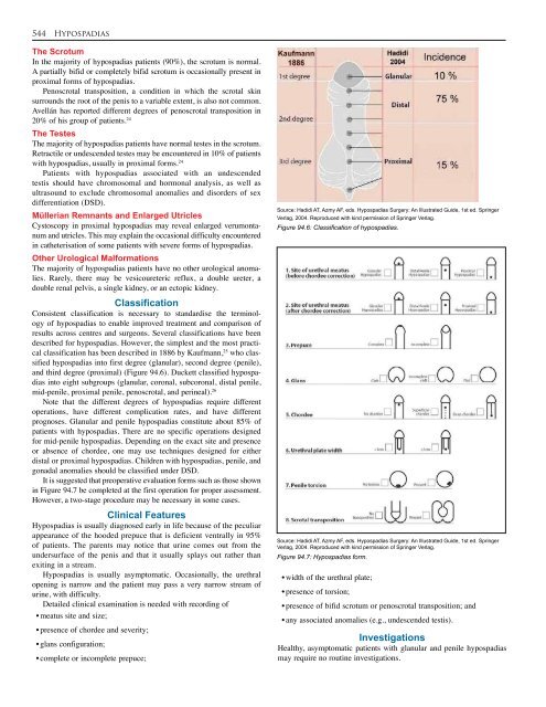
described for hypospadias. However, the simplest and the most practical<br />
classification has been described in 1886 by Kaufmann, 25 who classified<br />
hypospadias into first degree (glanular), second degree (penile),<br />
and third degree (proximal) (Figure <strong>94.</strong>6). Duckett classified hypospadias<br />
into eight subgroups (glanular, coronal, subcoronal, distal penile,<br />
mid-penile, proximal penile, penoscrotal, and perineal). 26<br />
Note that the different degrees of hypospadias require different<br />
operations, have different complication rates, and have different<br />
prognoses. Glanular and penile hypospadias constitute about 85% of<br />
patients with hypospadias. There are no specific operations designed<br />
for mid-penile hypospadias. Depending on the exact site and presence<br />
or absence of chordee, one may use techniques designed for either<br />
distal or proximal hypospadias. Children with hypospadias, penile, and<br />
gonadal anomalies should be classified under DSD.<br />
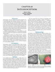
It is suggested that preoperative evaluation forms such as those shown<br />
in Figure <strong>94.</strong>7 be completed at the first operation for proper assessment.<br />
However, a two-stage procedure may be necessary in some cases.<br />
Clinical Features<br />
<strong>Hypospadias</strong> is usually diagnosed early in life because of the peculiar<br />
appearance of the hooded prepuce that is deficient ventrally in 95%<br />
of patients. The parents may notice that urine comes out from the<br />
undersurface of the penis and that it usually splays out rather than<br />
exiting in a stream.<br />
<strong>Hypospadias</strong> is usually asymptomatic. Occasionally, the urethral<br />
opening is narrow and the patient may pass a very narrow stream of<br />
urine, with difficulty.<br />
Detailed clinical examination is needed with recording of<br />
• meatus site and size;<br />
• presence of chordee and severity;<br />
• glans configuration;<br />
• complete or incomplete prepuce;<br />
Source: Hadidi AT, Azmy AF, eds. <strong>Hypospadias</strong> Surgery: An Illustrated Guide, 1st ed. Springer<br />
Verlag, 2004. Reproduced with kind permission of Springer Verlag.<br />
Figure <strong>94.</strong>6: Classification of hypospadias.<br />
Source: Hadidi AT, Azmy AF, eds. <strong>Hypospadias</strong> Surgery: An Illustrated Guide, 1st ed. Springer<br />
Verlag, 2004. Reproduced with kind permission of Springer Verlag.<br />
Figure <strong>94.</strong>7: <strong>Hypospadias</strong> form.<br />
• width of the urethral plate;<br />
• presence of torsion;<br />
• presence of bifid scrotum or penoscrotal transposition; and<br />
• any associated anomalies (e.g., undescended testis).<br />
Investigations<br />
Healthy, asymptomatic patients with glanular and penile hypospadias<br />
may require no routine investigations.

![Clubfoot: Ponseti Management [Vietnamese] - Global HELP](https://img.yumpu.com/51276842/1/184x260/clubfoot-ponseti-management-vietnamese-global-help.jpg?quality=85)


![Steenbeek Brace For Clubfoot [2nd Edition] - Global HELP](https://img.yumpu.com/46612972/1/190x245/steenbeek-brace-for-clubfoot-2nd-edition-global-help.jpg?quality=85)


![Basics Of Wound Care [Indonesia] - Global HELP](https://img.yumpu.com/41566370/1/190x245/basics-of-wound-care-indonesia-global-help.jpg?quality=85)