94. Hypospadias - Global HELP
94. Hypospadias - Global HELP
94. Hypospadias - Global HELP

You also want an ePaper? Increase the reach of your titles
YUMPU automatically turns print PDFs into web optimized ePapers that Google loves.
546 <strong>Hypospadias</strong><br />
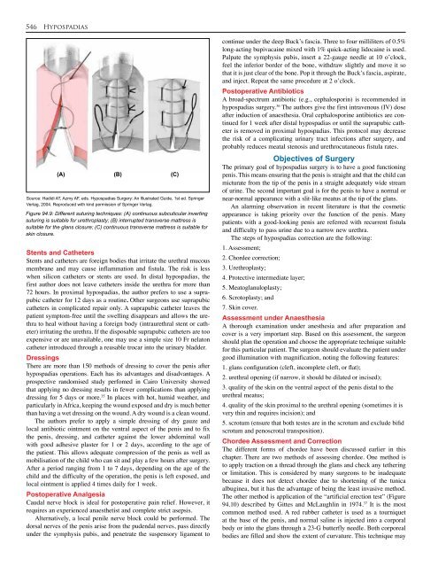
(A) (B) (C)<br />
Source: Hadidi AT, Azmy AF, eds. <strong>Hypospadias</strong> Surgery: An Illustrated Guide, 1st ed. Springer<br />
Verlag, 2004. Reproduced with kind permission of Springer Verlag.<br />
Figure <strong>94.</strong>9: Different suturing techniques: (A) continuous subcuticular inverting<br />
suturing is suitable for urethroplasty; (B) interrupted transverse mattress is<br />
suitable for the glans closure; (C) continuous transverse mattress is suitable for<br />
skin closure.<br />
Stents and Catheters<br />
Stents and catheters are foreign bodies that irritate the urethral mucous<br />
membrane and may cause inflammation and fistula. The risk is less<br />
when silicon catheters or stents are used. In distal hypospadias, the<br />
first author does not leave catheters inside the urethra for more than<br />
72 hours. In proximal hypospadias, the author prefers to use a suprapubic<br />
catheter for 12 days as a routine. Other surgeons use suprapubic<br />
catheters in complicated repair only. A suprapubic catheter leaves the<br />
patient symptom-free until the swelling disappears and allows the urethra<br />
to heal without having a foreign body (intraurethral stent or catheter)<br />
irritating the urethra. If the disposable suprapubic catheters are too<br />
expensive or are unavailable, one may use a simple size 10 Fr nelaton<br />
catheter introduced through a reusable trocar into the urinary bladder.<br />
Dressings<br />
There are more than 150 methods of dressing to cover the penis after<br />
hypospadias operations. Each has its advantages and disadvantages. A<br />
prospective randomised study perfomed in Cairo University showed<br />
that applying no dressing results in fewer complications than applying<br />
dressing for 5 days or more. 35 In places with hot, humid weather, and<br />
particularly in Africa, keeping the wound exposed and dry is much better<br />
than having a wet dressing on the wound. A dry wound is a clean wound.<br />
The authors prefer to apply a simple dressing of dry gauze and<br />
local antibiotic ointment on the ventral aspect of the penis and to fix<br />
the penis, dressing, and catheter against the lower abdominal wall<br />
with good adhesive plaster for 1 or 2 days, according to the age of<br />
the patient. This allows adequate compression of the penis as well as<br />
mobilisation of the child who can sit and play a few hours after surgery.<br />
After a period ranging from 1 to 7 days, depending on the age of the<br />
child and the difficulty of the operation, the penis is left exposed, and<br />
local ointment is applied 4 times daily for 1 week.<br />
Postoperative Analgesia<br />
Caudal nerve block is ideal for postoperative pain relief. However, it<br />
requires an experienced anaesthetist and complete strict asepsis.<br />
Alternatively, a local penile nerve block could be performed. The<br />
dorsal nerves of the penis arise from the pudendal nerves, pass directly<br />
under the symphysis pubis, and penetrate the suspensory ligament to<br />
continue under the deep Buck’s fascia. Three to four milliliters of 0.5%<br />
long-acting bupivacaine mixed with 1% quick-acting lidocaine is used.<br />
Palpate the symphysis pubis, insert a 22-gauge needle at 10 o’clock,<br />
feel the inferior border of the bone, withdraw slightly and move it so<br />
that it is just clear of the bone. Pop it through the Buck’s fascia, aspirate,<br />
and inject. Repeat the same procedure at 2 o’clock.<br />
Postoperative Antibiotics<br />
A broad-spectrum antibiotic (e.g., cephalosporin) is recommended in<br />
hypospadias surgery. 36 The authors give the first intravenous (IV) dose<br />
after induction of anaesthesia. Oral cephalosporine antibiotics are continued<br />
for 1 week after distal hypospadias or until the suprapubic catheter<br />
is removed in proximal hypospadias. This protocol may decrease<br />
the risk of a complicating urinary tract infections after surgery, and<br />
probably reduces meatal stenosis and urethrocutaneous fistula rates.<br />
Objectives of Surgery<br />
The primary goal of hypospadias surgery is to have a good functioning<br />
penis. This means ensuring that the penis is straight and that the child can<br />
micturate from the tip of the penis in a straight adequately wide stream<br />
of urine. The second important goal is for the penis to have a normal or<br />
near-normal appearance with a slit-like meatus at the tip of the glans.<br />
An alarming observation in recent literature is that the cosmetic<br />
appearance is taking priority over the function of the penis. Many<br />
patients with a good-looking penis are referred with recurrent fistula<br />
and difficulty to pass urine due to a narrow new urethra.<br />
The steps of hypospadias correction are the following:<br />
1. Assessment;<br />
2. Chordee correction;<br />
3. Urethroplasty;<br />
4. Protective intermediate layer;<br />
5. Meatoglanuloplasty;<br />
6. Scrotoplasty; and<br />
7. Skin cover.<br />
Assessment under Anaesthesia<br />
A thorough examination under anesthesia and after preparation and<br />
cover is a very important step. Based on this assessment, the surgeon<br />
should plan the operation and choose the appropriate technique suitable<br />
for this particular patient. The surgeon should evaluate the patient under<br />
good illumination with magnification, noting the following features:<br />
1. glans configuration (cleft, incomplete cleft, or flat);<br />
2. urethral opening (if narrow, it should be dilated or incised);<br />
3. quality of the skin on the ventral aspect of the penis distal to the<br />
urethral meatus;<br />
4. quality of the skin proximal to the urethral opening (sometimes it is<br />
very thin and requires incision); and<br />
5. scrotum (ensure that both testes are in the scrotum and exclude bifid<br />
scrotum and penoscrotal transposition).<br />
Chordee Assessment and Correction<br />
The different forms of chordee have been discussed earlier in this<br />
chapter. There are two methods of assessing chordee. One method is<br />
to apply traction on a thread through the glans and check any tethering<br />
or limitation. This is considered by many surgeons to be inadequate<br />
because it does not detect chordee due to shortening of the tunica<br />
albuginea, but it has the advantage of being the least invasive method.<br />
The other method is application of the “artificial erection test” (Figure<br />
<strong>94.</strong>10) described by Gittes and McLaughlin in 1974. 37 It is the most<br />
common method used. A red rubber catheter is used as a tourniquet<br />
at the base of the penis, and normal saline is injected into a corporal<br />
body or into the glans through a 23-G butterfly needle. Both corporeal<br />
bodies are filled and show the extent of curvature. This technique may

![Clubfoot: Ponseti Management [Vietnamese] - Global HELP](https://img.yumpu.com/51276842/1/184x260/clubfoot-ponseti-management-vietnamese-global-help.jpg?quality=85)

![Steenbeek Brace For Clubfoot [2nd Edition] - Global HELP](https://img.yumpu.com/46612972/1/190x245/steenbeek-brace-for-clubfoot-2nd-edition-global-help.jpg?quality=85)

![Basics Of Wound Care [Indonesia] - Global HELP](https://img.yumpu.com/41566370/1/190x245/basics-of-wound-care-indonesia-global-help.jpg?quality=85)