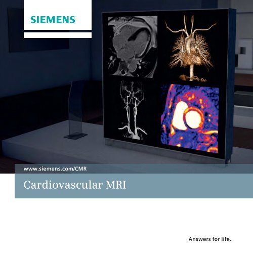
Cardiovascular MRI Brochure 594kB - Siemens Healthcare
Cardiovascular MRI Brochure 594kB - Siemens Healthcare
Cardiovascular MRI Brochure 594kB - Siemens Healthcare

You also want an ePaper? Increase the reach of your titles
YUMPU automatically turns print PDFs into web optimized ePapers that Google loves.
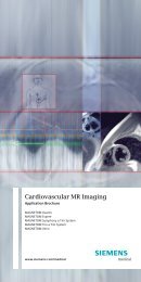
www.siemens.com/CMR<br />
<strong>Cardiovascular</strong> <strong>MRI</strong><br />
Answers for life.
How can I make<br />
cardiovascular<br />
<strong>MRI</strong> imaging part<br />
of my routine?<br />
How can I perform a<br />
comprehensive <strong>MRI</strong><br />
stress test including<br />
function, perfusion,<br />
and viability in under<br />
30 minutes?<br />
How can I achieve<br />
consistent image<br />
quality in cardiac <strong>MRI</strong><br />
and MR angiography?<br />
<strong>Cardiovascular</strong> <strong>MRI</strong><br />
Cardiac <strong>MRI</strong> is widely regarded as a highly complex<br />
examination, but also as one of the most comprehensive<br />
diagnostic tools in cardiology. With easier tools and better<br />
technology available, cardiac <strong>MRI</strong> is spreading more<br />
into routine use. The Cardiac Dot Engine is at the center<br />
of this development, as it makes examinations faster and<br />
more robust. At the same time, it offers new processing<br />
capabilities, helping make diagnoses of perfusion images<br />
easier and more standardized.<br />
Cardiac <strong>MRI</strong> offers excellent soft tissue differentiation,<br />
high spatial and temporal resolution, and 3D as well as<br />
4D data acquisition. These and other sophisticated<br />
capabilities make cardiovascular <strong>MRI</strong> ideally suited for an<br />
array of studies, such as evaluation of cardiac anatomy<br />
and morphology, ventricular and valvular function,<br />
myocardial perfusion and viability, flow, and angiography.<br />
Furthermore, cardiac <strong>MRI</strong> is a useful tool for monitoring<br />
myocardial tissue to differentiate between reversible and<br />
irreversible as well as acute from healed myocarditis. <strong>MRI</strong><br />
assessment of myocardial iron and function has modified<br />
the treatment of thalassemia.<br />
MR angiography (MRA) is performed at almost every <strong>MRI</strong><br />
system, but very often the quality of the results varies<br />
depending on the user and the patient.<br />
With the broad spectrum of contrast and non-contrast<br />
applications such as the TimCT Angio Dot Engine, the<br />
Angio Dot Engine, syngo TWIST, and syngo NATIVE,<br />
<strong>Siemens</strong> provides excellent tools to achieve consistently<br />
excellent results.<br />
2
Cardiac Function, TrueFISP cine retro, GRAPPA 2;<br />
MAGNETOM Verio<br />
EKO <strong>MRI</strong> Centre, Kolkata, India<br />
Cardiac Function, TrueFISP cine retro, GRAPPA 2;<br />
MAGNETOM ESSENZA<br />
Medical Center Pro Vita, Sofia, Bulgaria<br />
Cardiac Function, TrueFISP cine retro,<br />
GRAPPA 3; MAGNETOM Skyra<br />
Cardiac Function, FLASH cine tagging;<br />
MAGNETOM Espree<br />
Cardiac Morphology, T2 TSE Dark Blood, GRAPPA<br />
2; MAGENTOM Aera<br />
Cardiac Moprhology, T2 TSE SPAIR Dark<br />
Blood, GRAPPA 2; MAGNETOM Aera<br />
3
Cardiac flow FLASH cine retro phased;<br />
MAGNETOM Avanto<br />
Cardiac Function, TrueFISP cine radial,<br />
32-channel Body coil; MAGNETOM Verio<br />
Cardiac Function, TrueFISP cine retro,<br />
GRAPPA 2; MAGNETOM Skyra<br />
AKH, Linz, Austria<br />
Cardiac Viability, FLASH IR post contrast,<br />
GRAPPA 2; MAGNETOM Aera<br />
Centre Cardio Thoracique de Monaco,<br />
Monaco<br />
Cardiac Function, TrueFISP cine retro, GRAPPA 2;<br />
MAGNETOM Avanto<br />
BC Children’s Hospital, British Columbia, Canada<br />
Cardiac Non-contrast Cornary Angiography,<br />
T2 3D TrueFISP, thin MIP; MAGNETOM Aera<br />
4
1a<br />
1b<br />
2a<br />
2b<br />
1c<br />
1d<br />
2c<br />
2d<br />
1a and 1b: Cardiac Morphology, TIRM Dark Blood<br />
1c and 1d: Cardiac Function, TrueFISP cine retro, GRAPPA 2<br />
2a – 2d: Cardiac Perfusion, Turbo FLASH post contrast, GRAPPA 2<br />
3a<br />
3b<br />
4a<br />
4b<br />
3c<br />
3d<br />
4c<br />
4d<br />
3a and 3b: Cardiac Flow, FLASH cine retro, GRAPPA 2<br />
3c and 3d: Cardiac Flow, FLASH cine retro, GRAPPA 2, phased<br />
4a and 4b: Cardiac Viability, IR Flash post contrast, GRAPPA 2<br />
4c and 4d: Cardiac Viability, IR TrueFISP FatSat post contrast, GRAPPA 2<br />
Clinical case from one patient: acute myocardial infarction,<br />
MAGNETOM Aera<br />
Centre Cardio Thoracique de Monaco, Monaco<br />
5
Cardiac Dot Engine<br />
To bring cardiac <strong>MRI</strong> to the routine, the Cardiac Dot Engine<br />
guides users through the examination step-by-step to help<br />
achieve excellent results, consistently. In addition, patientspecific<br />
adaptation to the individual condition and<br />
workflow automation helps the user achieve fast and<br />
consistent results.<br />
Personalized:<br />
parameter adaption to heart rate<br />
and breath hold capability<br />
Guided:<br />
guidance through placing five anatomical<br />
landmarks for automated slice positioning<br />
Automated:<br />
slice positioning is matched for the entire exam and<br />
standard cardiac views are obtained automatically<br />
6
One of today’s most important clinical needs is being able<br />
to perform a comprehensive ischemic heart evaluation<br />
within 30 minutes. This includes function, perfusion, and<br />
viability analysis – a comprehensive examination that only<br />
<strong>MRI</strong> can offer as a single modality. The Cardiac Dot Engine<br />
supports complete exams for ischemic and non-ischemic<br />
diseases. Inline technology speeds up the workflow even<br />
more with Inline VF (Ventricular Function) and Inline Time<br />
Course Evaluation (Inline Perfusion).<br />
Inline Function<br />
Cardiac Function, TrueFISP cine retro,<br />
GRAPPA 3, Argus; MAGNETOM Aera<br />
Northwestern Memorial Hospital, Chicago,<br />
USA<br />
Inline Perfusion<br />
Cardiac Perfusion, Turbo FLASH post contrast,<br />
Upslope Map, GRAPPA 2; MAGNETOM Aera<br />
Northwestern Memorial Hospital, Chicago,<br />
USA<br />
Viability<br />
Cardiac Viability, FLASH IR post contrast;<br />
MAGNETOM Avanto<br />
Helios Klinikum Berlin-Buch, Berlin,<br />
Germany<br />
7
syngo.via*<br />
Working with large amounts of cardiac <strong>MRI</strong> data like<br />
morphology, cine, perfusion or viability on a conventional<br />
workstation can be a challenge. Multiple applications are<br />
needed; synchronization and navigation capabilities may<br />
be insufficient. syngo.MR General Engine offers a rich<br />
suite of functionality to cover all routine reading and post<br />
processing needs. Be it managing images from different<br />
cardiac views or phases; or searching for useful<br />
information from<br />
other acquisitions, the syngo.MR Cardiac Reading<br />
workflow in our syngo.MR General Engine already links<br />
all the data and images to the relevant series.<br />
Features include guided workflows, synchronization<br />
of data through space and time and at the end of the<br />
analysis, a dedicated structured Cardiac Report<br />
is generated.<br />
* syngo.via can be used as a standalone device or<br />
together with a variety of syngo.via-based<br />
software options, which are medical devices in<br />
their own rights. syngo.via and the syngo.via<br />
based software options are pending in some<br />
countries the necessary clearances and thus are<br />
not available for sales in all countries.<br />
** syngo.MR Cardiac Flow is not commercially<br />
available. Due to regulatory reasons its future<br />
availability cannot be guaranteed.<br />
8
syngo. MR Cardiac<br />
4D Ventricular<br />
Function<br />
Functional and volumetric evaluation is the cornerstone<br />
of every cardiac MR examination in ischemic as well as<br />
non ischemic cardiomyopathies. syngo.MR Cardiac 4D<br />
Ventricular Function offers precise analysis of all relevant<br />
volumetric parameters such as ejection fraction, stroke<br />
volume, and segmental analysis of wall thickening.<br />
syngo. MR<br />
Cardiac Flow**<br />
With syngo.MR Cardiac Flow, the evaluation of blood<br />
flow dynamics is quick and easy. The flow analysis<br />
determines the mean and maximum velocity of blood<br />
flow.<br />
Special Features include Velocity encoding gradient (venc)<br />
correction and one-click vessel segmentation.<br />
9
MR Angiography<br />
MR angiography is one of the most, performed exams in<br />
<strong>MRI</strong>. In addition to diseases of the heart’s vessels, MRA<br />
also helps diagnose large vessels disease, such as aortic<br />
dissection or pulmonary embolism. Furthermore,<br />
cerebrovascular diseases or peripheral artery diseases are<br />
often manifest in CAD patients. <strong>MRI</strong> offers not only<br />
visualization of vessel lumen like conventional<br />
angiography does, but also analysis of the vessel wall.<br />
MR angiography is increasingly playing an important role<br />
for planning and guidance of vascular interventions<br />
offering high diagnostic accuracy, such as the 3D wholeheart<br />
coronary MRA, which enables a CT-like exam. The<br />
vascular information acquired through an <strong>MRI</strong> scan is used<br />
to plan or guide vascularintervention, which comprises<br />
balloon angioplasty and other endovascular therapies like<br />
implantation of stent grafts and EP ablation planning.<br />
<strong>MRI</strong> delivers fast and accurate angiography results,<br />
offering several advantages; visualization and<br />
quantification of flow, dynamic information through<br />
syngo TWIST and non-contrast angiography using<br />
syngo NATIVE.<br />
Due to today’s possibilities of scanning during continuous<br />
table movement (syngo TimCT), a complete angiographic<br />
study is now done in a matter of minutes. Vascular<br />
interventions under <strong>MRI</strong> guidance is a relatively new and<br />
emerging field, which holds great promise.<br />
1 2<br />
10
4a<br />
4b<br />
1: Carotid Angiography, 3D FLASH ce-MRA, VRT,<br />
GRAPPA 3; MAGNETOM Verio<br />
MR Bremen Mitte, Bremen, Germany<br />
2: Coronary Whole Heart Angiography, T2 3D TrueFISP<br />
FatSat VRT, GRAPPA 2; MAGNETOM Avanto<br />
University Hospital UCLA, Los Angeles, USA<br />
3: Non-contrast Pulmonary MRA, T2 SPACE IR, thin mIP,<br />
inverted, GRAPPA 3; MAGNETOM Aera<br />
4a and 4b: Peripheral Angiography, 3D FLASH ce-MRA,<br />
3 steps, MIP and MIP inverted, GRAPPA 2;<br />
MAGNETOM Espree<br />
Battlefield Imaging, Ringgold, USA<br />
3<br />
11
syngo TWIST<br />
For a better detection of various congential diseases<br />
syngo TWIST provides dynamic information. syngo TWIST<br />
is a time-resolved 3D MRA technique that achieves both<br />
a high temporal and spatial resolution. syngo TWIST<br />
offers a practical, flexible, and elegant way to perform<br />
sub-second, time-sequential 3D measurements. syngo<br />
TWIST provides further clinical information, including the<br />
evaluation of abnormal anatomy as well as vascular<br />
heamodynamics.<br />
Throacic Angiography, 3D FLASH ce-MRA TWIST,<br />
MIP dynamic, GRAPPA 3; MAGNETOM Aera<br />
University Hospital of Saarland, Homburg, Germany<br />
12
syngo NATIVE<br />
syngo NATIVE is a contrast-free MR angiography technique<br />
for visualizing the vessels. The package contains protocols<br />
tailored for use in different body regions (e.g., renal<br />
arteries, peripheral vessels). Inline subtraction and inline<br />
maximum intensity projection (MIP) further simplify the<br />
workflow.<br />
For non-contrast MR angiography, <strong>Siemens</strong> currently<br />
offers two techniques under the umbrella syngo NATIVE –<br />
syngo NATIVE TrueFISP and syngo NATIVE SPACE.<br />
Renal Angiography,<br />
3D NATIVE TrueFISP FatSat,<br />
MIP and VRT, GRAPPA 2;<br />
MAGNETOM ESSENZA<br />
Tateyama Hospital, Tateyama, Japan<br />
13
Angio Dot Engine<br />
The Angio Dot Engine is designed to empower the user to<br />
perform high spatial resolution contrast-enhanced MR<br />
angiography. This can often even be achieved using<br />
optimized protocols for low dosed contrast medium<br />
acquisitions. contrast medium. The timing of the contrast<br />
medium in MRA is a particular challenge. The Angio Dot<br />
Engine assists the user in achieving the optimal timing for<br />
contrast arrival, easily and consistently, and provides an<br />
interactive approach to contrast timing.<br />
After a simple click the<br />
localizer and vessel scout<br />
are acquired and loaded<br />
into the Graphical Slice<br />
Positioning (GSP). The<br />
next workflow step opens<br />
a customizable Guidance<br />
View that visually explains<br />
how to set up the test<br />
bolus measurement<br />
correctly. According to<br />
the instructions, the test<br />
bolus slice can be<br />
adjusted in the GSP. The<br />
planning of the test bolus<br />
is completed by simply<br />
clicking on apply.<br />
14
TimCT Angio Dot Engine<br />
TimCT Angio Dot Engine employs the revolutionary<br />
TimCT – Continuous Table move – technology for a large<br />
field of view in angiography examinations with the<br />
smooth workflow and the most homogeneous image<br />
quality. Thanks to the streamlined and automated<br />
workflow, TimCT Angio Dot Engine supports short<br />
acquisition times. A complete peripheral vessel runoff<br />
exam can be performed in less than 15 minutes with<br />
optimal bolus timing.<br />
The TimCT Angio Dot Engine offers optimized protocols<br />
for peripheral vessel runoff exams. It allows CT-like<br />
scanning with <strong>MRI</strong>. There is no need to plan multiple steps<br />
and no need to plan overlapping sections.<br />
TimCT Angiography,<br />
3D FLASH ce-MRA,<br />
MIP, GRAPPA 3;<br />
MAGNETOM Aera<br />
University Hospital,<br />
Essen, Germany<br />
15
<strong>Cardiovascular</strong> sequences<br />
syngo Beat<br />
syngo BEAT can simplify cardiac <strong>MRI</strong> significantly. Now you<br />
can diagnose cardiovascular disease faster and more easily. Achieve<br />
excellent results, even in difficult patients, from the smallest to<br />
the tallest. Improves treatment planning and introduce CT-like<br />
scanning to CMR with 3D and 4D exams.<br />
Advanced Cardiac Package<br />
Special sequences and protocols for advanced cardiac imaging,<br />
including 3D Cine, 3D Viability, Tagging, 3D Coronaries, and Multi Echo<br />
imaging among other specialized techniques as well as 4D<br />
functionalities. Allows comprehensive exams for cardiac experts.<br />
syngo Interactive RT<br />
Interactive cardiac real-time imaging.<br />
syngo Flow Quantification<br />
Special sequences for quantitative flow exams.<br />
syngo TWIST<br />
3D MRA to achieve high spatial and temporal resolution with minimal<br />
amount of contrast agent and no venous contamination.<br />
syngo NATIVE<br />
Contrast-free MRA with tailored protocols for different body regions.<br />
syngo TimCT Angiography<br />
Revolutionary TimCT Continuous Table movement technology, for<br />
smooth workflow and homogenous image quality of vascular<br />
examinations covering extended FoV’s, up to the whole body.<br />
Don’t miss the wealth of clinical information<br />
on cardiovascular <strong>MRI</strong> at<br />
www.siemens.com/magnetom-world<br />
16
<strong>Cardiovascular</strong> applications<br />
syngo.MR General Engine<br />
Software for professional and routine <strong>MRI</strong> reading usage.<br />
Includes workflows for dedicated <strong>MRI</strong> examinations that load and<br />
structure examination results automatically into meaningful layouts<br />
including user support to make sure that no data is missed. Contains<br />
several <strong>MRI</strong> radiology workflows, cardiovascular workflows and<br />
MR evaluation features.<br />
Includes: syngo.MR Cardiac Reading<br />
syngo.MR Angio Reading<br />
syngo.MR Cardiac 4D<br />
Ventricular Function<br />
Automated evaluation and quantification of cardiac function<br />
for the left and right ventricle.<br />
syngo.MR Cardiac Flow*<br />
Automated blood flow evaluation for calculation of parameters,<br />
color-coded display and improved background phase correction.<br />
syngo.MR composing*<br />
Composing of images from different body regions to see the complete<br />
anatomy (also automated as Inline functionality).<br />
Dedicated offline application for creation of full-format images from<br />
overlapping MR volume data sets acquired at multiple stages. Can be<br />
used to compose images in any of the other syngo.via workflows.<br />
syngo Vessel View<br />
Automated analysis tool for MR angiography data for small and large<br />
vessels. Supports automatic quantification of stenoses and evaluation of<br />
aneurysms – for potentially faster diagnosis of vascular disease.<br />
syngo Argus Dynamic Signal<br />
Evaluation software for automated image analysis of dynamic studies in<br />
the body. Automated contouring, motion correction and easy-to-use<br />
editing tools to quantify regional changes in the tissue based on signaltime<br />
curves.<br />
* syngo.MR Cardiac Flow and syngo.MR Composing are not commercially available.<br />
Due to regulatory reasons their future availability cannot be guaranteed.<br />
17
Not for distribution in USA.<br />
On account of certain regional limitations<br />
of sales rights and service availability, we cannot<br />
guarantee that all products included in this<br />
brochure are available through the <strong>Siemens</strong> sales<br />
organization worldwide. Availability and<br />
packaging may vary by country and are subject<br />
to change without prior notice. Some/All of the<br />
features and products described herein may not<br />
be available in the United States.<br />
All devices listet herein may not be licensed<br />
according to Canadian Medical Devices<br />
Regulations. The information in this document<br />
contains general technical descriptions of<br />
Local Contact Information<br />
In the USA<br />
<strong>Siemens</strong> Medical Solutions USA, Inc.<br />
51 Valley Stream Parkway<br />
Malvern, PA 19355<br />
Phone: +1 888-826-9702<br />
Phone: +1 610-448-4500<br />
Fax: +1 610-448-2254<br />
In China<br />
<strong>Siemens</strong> Medical Park, Shanghai<br />
278, Zhouzhu Road<br />
SIMZ, Nanhui District<br />
Shanghai, 201318, P.R. China<br />
Phone: +86-21-38895000<br />
Fax: +86-10-28895001<br />
specifications and options as well as standard<br />
and optional features which do not always have<br />
to be present in individual cases.<br />
<strong>Siemens</strong> reserves the right to modify the design,<br />
packaging, specifications, and options described<br />
herein without prior notice. Please contact your<br />
local <strong>Siemens</strong> sales representative for the most<br />
current information.<br />
Note: Any technical data contained in this<br />
document may vary within defined tolerances.<br />
Original images always lose a certain amount<br />
of detail when reproduced.<br />
Please find fitting accessories:<br />
www.siemens.com/medical-accessories<br />
In Japan<br />
<strong>Siemens</strong>-Asahi<br />
Medical Technologies Ltd.<br />
Takanawa Park Tower 14F<br />
20-14, Higashi-Gotanda 3-chome<br />
Shinagawa-ku, Tokyo 141-8644<br />
Phone: +81 3 5423 8411<br />
In Asia<br />
<strong>Siemens</strong> Pte Ltd<br />
<strong>Healthcare</strong> Sector<br />
Regional Headquarters<br />
The <strong>Siemens</strong> Center<br />
60 MacPherson Road,<br />
Singapore 348615<br />
Phone: +65 6490-6000<br />
Fax: +65 6490-6001<br />
Titel image courtesies, from left to right:<br />
Cardio Viability, FLASH IR post contrast,<br />
GRAPPA 2; MAGNETOM Aera<br />
Centre Cardio Thoracique de Monaco,<br />
Monaco<br />
Thoracic Angiography, 3D FLASH ce-MRA<br />
TWIST, VRT, GRAPPA 3; MAGNETOM Skyra<br />
Carotids Angiography, 3D FLASH ce-MRA,<br />
MIP, GRAPPA 2; MAGNETOM Verio<br />
Seoul National University Hospital,<br />
Seoul, Republic Korea<br />
Cardio Pefusion, Turbo FLASH post<br />
contrast, Map Slope, GRAPPA 2;<br />
MAGNETOM Avanto<br />
Northwestern Memorial Hospital,<br />
Chicago, USA<br />
Global Business Unit<br />
<strong>Siemens</strong> AG<br />
<strong>Healthcare</strong> Sector<br />
Magnetic Resonance<br />
Henkestr. 127<br />
91052 Erlangen<br />
Germany<br />
Phone: +49 9131 84-0<br />
Global <strong>Siemens</strong> Headquarters<br />
<strong>Siemens</strong> AG<br />
Wittelsbacherplatz 2<br />
80333 Muenchen<br />
Germany<br />
Global <strong>Siemens</strong> <strong>Healthcare</strong> Headquarters<br />
<strong>Siemens</strong> AG<br />
<strong>Healthcare</strong> Sector<br />
Henkestrasse 127<br />
91052 Erlangen<br />
Germany<br />
Phone: +49 9131 84-0<br />
www.siemens.com/healthcare<br />
Legal Manufacturer<br />
<strong>Siemens</strong> AG<br />
Wittelsbacherplatz 2<br />
DE-80333 Munich<br />
Germany<br />
Order No. A91MR-1100-43X-7600 | Printed in Germany | CC MR WS 08111. | © 08.2011, <strong>Siemens</strong> AG<br />
www.siemens.com/healthcare


![WalkAway plus Technical Specifications [41 KB] - Siemens Healthcare](https://img.yumpu.com/51018135/1/190x253/walkaway-plus-technical-specifications-41-kb-siemens-healthcare.jpg?quality=85)