Sura F
Sura F
Sura F

Create successful ePaper yourself
Turn your PDF publications into a flip-book with our unique Google optimized e-Paper software.
J Bagh College Dentistry Vol. 23(special issue), 2011 Morphometric analysis of<br />
Morphometric analysis of mandibular canal course and<br />
position in relation to gender and age of Iraqi sample using<br />
digital panoramic imaging<br />
<strong>Sura</strong> A. Rashid, B.D.S., M.Sc. (1)<br />
Jamal Ali B.D.S., M.Sc. (2)<br />
ABSTRACT<br />
Background: The knowledge of the course and position of the mandibular canal in relation to anatomical landmarks<br />
of the jaw is of great importance in certain dental interventions; therefore it involves preservation of the anatomic<br />
structures that pass through it. Morphometric study by means of digital panoramic radiography reveals the<br />
differences and inherent alterations between genders or ages, it has become a useful tool in research that analyzes<br />
the craniofacial complex development process, which are important for planning the dental, clinical and surgical<br />
procedures. The aim of the study was undertaken to analyze mandibular canal morphology and position along its<br />
course and its relation to mandibular anatomical landmarks by using digital panoramic radiography.<br />
Materials and Methods: 300 subjects, (150 male and 150 female) were examined, age distribution of them was<br />
ranging between (20-49) years old, factors considered included the age and sex of the patients. Ten linear vertical<br />
measurements were performed on the radiographic image of each subject on both right and left sides of the<br />
mandible (600) sides, to evaluate the position of the mandibular canal along its course. The relation of proximity<br />
between the mandibular canal and the roots of the mandibular posterior teeth were also evaluated.<br />
Results: Statistically significant differences were observed in six of the linear measurements between genders, males<br />
almost have higher measurements than females. However, no significant linear trend was evident between age<br />
groups of the same gender for any of the measurements. The most frequent position of the mental foramen was<br />
between the two mandibular premolars, the mandibular canal presented a relation of proximity to the roots of the<br />
mandibular third molar, moving gradually away from the roots of the other posterior teeth.<br />
Conclusion: the measurements related to the mandibular canal performed in this study can be influenced by the<br />
gender but are independent of age.<br />
Key words: Mandibular canal, position, morphometry. (J Bagh Coll Dentistry 2011; 23(sp. issue):92-99).<br />
INTRODUCTION<br />
The mandibular canal (MC), or the inferior<br />
alveolar canal (IAC), is a canal within the<br />
mandible that is beginning in mandibular<br />
foramen on the medial surface of the ascending<br />
mandibular ramus. It runs obliquely downward<br />
and forward in the ramus, and then horizontally<br />
forward in the body till mental foramen. It<br />
transmits the inferior alveolar nerve, a branch of<br />
the third division of the trigeminal nerve, and the<br />
associated vessels (1,2) The MC is of particular<br />
importance to the dentist and dental specialist as<br />
it carries both the dental division of the<br />
mandibular nerve and the nerve supply for the<br />
lower lip (2,3)<br />
The knowledge of the morphology and<br />
topography of the MC is important for<br />
performing dental interventions in the jaw;<br />
therefore it involves preservation of the anatomic<br />
structures that pass through it. In the surgical<br />
approach to the jaw, the MC isomical structure<br />
used as reference (4) .<br />
(1) Oral and Maxillofacial Radiologist, Ministry of Health.<br />
(2) Oral and Maxillofacial Radiology, Department of Oral<br />
Diagnosis, College of Dentistry, University of Baghdad<br />
Extraction of mandibular third molars, implant<br />
surgeries, orthognathic surgeries and those to<br />
reduce and to fix of fractures in the different<br />
regions of the jaw, are examples of the<br />
procedures that can be done close to the<br />
mandibular canal, increasing the risks of injuries<br />
to the inferior alveolar nerve (5,6)<br />
The mandibular foramen is a useful anatomic<br />
point for positioning the needle in inferior<br />
alveolar nerve anesthesia, and for planning<br />
surgical procedures in the mandibular ramus<br />
region. Injecting anesthetic solution in the<br />
pterigomandibular space, region where the<br />
inferior alveolar nerve is found in the mandibular<br />
foramen, is an anesthetic technique frequently<br />
used in the dental clinic. The failure indices of<br />
this technique range from 29% to 35% because<br />
of the lack of precision in locating the anatomy<br />
of this foramen (7)<br />
The mental foramen, the place where the mesial<br />
portion of the mandibular canal is exposed, is a<br />
reference structure with a great clinical<br />
applicability, being the place where the mental<br />
nerve passes through. Injecting anesthetic<br />
solution in the region of this foramen allows the<br />
mental nerve and inferior alveolar nerve to be<br />
Oral Diagnosis 92
J Bagh College Dentistry Vol. 23(special issue), 2011 Morphometric analysis of<br />
effectively blocked in the mandibular premolar<br />
region (8)<br />
Panoramic radiography is a widely used<br />
technique because it has the advantage of<br />
providing, in a single film, the image of both<br />
jaws, with a relatively low radiation dose, in a<br />
short period of time, and at lower cost if<br />
compared to more sophisticated techniques. This<br />
technique can offer information about the<br />
localization of anatomic structures vertical and<br />
(9)<br />
horizontal bony diamentions. However, a<br />
panoramic radiograph is a two-diamentional<br />
image, lacking information in the bucco-lingual<br />
direction and magnifying in both vertical and<br />
horizontal directions. The magnification factors<br />
are different to each unite so the resultant<br />
magnification is specific for each panoramic x-<br />
ray machine. (10) Nevertheless, the fact that the<br />
availability of panoramic imaging in the dental<br />
office and its widely use for evaluation of the<br />
jaws, justifies the interest in determining the<br />
visibility of anatomical structures on these films.<br />
(11)<br />
SUBJECTS AND METHODS<br />
The study sample was 300 Iraqi patients from<br />
both genders, (150 male & 150 female),<br />
attending to the Maxillofacial Radiology<br />
Department at Al-Karkh Hospital in Baghdad,<br />
referred for digital panoramic radiographs for<br />
various purposes, These participants were<br />
evaluated according to age and gender, they were<br />
divided to 3 groups according to age: first group:<br />
Subjects from (20-29) years of age (50 male &<br />
50 female); second group: Subjects from (30-39)<br />
years of age (50 male & 50 female); and third<br />
group: Subjects from (40-49) years of age (50<br />
male & 50 female). Sample individuals should<br />
have no history of any systemic disease that<br />
might affect bone metabolism, Alveolar crest<br />
resorption would have to be absent, Patients with<br />
extracted mandibular second molar and/or third<br />
molar, presence of crowding and spacing in the<br />
mandibular arch, past history of orthodontic<br />
treatment for mandibular arch, any pathology or<br />
congenital anomaly in the mandible that could<br />
affect the interpretation of radiographic image<br />
were excluded.<br />
Methods<br />
Panoramic image was taken for each subject<br />
using Dimax-3 digital X-ray machine, all<br />
subjects were positioned in the machine<br />
according to the manufacturer manual.<br />
All images were examined on the monitor and<br />
the resolution was enhanced to what was<br />
considered optimum. The selected radiographic<br />
images are imported by (the DIMAX3 digital<br />
software) with specific tools for making linear<br />
measurements on images of the mandibular jaw.<br />
Six linear vertical measurements (D1, D2, D3,<br />
D4, D9 & D10) were performed on all<br />
radiographic images to describe the course of the<br />
mandibular canal and its relations with the<br />
anatomic structures of jaw according to Amorim<br />
et al, (12) , and four linear vertical measurements<br />
(D5, D6, D7 & D8) were performed to describe<br />
the vertical position of the mandibular canal in<br />
relation to the root apecis of the mandibular<br />
second molar tooth and mandible base according<br />
to Sato et al, (13) . These measurements were done<br />
on the right and left sides of the mandible image.<br />
The following measurements were taken, figure<br />
(1):<br />
D1 – Vertical distance of the most inferior point<br />
of the image of the inferior edge of the mental<br />
foramen to the image of the inferior limit of the<br />
mandible base at the shortest line connecting the<br />
alveolar crest and the inferior limit of mandible<br />
base, passing through the center of the mental<br />
foramen.<br />
D2 – Vertical distance of the most superior point<br />
of the image of the superior edge of the mental<br />
foramen to the image of the superior limit of the<br />
alveolar crest at the shortest line connecting the<br />
alveolar crest and the inferior limit of mandible<br />
base, passing through the center of the mental<br />
foramen.<br />
D3 – Vertical distance of the image of the<br />
inferior cortical wall of the mandibular canal to<br />
the inferior limit of the mandible base, in the<br />
height of the image of the anterior edge of the<br />
mandibular ramus.<br />
D4 - Vertical distance of the image of the<br />
superior cortical wall of the mandibular canal to<br />
the inferior limit of the image of the oblique line<br />
in the height of the image of the anterior edge of<br />
the mandibular ramus.<br />
D5 - Vertical distance of the most inferior point<br />
of the image of the inferior cortical wall of the<br />
mandibular canal to the inferior limit of the<br />
mandible base, at the shortest vertical line<br />
connecting the inferior point of the apex of<br />
mesial root of mandibular second molar and the<br />
inferior limit of the mandible base.<br />
D6- Vertical distance of the image of the superior<br />
cortical wall of the mandibular canal to the<br />
inferior point of the apex of mesial root of<br />
mandibular second molar at the shortest vertical<br />
line connecting the inferior point of the apex of<br />
mesial root of mandibular second molar and the<br />
inferior limit of the mandible base.<br />
D7 - Vertical distance of the most inferior point<br />
of the image of the inferior cortical wall of the<br />
mandibular canal to the inferior limit of the<br />
Oral Diagnosis 93
J Bagh College Dentistry Vol. 23(special issue), 2011 Morphometric analysis of<br />
mandible base at the shortest vertical line<br />
connecting the inferior point of the apex of distal<br />
root of mandibular second molar and the inferior<br />
limit of the mandible base.<br />
D8 - Vertical distance of the image of the<br />
superior cortical wall of the mandibular canal to<br />
the inferior point of the apex of distal root of<br />
mandibular second molar at the shortest vertical<br />
line connecting the inferior point of the apex of<br />
mesial root of mandibular second molar and the<br />
inferior limit of the mandible base.<br />
D9 - Vertical distance of the most inferior point<br />
of the image of the mandibular notch to the<br />
image of the most superior point of the image of<br />
the superior edge of mandibular foramen at the<br />
line connecting the most inferior point of the<br />
image of the mandibular notch and the inferior<br />
edge of the mandibular ramus, passing through<br />
the center of the mandibular foramen.<br />
D10 - Vertical distance of the most inferior point<br />
of the image of the mandibular notch to the<br />
image of the inferior edge of the mandibular<br />
ramus, passing through the center of the<br />
mandibular foramen.<br />
Figure 1: Measurements used to<br />
describe mandibular canal course and<br />
position.<br />
Based on these ten measurements, the following<br />
ratios were calculated: R1 - Ratio between the<br />
measurements D1 and D2; R2 - Ratio between<br />
the measurements D3 and D4; R3 - Ratio<br />
between the measurements D5 and D6;R4 - Ratio<br />
between the measurements D7 and D8; and R5 -<br />
Ratio between the measurements D9 and D10.<br />
The proximity of the image of the MC to the<br />
roots of mandibular posterior teeth was classified<br />
according to Madeira (14) in to three types of<br />
relation: a. There is a relation of proximity of the<br />
MC image only with the image of the root of the<br />
mandibular third molar, and from this point,<br />
there is a gradual removal of the MC in relation<br />
to the roots of other mandibular posterior teeth;<br />
b. There is an absence of proximity between the<br />
MC and the roots of mandibular posterior teeth;<br />
c. There is a relation of proximity between the<br />
MC and the roots of all the mandibular posterior<br />
teeth. This relation was determined by visual<br />
examination for both the right and left sides of<br />
the mandible image.<br />
Statistical analysis:<br />
Statistical analyses were computer assisted using<br />
SPSS (Statistical Package for Social Sciences).<br />
The bony measurements were normally<br />
distributed variables and thus conveniently<br />
described by mean, while ratios of two bony<br />
measurements are non-normally distributed and<br />
such variables are described by median and<br />
interquartile range (25 th to 75 th percentile).<br />
Independent samples t-test was used to test the<br />
statistical significance of difference in mean<br />
between 2 groups, Kappa statistics was used to<br />
assess the magnitude of agreement beyond<br />
chance. The statistical significance, direction and<br />
strength of linear correlation between age and a<br />
quantitative normally distributed variable was<br />
measured by Pearson’s linear correlation<br />
coefficient. P- value less than the 0.05 level of<br />
significance was considered statistically<br />
significant.<br />
RESULTS<br />
Inspit of slight discrepancies in measurements, it<br />
was revealed that the mean of total values on the<br />
right side of the mandible was almost concordant<br />
with that on the left side for each linear<br />
measurement, no statistically significant<br />
differences existed between sides measurements,<br />
which justify using sides instead of subjects as<br />
the original unit of sampling (600 sides).<br />
The difference in the mean of total values of<br />
linear measurements between the right and the<br />
left sides of the mandible was illustrated in table<br />
1. Also, There was a bilateral concordance in the<br />
mandibular canal morphology in relation to the<br />
roots of posterior teeth, as illustrated in table 2.<br />
The mandibular canal was proximal to the<br />
mandibular third molar tooth in most of the<br />
subjects (83.7 %), both for male and female and<br />
there were gradual removal of the canal from<br />
other molar teeth, as shown in table 3.<br />
Details on the ten linear vertical measurements<br />
and their ratios variations in relation to age<br />
groups and gender are illustrated in tables 4,5&6.<br />
Age had no evident linear correlation with any of<br />
study measurements and their ratios. The means<br />
of overall values for D1, D2, D3, D4, D9 & D10<br />
were significantly higher in male in comparison<br />
to female. The measurements D5, D6, D7 and D8<br />
were almost higher in male than female<br />
measurements, however, these differences have<br />
no evident statistical significance. The position<br />
Oral Diagnosis 94
J Bagh College Dentistry Vol. 23(special issue), 2011 Morphometric analysis of<br />
of the canal varies with respect to the lower<br />
border of the mandible and the apices of the roots<br />
of mandibular molar teeth. The MC relative<br />
vertical position tends to be closer to the<br />
mandibular teeth roots apices than to the<br />
mandible lower border.<br />
The median value of R1 seem to be equal to that<br />
of R2, both for male and female, although the<br />
median of overall values for R1 & R2 were<br />
slightly higher in male (0.6) in comparison to<br />
female (0.58), this difference had no evident<br />
statistical value. R3 & R4 median values were<br />
significantly higher in male than female that<br />
means the MC in female tends to be relatively<br />
lower than that in male independent on age.<br />
There was no statistically significant difference<br />
between male and female in the median value of<br />
R5 as it was almost (0.5), that means that the<br />
mandibular foramen was almost located in the<br />
middle of the mandibular ramus independent on<br />
age or gender.<br />
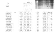
Table 1: Difference in the mean of total values between the right and the left sides of the jaw for<br />
the vertical linear measurements related to the MC<br />
Difference between right<br />
Distance-1 Right(N=300) Left(N=300) and left P (paired t-test)<br />
Mean 9.63 9.68 -0.05 0.1[NS]<br />
Distance-2<br />
Mean 16.87 16.89 -0.02 0.55[NS]<br />
Distance-3<br />
Mean 9.34 9.3 0.04 0.65[NS]<br />
Distance-4<br />
Mean 15.27 15.33 -0.06 0.35[NS]<br />
Distance-5<br />
Mean 5.49 5.49 0 0.98[NS]<br />
Distance-6<br />
Mean 4.11 4.07 0.04 0.54[NS]<br />
Distance-7<br />
Mean 5.85 5.79 0.06 0.41[NS]<br />
Distance-8<br />
Mean 3.56 3.54 0.02 0.64[NS]<br />
Distance-9<br />
Mean 24.15 24.16 -0.01 0.58[NS]<br />
Distance-10<br />
Mean 48.28 48.29 -0.01 0.85[NS]<br />
Table 2: Agreement in mandibular canal morphology between the right & left sides<br />
Left Canal<br />
Right Canal<br />
Proximal to Away from all Proximal to all<br />
third Molar Molars Molars<br />
Total<br />
Proximal to Third Molar 250 0 1 251<br />
Away from All Molars 1 19 0 20<br />
Proximal to All Molars 0 0 29 29<br />
Total 251 19 30 300<br />
Observed agreement = 99.3%<br />
Kappa= 97.7% P=0.017<br />
Table 3: Mandibular Canal Morphology<br />
Gender<br />
Male<br />
Female<br />
Mandibular Canal Morphology N % N % P<br />
overall 0.001<br />
Proximal to Third Molar 251 83.7 251 83.7<br />
Away from All Molars 20 6.7 39 13<br />
Proximal to All Molars 29 9.7 10 3.3<br />
Total 300 100 300 100<br />
P (Chi-square) for difference<br />
between age groups = 0.75[NS] 0.12[NS]<br />
Oral Diagnosis 95
J Bagh College Dentistry Vol. 23(special issue), 2011 Morphometric analysis of<br />
Table 4: Difference in the mean values of the vertical linear measurements related to the MC<br />
between males and females stratified by age groups.<br />
D1 Gender P (t-test)<br />
Male (Mean+/-SE)<br />
Female (Mean+/-SE)<br />
(20-29) years of age 9.63+/-0.172 8.81+/-0.168 0.001<br />
(30-39) years of age 10.6+/-0.177 9.84+/-0.134 0.001<br />
(40-49) years of age 9.96+/-0.164 9.07+/-0.173
J Bagh College Dentistry Vol. 23(special issue), 2011 Morphometric analysis of<br />
Table 5: Difference in overall mean values of the vertical linear measurements between males<br />
and females<br />
Distance Gender P (t-test)<br />
D1 Male (N = 300) Female (N = 300)<br />
Mean+/-SE 10.06+/-0.101 9.24+/-0.095
J Bagh College Dentistry Vol. 23(special issue), 2011 Morphometric analysis of<br />
values of both R3 & R4 had no evident linear<br />
correlation with age in the studied groups, but<br />
there was a statistical significant difference<br />
(P
J Bagh College Dentistry Vol. 23(special issue), 2011 Morphometric analysis of<br />
10. Al-Nakib LH. Magnification in panoramic<br />
radiography. J. Bagh Coll Dentistry 2005; 17(3):45-7.<br />
11. Jacobs R, Mraiwa N, Steenberghe D, Sanderink<br />
G and Quirynen M. Appearance of the mandibular<br />
incisive canal on panoramic radiographs. Surgical &<br />
Radiological anatomy 2004; 26(4): 329-33.<br />
12. Amorim MM, Borini CB, Lopes SLP, Neto FH<br />
& Caria PHF. Morphological Description of Mandibular<br />
Canal in Panoramic Radiographs of Brazilian Subjects:<br />
Association Between Anatomic Characteristic and<br />
Clinical Procedures. Int J Morphol 2009; 27(4):1243-8.<br />
13. Sato I, Ueno R, Kawai T & Yosue T. Rare<br />
courses of the mandibular canal in the molar regions of<br />
the human mandible: a cadaveric study. Okajimas Folia<br />
Anat Jpn 2005; 82(3):95-101.<br />
14. Madeira MC. Anatomy of the Face:<br />
Anatomical-Functional Bases for the Dental Practice. 3<br />
ed. 2003, São Paulo.<br />
15. Şahin S, Kaya Y, Şençimen M, Saygun I,<br />
Altuğ HA. Retrospective radiographic evaluation of the<br />
interforaminal region with spiral computerized<br />
tomography: adequacy for dental implant placement<br />
related to age and dental status. Gülhane Med J 2010;<br />
52(2): 69-75.<br />
16. Spaltenholz W, Tortella EP & Pedrals JV. Atlas<br />
of Human Anatomy. 3rd ed. 1967 Barcelona ©Editorial<br />
Labor.<br />
17. Neder AC & Arruda JV. Dental<br />
Anaestheology.1 st ed. 1977, São Paulo, Artes Médicas.<br />
18. Wang, TM, Shih C, Liu JC & Kuo KJ. A<br />
clinical and anatomical study of the location of the<br />
mental foramen in adult Chinese mandibles. Acta Anat<br />
1986; 126(1):29-33.<br />
19. Oguz O & Bozkir MG. Evaluation of location<br />
of mandibular and mental foramina in dry, young, adult<br />
human male, dentulous mandibles. West Indian Med J<br />
2002; 51(1):14-6.<br />
20. Souaga K, Adou A & Angoh Y. Topographical<br />
and morphological study of the mandibular foramen in<br />
black Africans from the Ivory Coast. Odontostomatol<br />
Trop 2004; 27(105):17-21.<br />
21. Freitas R. Oral and Maxillofacial Surgery 2006.<br />
São Paulo, Santos.<br />
22. Yesilyurt H, Aydlnlloglu A, Kavakll A, Ekinci<br />
N, Eroglu C, Haclaliogullarl M and Diyarbaklrll S. Local<br />
differences in the position of the mental foramen. Folia<br />
Morphol 2008; 67(1): 32–5.<br />
23. Junior OEM, Araújo ALD, Da Silva CMF,<br />
Rodrigues SCF & Lima FJC. Morphological and<br />
morphometric study of the mental foramen on the M-CP-<br />
18 jiachenjiang point. Int J Morphol 2009; 27(1):231-8.<br />
24. Nortjé CJ, Farman AG & Grotepass FW.<br />
Variations in the normal anatomy of the inferior dental<br />
(mandibular) canal: a retrospective study of panoramic<br />
radiographs from 3612 routine dental patients. Br J Oral<br />
Surg 1977; 15(1):55-63.<br />
25. Littner MM, Kaffe I, Tamse A & Dicapua P.<br />
Relationship between the apices of the lower molars and<br />
mandibular canal--a radiographic study. Oral Surg Oral<br />
Med Oral Pathol 1986; 62(5):595-602.<br />
26. Denio DT, Torabinejad M & Bakland LK.<br />
Anatomical relationship of the mandibular canal to its<br />
surrounding structures in mature mandibles. J Endod<br />
1992; 18(4):161-5.<br />
27. Kovisto T, Ahmad M, Bowles WR. Proximity<br />
of the Mandibular Canal to the Tooth Apex. Journal of<br />
Endodontics 2011; 37(3):311-5.<br />
28. Jalili MR. The research of mandibular foramen<br />
in panorex X-ray. Pak J Biol Sci 2010; 13: 1062-5.<br />
29. Enlow DH & Hans MG. Understanding Facial<br />
Growth. 2nd ed. 2002, São Paulo, Santos.<br />
30. Wowern N & Stoltze K. Pattern of age related<br />
bone loss in mandibles. Scand J Dent Res 1980;<br />
88(2):134-46.<br />
Oral Diagnosis 99