Moseley's Law and Compton Effect
Moseley's Law and Compton Effect
Moseley's Law and Compton Effect

You also want an ePaper? Increase the reach of your titles
YUMPU automatically turns print PDFs into web optimized ePapers that Google loves.
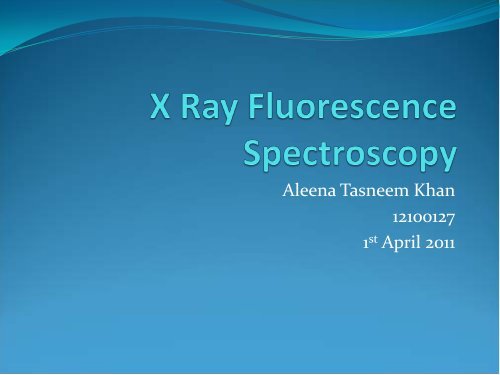
Aleena Tasneem Khan<br />
12100127<br />
1 st April 2011
Outline<br />
• What is X-Ray Fluorescence<br />
– Characteristic X Rays<br />
– Bremsstrahlung Rays<br />
– XRF Analysis – EDXRF <strong>and</strong> WDXRF<br />
• Sample Analysis<br />
– Energy Calibration<br />
– St<strong>and</strong>ard less Analysis<br />
– St<strong>and</strong>ard Analysis<br />
• Moseley’s <strong>Law</strong><br />
– Theory<br />
– Experimental Setup<br />
– Verified Results<br />
• <strong>Compton</strong> Scattering<br />
– Theory<br />
– Experimental Setup<br />
– Verified Results<br />
– Proposed Hardware
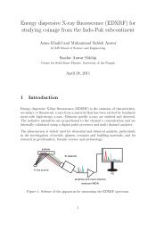
X Ray Fluorescence<br />
• X-Ray Fluorescence: Emission of characteristic secondary X<br />
rays from a material that has been excited by bombarding<br />
with high energy X-Rays.<br />
• Characteristic X-Rays:<br />
X-Rays emitted from heavy<br />
elements when their electrons make<br />
transitions from a higher to a lower<br />
atomic energy level.<br />
• Characteristic X Rays are unique<br />
for each element as energy emitted is<br />
proportional to the binding energy of<br />
the element.
• Bremsstrahlung Radiation: The<br />
electromagnetic radiation<br />
produced due to deceleration of a<br />
charged particle (electron) when<br />
deflected by another charged<br />
particle (the atomic nucleus).<br />
• Bremsstrahlung radiations have a<br />
continuous spectrum where as<br />
intense peaks of the characteristic<br />
x rays can be seen on the<br />
spectrum.
Bremsstrahlung <strong>and</strong> Characteristic<br />
X Rays - Spectrums
XRF Analysis<br />
• XRF Analysis is of two types<br />
• Energy Dispersive XRF (EDXRF)<br />
• Uses a solid state detector <strong>and</strong> distinguishes each peak<br />
according to its energy<br />
• Wave Dispersive XRF (WDXRF)<br />
• Uses the scanning crystal as the dispersive element <strong>and</strong><br />
distinguishes each peak according to its wavelength<br />
• We have used the EDXRF spectrometer.
EDXRF Spectrometer<br />
Working Principle
Sample Analysis<br />
• Obtaining A Spectrum<br />
• Calibration of Curve for Energy<br />
• XRF-FP Corrections<br />
• Sample Analysis<br />
• Calibration using St<strong>and</strong>ards – Intensities obtained<br />
• Calculation of Calibration Coefficients – Concentrations<br />
Obtained
Obtaining A Spectrum<br />
• We use ADMCA along with<br />
the PX4 MCA to obtain the<br />
spectrum for the sample<br />
• To Calibrate: We identify at<br />
least two peaks in the spectrum<br />
<strong>and</strong> enter the characteristic<br />
energy value for each peak.<br />
Using the “Calibrate” function of<br />
ADMCA we calibrate the spectrum in<br />
terms of energy<br />
• This Energy calibration is saved in the MCA <strong>and</strong> loaded<br />
automatically each time a new spectrum needs to be obtained.
XRF-FP Analysis<br />
• The XRF-FP software loads the spectrum obtained by<br />
ADMCA <strong>and</strong> using the initial parameters etc, corrects<br />
the spectrum.<br />
• Three steps for correction:<br />
• Corrections for escape peaks, sum peaks, background<br />
continuum, background peaks etc.<br />
• Deconvolution<br />
• Accounting for attenuation <strong>and</strong> matrix effects<br />
• Using the corrected spectrum, it then calculates the<br />
relative intensities <strong>and</strong> concentrations of each element<br />
in the sample.
XRF-FP Analysis<br />
• The following tables show the intensities <strong>and</strong><br />
concentrations of various elements in a Steel Sample.<br />
Element<br />
Concentration<br />
(wt (%))<br />
Cr 19.055 18.45<br />
Mn 1.281 1.63<br />
Fe 65.939 65.19<br />
Ni 8.67 12.18<br />
Cu 0.294 0.169<br />
Mo 2.902 2.38<br />
Proposed<br />
Concentration<br />
(wt(%))
Moseley’s <strong>Law</strong><br />
• <strong>Moseley's</strong> law is an empirical law concerning the<br />
characteristic x-rays that are emitted by atoms.<br />
• Moseley was able to show that the frequencies of<br />
characteristic X-rays emitted from chemical elements are<br />
proportional to the square of the element's atomic number<br />
• A finding which supported Bohr's model of the atom in<br />
which the atomic number is the same as the number of<br />
positive charges in the nucleus of the atom.
Verification of Results<br />
• Two different samples used for the verification of<br />
Moseley’s <strong>Law</strong>:<br />
• Steel<br />
• Silicon-Brass<br />
• A spectrum of the characteristic X Rays was obtained<br />
for each sample <strong>and</strong> calibrated. Using these energies,<br />
a graph between square root of energy <strong>and</strong> atomic<br />
number of the element was plotted.<br />
• The slope of the resulting graph gave the Rydberg<br />
Constant.
Silicon – Brass<br />
Element<br />
Atomic<br />
Number<br />
Chromium 24 5.41<br />
Energy<br />
(keV)<br />
Manganese 25 5.90<br />
Iron 26 6.40<br />
Cobalt 27 6.93<br />
Nickel 28 7.48<br />
Copper 29 8.05<br />
Zinc 30 8.64<br />
Antimony 51 26.36<br />
Lead 82 74.95<br />
Bismuth 83 77.1
Silicon - Brass
Steel<br />
Element<br />
Atomic<br />
Number<br />
Energy (keV)<br />
Cr 24 5.41<br />
Mn 25 5.90<br />
Fe 26 6.40<br />
Ni 28 7.48<br />
Cu 29 8.05<br />
Mo 42 17.48
Steel Sample
<strong>Compton</strong> Scattering<br />
• <strong>Compton</strong> scattering is the interaction of a high energy<br />
photon with an electron, <strong>and</strong> the resulting “scattered”<br />
photon which has a reduced frequency, <strong>and</strong> therefore<br />
reduced energy introducing a shift in its wavelength.<br />
m
Experimental Setup
Experimental Setup<br />
• Scattering of X Rays was observed by varying the angle<br />
at which the detector was placed. The distance<br />
between x-ray tube <strong>and</strong> the detector was kept larger<br />
than 25cm for the detector’s safety.<br />
An appreciable shift in wavelengths<br />
was seen when the angle was<br />
changed.
Results
Results
Analysis<br />
• A graph between shift in wavelength (delta lambda)<br />
<strong>and</strong> cosine of angle was plotted.
Proposed Assembly