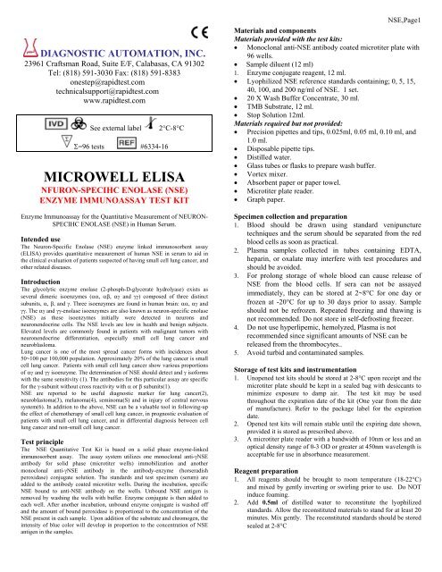
MICROWELL ELISA - Diagnostic Automation : Cortez Diagnostics
MICROWELL ELISA - Diagnostic Automation : Cortez Diagnostics
MICROWELL ELISA - Diagnostic Automation : Cortez Diagnostics

Create successful ePaper yourself
Turn your PDF publications into a flip-book with our unique Google optimized e-Paper software.
DIAGNOSTIC AUTOMATION, INC.<br />
23961 Craftsman Road, Suite E/F, Calabasas, CA 91302<br />
Tel: (818) 591-3030 Fax: (818) 591-8383<br />
onestep@rapidtest.com<br />
technicalsupport@rapidtest.com<br />
www.rapidtest.com<br />
See external label<br />
Σ=96 tests #6334-16<br />
2°C-8°C<br />
<strong>MICROWELL</strong> <strong>ELISA</strong><br />
NFURON-SPECIHC ENOLASE (NSE)<br />
ENZYME IMMUNOASSAY TEST KIT<br />
Enzyme Immunoassay for the Quantitative Measurement of NEURON-<br />
SPECIHC ENOLASE (NSE) in Human Serum.<br />
Intended use<br />
The Neuron-Specific Enolase (NSE) enzyme linked immunosorbent assay<br />
(<strong>ELISA</strong>) provides quantitative measurement of human NSE in serum to aid in<br />
the clinical evaluation of patients suspected of having small cell lung cancer, and<br />
other related diseases.<br />
Introduction<br />
The glycolytic enzyme enolase (2-phosph-D-glycerate hydrolyase) exists as<br />
several dimeric isoenzymes (αα, αβ, αγ and γγ) composed of three distinct<br />
subunits, α, β, and γ. Three isoenzymes are found in human brain: αα, αγ and<br />
γγ. The αγ and γγ-enolase isoenzymes are also known as neuron-specific enolase<br />
(NSE) as these isoenzymes initially were detected in neurons and<br />
neuronendocrine cells. The NSE levels are low in health and benign subjects.<br />
Elevated levels are commonly found in patients with malignant tumors with<br />
neuronendocrine differentiation, especially small cell lung cancer and<br />
neuroblasloma.<br />
Lung cancer is one of the most spread cancer forms with incidences about<br />
50~100 per 100,000 population. Approximately 20% of the lung cancer is small<br />
cell lung cancer. Patients with small cell lung cancer show various proportions<br />
of αγ and γγ isoenzyme. The determination of NSE should detect and γ isoforms<br />
with the same sensitivity (1). The antibodies for this particular assay are specific<br />
for the γ-subunit without cross reactivity with α or β subunits(1).<br />
NSE are reported to be useful diagnostic marker for lung cancer(2),<br />
neuroblastoma(3), melanoma(4), seminoma(S) and in injury of central nervous<br />
system(6). In addition to the above, NSE can be a valuable tool in following-up<br />
the effect of chemotherapy of small cell lung cancer, in prognostic evaluation of<br />
patients with small cell lung cancer, and in differential diagnosis between cell<br />
lung cancer and non-small cell lung cancer.<br />
Test principle<br />
The NSE Quantitative Test Kit is based on a solid phase enzyme-linked<br />
immunosorbent assay. The assay system utilizes one monoclonal anti-γNSE<br />
antibody for solid phase (microtiter wells) immobilization and another<br />
monoclonal anti-γNSE antibody in the antibody-enzyme (horseradish<br />
peroxidase) conjugate solution. The standards and test specimen (serum) are<br />
added to the antibody coated microtiter wells. During the incubation, specific<br />
NSE bound to anti-NSE antibody on the wells. Unbound NSE antigen is<br />
removed by washing the wells with buffer. Enzyme conjugate is then added to<br />
each well. After another incubation, unbound enzyme conjugate is washed off<br />
and the amount of bound peroxidase is proportional to the concentration of the<br />
NSE present in each sample. Upon addition of the substrate and chromogen, the<br />
intensity of blue color will develop in proportion to the concentration of NSE<br />
antigen in the samples.<br />
NSE,Page1<br />
Materials and components<br />
Materials provided with the test kits:<br />
• Monoclonal anti-NSE antibody coated microtiter plate with<br />
96 wells.<br />
• Sample diluent (12 ml)<br />
1. Enzyme conjugate reagent, 12 ml.<br />
• Lyophilized NSE reference standards containing; 0, 5, 15,<br />
40, 100, and 200 ng/ml of NSE. 1 set.<br />
• 20 X Wash Buffer Concentrate, 30 ml.<br />
• TMB Substrate, 12 ml.<br />
• Stop Solution 12ml.<br />
Materials required but not provided:<br />
• Precision pipettes and tips, 0.025ml, 0.05 ml, 0.10 ml, and<br />
1.0 ml.<br />
• Disposable pipette tips.<br />
• Distilled water.<br />
• Glass tubes or flasks to prepare wash buffer.<br />
• Vortex mixer.<br />
• Absorbent paper or paper towel.<br />
• Microtiter plate reader.<br />
• Graph paper.<br />
Specimen collection and preparation<br />
1. Blood should be drawn using standard venipuncture<br />
techniques and the serum should be separated from the red<br />
blood cells as soon as practical.<br />
2. Plasma samples collected in tubes containing EDTA,<br />
heparin, or oxalate may interfere with test procedures and<br />
should be avoided.<br />
3. For prolong storage of whole blood can cause release of<br />
NSE from the blood cells. If sera can not be assayed<br />
immediately, they can be stored at 2~8°C for one day or<br />
frozen at -20°C for up to 30 days prior to assay. Sample<br />
should not be refrozen. Repeated freezing and thawing is<br />
not recommended. Do not store in self-defrosting freezer.<br />
4. Do not use hyperlipemic, hemolyzed, Plasma is not<br />
recommended since significant amounts of NSE can be<br />
released from the thrombocytes..<br />
5. Avoid turbid and contaminated samples.<br />
Storage of test kits and instrumentation<br />
1. Unopened test kits should be stored at 2-8°C upon receipt and the<br />
microtiter plate should be kept in a sealed bag with desiccants to<br />
minimize exposure to damp air. The test kit may be used<br />
throughout the expiration date of the kit (One year from the date<br />
of manufacture). Refer to the package label for the expiration<br />
date.<br />
2. Opened test kits will remain stable until the expiring date shown,<br />
provided it is stored as prescribed above.<br />
3. A microtiter plate reader with a bandwidth of 10nm or less and an<br />
optical density range of 0-3 OD or greater at 450nm wavelength is<br />
acceptable for use in absorbance measurement.<br />
Reagent preparation<br />
1. All reagents should be brought to room temperature (18-22°C)<br />
and mixed by gently inverting or swirling prior to use. Do NOT<br />
induce foaming.<br />
2. Add 0.5ml of distilled water to reconstitute the lyophilized<br />
standards. Allow the reconstituted materials to stand for at least 20<br />
minutes. Mix gently. The reconstituted standards should be stored<br />
sealed at 2-8°C
3. To prepare 1x wash buffer, make a 20x dilution of Wash Buffer<br />
Concentrate. Mix gently to ensure complete mixing.<br />
Assay procedure<br />
1. Secure the desired number of coated wells in the holder.<br />
2. Dispense 25μl of standard, specimens, and controls into<br />
appropriate wells.<br />
3. Dispense 100μl of Sample diluent into each well.<br />
4. Thoroughly mix for 10 seconds. It is very important to<br />
have complete mixing in this setup.<br />
5. Incubate at room temperature (18-22°C) for 30 minutes.<br />
6. Remove the incubation mixture by flicking plate content<br />
into a waste container.<br />
7. Rinse and flick the microtiter wells 5 times with wash<br />
buffer.<br />
8. Strike the wells sharply onto absorbent paper or paper<br />
towels to remove all residual water droplets.<br />
9. Dispense 100μl of Enzyme Conjugate Reagent into each<br />
well. Gently mix for 5 seconds.<br />
10. Incubate at room temperature for 30 minutes.<br />
11. Remove the incubation mixture by flicking plate contents<br />
into a waste container.<br />
12. Rinse and flick the microtiter wells 4 times with wash<br />
buffer and distilled water 1 time.<br />
13. Strike the wells sharply onto absorbent paper to remove<br />
residual water droplets.<br />
14. Dispense 100μl TMB substrate into each well. Gentle mix<br />
for 5 seconds.<br />
15. Incubate at room temperature for 20 minutes.<br />
16. Stop the reaction by adding 100μl of stop solution into each<br />
well.<br />
17. Gently mix for 30 seconds to make sure that the blue color<br />
changes to yellow color completely.<br />
18. Read optical density at 450nm with a microtiter reader<br />
within 30 minutes.<br />
Important Note:<br />
1. The wash procedure is critical. Insufficient washing will result in<br />
poor precision and falsely elevated absorbance readings.<br />
2. It is recommended that no more than 32 wells be used for each<br />
assay run if manual pipetting is used since pipetting of all<br />
standards, specimens and controls should be completed within 5<br />
minutes. A full plate of 96 wells may be used if automated<br />
pipetting is available.<br />
3. Duplication of all standards and specimens, although not<br />
required, is recommended.<br />
4. If a serum specimen contains greater than 180 ng/ml of<br />
NSE the sample must be diluted with sample diluent and reassayed<br />
as described in the assay procedure<br />
Calculation of results<br />
Calculate the mean absorbance value for each set of NSE<br />
reference standards, specimens and controls. Construct a<br />
standard curve by plotting the mean absorbance obtained from<br />
each reference standard against its concentration in units per ml<br />
on linear graph paper, with absorbance values on the vertical or<br />
Y axis and concentrations on the horizontal or X axis. Use the<br />
mean absorbance values for each specimen to determine the<br />
corresponding concentration of NSE in units per ml from the<br />
standard curve. Any diluted specimens must be corrected by<br />
the appropriate dilution factor.<br />
NSE,Page2<br />
Example of standard curve<br />
Results of a typical standard run with optical density reading at 450nm<br />
shown in the Y axis against NSE concentrations shown in the X axis.<br />
NSE Values (ng/ml) Absorbance (450nm)<br />
0 0.010<br />
5 0.195<br />
15 0.418<br />
40 0.928<br />
100 1.980<br />
200 3.309<br />
4<br />
3<br />
2<br />
1<br />
0<br />
0 50 100 150 200<br />
This standard curve is for the purpose of illustration only, and<br />
should not be used to calculate unknowns. Each user should<br />
obtain his or her own standard curve and data.<br />
Expected values and sensitivity<br />
1. It is recommended that each laboratory should determine its<br />
own normal and abnormal ranges as to account for its<br />
environmental factors such as diet, climate etc.<br />
2. A clinical study of the NSE Quantitative kit was conducted<br />
and results are summarized as follows: Nearly all the<br />
individuals have NSE values below 15 ng/ml (95 th<br />
percentile).<br />
3. The expect ranges are representative only, and do not<br />
necessarily reflect the ranges that will be observed in a<br />
particular clinical laboratory.<br />
Limitations and applications<br />
1. For diagnostic purposes, the NSE test results must be used<br />
in conjunction with other data available to the physician.<br />
2. The NSE test should not be used in cancer screening and<br />
should not replace any established clinical examination.<br />
3. Samples with NSF level above 180 ng/ml should be diluted<br />
to obtain accurate value.<br />
4. High NSE values may be found in dialysis patients with<br />
leukaemic diseases.<br />
5. Serum should not contain visible hemolysis since<br />
erythrocytes contai significant amounts of NSE.<br />
6. Prolonged storage of whole blood can cause release of NSE<br />
from the blood cells.<br />
References<br />
1. Paus E. Nustad K. Immunoradiometric assay for αγ and γγ--<br />
Enolase(Neuron Specific Enolase), with use of Monoclonal antibodies and<br />
Magnetizable Polymer Particles. Clin.Chem, 35:2034, 1989.<br />
2. Paus E., Risberg T., Establishment and Evaluation of A Radioimmunoassy<br />
for Neuron-Specific Enolase, Tumour Biol 10:23-30, 1989.<br />
3. Cooper E.H., Pritchard J, bailey CC, Serum Neuron-Specific Enolase in<br />
children's cancer. Br J Cancer. 56:65-67,1987.
4. Wibe E., Paus E., Aamdal S., Neuron Specific Enolase (NSE) in serum of<br />
Patients with Malignant Melanoma, Cancer Letters, 52:29-31,1990.<br />
5. Fossa S.D., Kiepp O., Paus E., Neuron-Specific (NSE) a serum tumor<br />
marker in seminoma. Br. J. Cancer, 65, 297-299,1992.<br />
6. Skogseid I.M., nordby H.K., Urdal P., Paus E., Lilleaas F., Increased serum<br />
creatine kinase BB and Neuron-Specific Enolase following Head Injury<br />
indicates brain damage, Acta Neuronchir(Wien) 115:106-111,1992.<br />
7. Pahlman S., Esscher T., Bergvall p. and Odelstad L. Purification and<br />
Characterization of human -specific Enolase, Radioimmunoassay<br />
Development Tumor Biol. 5,127-139, 1984.<br />
NSE,Page3<br />
DIAGNOSTIC AUTOMATION, INC.<br />
23961 Craftsman Road, Suite E/F, Calabasas, CA 91302<br />
Tel: (818) 591-3030 Fax: (818) 591-8383<br />
ISO 13485-2003<br />
Revision Date: 4/6/06