Case 136: Intraductal Papillary Mucinous Tumor (Main ... - RIHUC
Case 136: Intraductal Papillary Mucinous Tumor (Main ... - RIHUC
Case 136: Intraductal Papillary Mucinous Tumor (Main ... - RIHUC

You also want an ePaper? Increase the reach of your titles
YUMPU automatically turns print PDFs into web optimized ePapers that Google loves.
Note: This copy is for your personal, non-commercial use only. To order presentation-ready copies for<br />
distribution to your colleagues or clients, use the Radiology Reprints form at the end of this article.<br />
<strong>Case</strong> <strong>136</strong>: <strong>Intraductal</strong> <strong>Papillary</strong><br />
<strong>Mucinous</strong> <strong>Tumor</strong> (<strong>Main</strong> Duct Type)<br />
of the Pancreas 1<br />
DIAGNOSIS PLEASE CASE <strong>136</strong><br />
Ana M. Ferreira, MD<br />
Ana Vaz, MD<br />
Morgado Viana, MD<br />
Paulo Gil-Agostinho, MD<br />
Filipe <strong>Case</strong>iro-Alves, PhD<br />
History<br />
A 57-year-old man presented with a 3-month history of<br />
abdominal pain. Physical examination revealed a large firm<br />
palpable mass in the epigastric region. There was no history<br />
of alcoholism, trauma, or previous pancreatitis. Laboratory<br />
findings were unremarkable. The patient underwent<br />
abdominal upright radiography, upper gastrointestinal<br />
barium examination, abdominal ultrasonography (US),<br />
and abdominal computed tomography (CT).<br />
Imaging Findings<br />
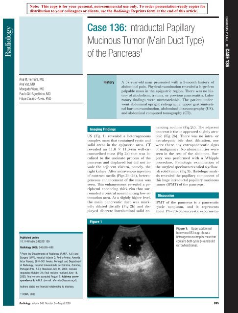
US (Fig 1) revealed a heterogeneous<br />
complex mass that contained cystic and<br />
solid areas in the epigastric area. CT<br />
revealed an 11.6 11.5-cm well-circumscribed<br />
mass (Fig 2a) that was localized<br />
to the uncinate process of the<br />
pancreas and displaced but did not invade<br />
the adjacent viscera, namely, the<br />
right kidney. After intravenous injection<br />
of contrast media (Figs 2b–2d), heterogeneous<br />
enhancement of the mass was<br />
seen. This enhancement revealed a peripheral<br />
enhancing thick rim that surrounded<br />
a central nonenhancing low-attenuation<br />
area. At a slightly higher level,<br />
the main pancreatic duct was markedly<br />
dilated distally (Fig 2b) and displayed<br />
discrete intraluminal solid enhancing<br />
nodules (Fig 2c). The adjacent<br />
pancreatic tissue appeared slightly atrophic<br />
(Fig 2b). There was no intra- or<br />
extrahepatic bile duct dilatation, nor<br />
were there any extrapancreatic signs<br />
of malignancy. No abnormalities were<br />
seen in the rest of the abdomen. Surgery<br />
was performed with a Whipple<br />
procedure. Pathologic examination of<br />
the surgical specimen revealed a yellowish<br />
solid tumor (Fig 3). Histologic analysis<br />
revealed the papillary component of<br />
this huge intraductal papillary mucinous<br />
tumor (IPMT) of the pancreas.<br />
Discussion<br />
IPMT of the pancreas is a pancreatic<br />
cystic neoplasm, and it represents<br />
about 1%–2% of pancreatic exocrine tu-<br />
Figure 1<br />
Published online<br />
10.1148/radiol.2482031139<br />
Radiology 2008; 248:695–698<br />
Figure 1: Upper abdominal<br />
transverse US image shows a<br />
heterogeneous complex mass that<br />
contains both cystic () and solid<br />
(arrowhead) areas.<br />
1 From the Departments of Radiology (A.M.F., A.V.) and<br />
Surgery (M.V.), Hospital Infante D. Pedro-Aveiro, Avenida<br />
Artur Ravara, 3814-501 Aveiro, Portugal; and Department<br />
of Radiology, Hospital Universidade de Coimbra, Coimbra,<br />
Portugal (P.G., F.C.). Received July 31, 2003; revision<br />
requested October 21; final revision received June 18,<br />
2005; final version accepted August 5. Address correspondence<br />
to A.M.F. (e-mail: aferreira@essua.ua.pt).<br />
Authors stated no financial relationship to disclose.<br />
RSNA, 2008<br />
Radiology: Volume 248: Number 2—August 2008 695
DIAGNOSIS PLEASE: <strong>Intraductal</strong> <strong>Papillary</strong> <strong>Mucinous</strong> <strong>Tumor</strong> (<strong>Main</strong> Duct Type) of the Pancreas<br />
Ferreira et al<br />
mors (1). IPMTs occur with equal frequency<br />
in male and female subjects, and<br />
the peak time of occurrence is the 6th<br />
decade of life, regardless of sex (2). Approximately<br />
75% of IPMTs originate<br />
from the main pancreatic duct within<br />
the head of the pancreas (3). Imaging of<br />
IPMTs is important not only to identify<br />
the tumor but also to determine the appropriate<br />
treatment strategy in relation<br />
to the site and size of the lesion (2).<br />
Although the presence of a dominant<br />
mass of considerable size (as in the<br />
present case) is a rare occurrence, it<br />
should not preclude the diagnosis of<br />
IPMT.<br />
Imaging findings vary according to<br />
the ductal involvement of the disease,<br />
which can be divided into three types:<br />
(a) the main duct type, (b) the branch<br />
duct type, and (c) the combined type<br />
Figure 2<br />
(4). In main duct IPMTs, the lesion is<br />
usually homogeneous and hypoechoic<br />
on US images and hypoattenuating on<br />
CT images. <strong>Intraductal</strong> filling defects<br />
can be seen in the main duct. These<br />
defects represent mucin globules, papillary<br />
projections of the tumor that are<br />
hyperechoic on US images, or both<br />
(2,5). At CT, unenhanced mucin deposits<br />
can be distinguished from contrastenhanced<br />
papillary proliferations (5).<br />
Sometimes, it is possible to identify<br />
amorphous intraductal calcifications,<br />
which are thought to represent calcium<br />
deposits within the mucinous collections<br />
and which may lead to an incorrect<br />
diagnosis of chronic pancreatitis<br />
(6). Although Fukushima et al (7) reported<br />
5.0–10.5-cm-diameter IPMTs,<br />
large size is an uncommon feature of<br />
this tumor.<br />
Branch duct IPMTs, which more<br />
frequently are located in the uncinate<br />
process, may have a macrocystic or<br />
microcystic pattern. The microcystic<br />
pattern is characterized by multiple<br />
thin septa separating fluid-filled spaces<br />
showing US, CT, and magnetic resonance<br />
cholangiopancreatography<br />
(MRCP) features, such as clustered<br />
small cysts with a lobulated contour<br />
(2,4). The macrocystic pattern, which<br />
is seen more frequently, is characterized<br />
by a unilocular or multilocular internal<br />
architecture (2). If the tumor is<br />
limited to one of the branch ducts, it<br />
has a unilocular cystic appearance (4).<br />
In later stages, the main duct may also<br />
be dilated because of mucin accumulation<br />
(2), and severe pancreatic atrophy<br />
may follow (4). Demonstration of communication<br />
between the lesion and the<br />
main duct leads to the correct diagnosis<br />
and can occasionally be shown by using<br />
thin-section spiral CT (2).<br />
In the combined type of IPMT, both<br />
the branch ducts and the main pancreatic<br />
duct are involved (4). The duodenal<br />
papilla may protrude into the duodenal<br />
lumen (4). This finding, which is more<br />
frequent in malignant forms (4), is virtually<br />
pathognomonic of IPMT (2).<br />
Endoscopically, the ampulla of Vater<br />
is often prominent, giving rise to an<br />
abundant discharge of mucinous fluid<br />
(8). Endoscopic retrograde cholangiopancreatography<br />
(ERCP) may be the<br />
Figure 3<br />
Figure 2: Transverse (a) unenhanced and (b–d) contrast material–enhanced (120 mL, 350 mg of iodine<br />
per mL) abdominal CT scans at the level of the palpable mass. In a, an 11.6 11.5-cm well-circumscribed<br />
mass () localized at the pancreatic head can be seen. The right kidney (arrowhead) is also visible. In b, the<br />
main pancreatic duct () is slightly dilated, and the adjacent pancreatic tissue (arrowhead) appears slightly<br />
atrophic. In c, a mass with a peripheral enhancing thick rim surrounding a central low-attenuation area is visible.<br />
A nodule (arrowhead) can also be seen. In d, there are some discrete intraluminal solid enhancing nodules<br />
(arrow).<br />
Figure 3: Gross solid tumor specimen containing<br />
yellowish papillary projections ()atthe<br />
periphery.<br />
696 Radiology: Volume 248: Number 2—August 2008
DIAGNOSIS PLEASE: <strong>Intraductal</strong> <strong>Papillary</strong> <strong>Mucinous</strong> <strong>Tumor</strong> (<strong>Main</strong> Duct Type) of the Pancreas<br />
Ferreira et al<br />
most direct method with which to diagnose<br />
IPMT when it shows the characteristic<br />
appearance of mucin discharge<br />
from a protruded papilla of Vater (9),<br />
the dilated pancreatic duct with intraluminal<br />
filling defects due to the presence<br />
of mucous plugs (10), and the direct<br />
communication of the cystically dilated<br />
ductal segment with the main pancreatic<br />
duct (2). The dilated duct may lead<br />
to a grapelike cluster of cysts with pooling<br />
of contrast material, or it may be<br />
obstructed by the mucin or the hyperplastic<br />
papillary epithelium (10). Sometimes,<br />
it demonstrates a fistulous tract<br />
leaking mucin between the lesion and<br />
the duodenal wall (6). The ERCP data<br />
are not constant because mucous plugs<br />
may occlude the patency of the ampullary<br />
orifice, obviating retrograde opacification<br />
by the contrast material (6).<br />
MRCP is more sensitive than ERCP<br />
in the detection of cystic dilatation of<br />
the side branches (11), and it enables<br />
one to avoid the complications associated<br />
with ERCP (9). Nevertheless, inspection<br />
of a patulous papilla is only<br />
possible with use of ERCP (9), as it is<br />
superior to MRCP for depicting direct<br />
communication of the lesion with the<br />
main pancreatic duct (2). Perhaps secretin-enhanced<br />
MRCP may have an<br />
added value in this regard (2).<br />
Despite its unusually large size,<br />
IPMT was a likely diagnosis because it<br />
appeared as a cystlike tumor of the pancreatic<br />
head in a middle-aged man, with<br />
CT features of main duct dilatation containing<br />
solid papillary projections. The<br />
findings in this patient suggest several<br />
diagnoses for a cystic pancreatic mass:<br />
(a) mucinous cystic neoplasm, (b) serous<br />
cystadenoma, (c) solid and papillary<br />
epithelial neoplasm, and (d) nonfunctioning<br />
islet cell tumor.<br />
<strong>Mucinous</strong> cystic neoplasms of the<br />
pancreas occur in middle-aged women,<br />
and the majority of these lesions are<br />
located in the pancreatic body or tail<br />
(12). They tend to be large; indeed,<br />
many are larger than 10 cm (13). They<br />
can manifest in the form of abdominal<br />
pain, a palpable mass, dyspepsia, anorexia,<br />
weight loss, nausea, or vomiting.<br />
Symptoms usually result from compression<br />
or displacement of neighboring<br />
organs and are commonly insidious and<br />
of long duration. When symptoms are<br />
located in the pancreatic head, they<br />
cause early jaundice, which is commonly<br />
seen in malignant neoplasms<br />
(12). Pathologically, mucinous cystic tumors<br />
are encapsulated and multiloculated,<br />
with fewer than six cysts being<br />
larger than 2 cm in diameter (14). The<br />
cystic walls are often thickened, and irregular<br />
septa or papillary projections<br />
may be seen (14). The amount of<br />
stroma varies and, when small, the<br />
septa may not be apparent (15). Contrary<br />
to IPMTs, mucinous cystic tumors<br />
tend to occur in female patients and are<br />
generally located in the body or tail of<br />
the pancreas, without accompanying<br />
ductal dilatation.<br />
In 1978, Compagno and Oertel (16)<br />
differentiated serous cystadenoma from<br />
mucinous cystic tumor. Serous cystadenomas<br />
are almost always benign; malignant<br />
forms are exceedingly rare. Benign<br />
serous cystadenomas occur predominantly<br />
in elderly women without predilection<br />
for a particular segment of the<br />
pancreatic gland. Benign serous cystadenomas<br />
are often an incidental finding<br />
or are seen in patients who present<br />
with nonspecific clinical features. The<br />
median size of these lesions ranges from<br />
a few millimeters to many centimeters.<br />
The macroscopic structure of these lesions<br />
is greatly variable. Although these<br />
lesions are characteristically microlacunar,<br />
tumors with a mixed structure consisting<br />
of a microlacunar core surrounded<br />
by cysts larger than 2 cm in<br />
diameter, as well as macrolacunar tumors<br />
with unilocular cysts usually larger<br />
than 2 cm in diameter, have been reported<br />
(10). The preponderance of lesions<br />
in women, the central calcification,<br />
and the microcystic appearance<br />
enable one to readily distinguish this entity<br />
from IPMT.<br />
A solid and papillary neoplasm is an<br />
uncommon low-grade malignancy that<br />
occurs predominantly in young women<br />
(mean age, 25 years). These tumors are<br />
typically large and encapsulated, have a<br />
mean size of 9 cm, and are most commonly<br />
located in the tail of the pancreas.<br />
The internal architecture shows<br />
variable proportions of solid and cystic<br />
areas, indicating cystic degeneration<br />
and necrosis (17,18). Some cases can<br />
be exclusively solid, whereas others<br />
may be entirely cystic (18). This entity<br />
could be ruled out not only because of<br />
the tumor location but also because this<br />
patient was a 57-year-old man.<br />
Nonfunctioning islet cell tumors<br />
should also be included in the differential<br />
diagnosis because they are clinically<br />
silent until they are large or until they<br />
metastasize, and they may grow to be<br />
large (3–24 cm in diameter), with cystic<br />
change and necrosis (19). However,<br />
contrary to IPMTs, nonfunctioning islet<br />
cell tumors do not have ductal dilatation<br />
and internal enhancing papillary projections<br />
as imaging features.<br />
Acknowledgments: We thank Abílio Brandão,<br />
MD, Department of Pathology, Hospital Infante<br />
D. Pedro-Aveiro, and Augusta Cipriano, MD,<br />
Department of Pathology, Hospital Universidade<br />
Coimbra, for having facilitated and labeled the<br />
histologic images.<br />
References<br />
1. Taouli B, Vilgrain V, Vullierme MP, et al.<br />
<strong>Intraductal</strong> papillary mucinous tumors of the<br />
pancreas: helical CT with histopathologic<br />
correlation. Radiology 2000;217(3):757–764.<br />
2. Procacci C, Megibow A, Carbognin G, et<br />
al. <strong>Intraductal</strong> papillary mucinous tumor of<br />
the pancreas: a pictorial essay. RadioGraphics<br />
1999;19(6):1447–1463.<br />
3. Klöppel G. Clinicopathologic view of intraductal<br />
papillary-mucinous tumor of the pancreas.<br />
Hepatogastroenterology 1998;45(24):<br />
1981–1985.<br />
4. Lim JH, Lee G, Oh YL. Radiologic spectrum of<br />
intraductal papillary mucinous tumor of the pancreas.<br />
RadioGraphics 2001;21(2):323–337.<br />
5. Procacci C, Schenal G, Chiara ED, Fuini A,<br />
Guarise A. <strong>Intraductal</strong> papillary mucinous<br />
tumors: imaging. In: Procacci C, Megibow AJ,<br />
eds. Imaging of the pancreas: cystic and rare<br />
tumors. New York, NY: Springer, 2003;97–135.<br />
6. Procacci C, Graziani R, Bicego E, et al. <strong>Intraductal</strong><br />
mucin-producing tumors of the pancreas: imaging<br />
findings. Radiology 1996;198(1):249–257.<br />
7. Fukushima N, Mukai K, Sakamoto M, et al.<br />
Invasive carcinoma derived from intraductal<br />
papillary-mucinous carcinoma of the pancreas:<br />
clinicopathologic and immunohistochemical<br />
study of eight cases. Virchows Arch 2001;<br />
439(1):6–13.<br />
8. Jyotheeswaran S, Zotalis G, Penmetsa P, Le-<br />
Radiology: Volume 248: Number 2—August 2008 697
DIAGNOSIS PLEASE: <strong>Intraductal</strong> <strong>Papillary</strong> <strong>Mucinous</strong> <strong>Tumor</strong> (<strong>Main</strong> Duct Type) of the Pancreas<br />
Ferreira et al<br />
vea CM, Schoeniger LO, Shah AN. A newly<br />
recognized entity: intraductal “oncocytic” papillary<br />
neoplasm of the pancreas. Am J Gastroenterol<br />
1998;93(12):2539–2543.<br />
9. Albert J, Schilling D, Breer H, Jungius KP,<br />
Riemann JF, Adamek HE. <strong>Mucinous</strong> cystadenomas<br />
and intraductal papillary mucinous<br />
tumors of the pancreas in magnetic resonance<br />
cholangiopancreatography. Endoscopy<br />
2000;32(6):472–476.<br />
10. Procacci C, Graziani R, Bicego E, et al. Serous<br />
cystadenoma of the pancreas: report of<br />
30 cases with emphasis on the imaging findings.<br />
J Comput Assist Tomogr 1997;21(3):<br />
373–382.<br />
11. Dunnick NR. Image interpretation session:<br />
1999. <strong>Intraductal</strong> mucin-producing tumor of<br />
the pancreas. RadioGraphics 2000;20(1):<br />
258–260.<br />
12. de Lima JE Jr, Javitt MC, Mathur SC. <strong>Mucinous</strong><br />
cystic neoplasm of the pancreas. Radio-<br />
Graphics 1999;19(3):807–811.<br />
13. Bluemke DA, Soyer P. CT of endocrine and<br />
cystic tumors of the pancreas. In: Terrier F,<br />
Grossholz M, Becker CD, eds. Spiral CT of<br />
the abdomen. New York, NY: Springer,<br />
2000;215–226.<br />
14. Jacobs J. Cystic pancreatic neoplasms. Acta<br />
Radiol Port 2002;53(14):35–36.<br />
15. Stanley RJ, Semelka RC. Pancreas. In: Lee<br />
JKT, Sagel SS, Stanley RJ, Heiken JP, eds.<br />
Computed body tomography with MRI correlation.<br />
3rd ed. Philadelphia, Pa: Lippincott-Raven,<br />
1998;907–913.<br />
16. Compagno J, Oertel JE. Microcystic adenomas<br />
of the pancreas (glycogen-rich cystadenomas):<br />
a clinicopathologic study of 34 cases. Am J Clin<br />
Pathol 1978;69(3):289–298.<br />
17. Stanley RJ, Semelka RC. Pancreas. In: Lee<br />
JKT, Sagel SS, Stanley RJ, Heiken JP, eds.<br />
Computed body tomography with MRI correlation.<br />
3rd ed. Philadelphia, Pa: Lippincott-Raven,<br />
1998;913–914.<br />
18. Zamboni G, Capelli P, Pesci A, Brighenti A.<br />
Pathology of cystic tumors. In: Imaging of<br />
the pancreas cystic and rare tumors. New<br />
York, NY: Springer, 2003;9–25.<br />
19. Stanley RJ, Semelka RC. Pancreas. In: Lee<br />
JKT, Sagel SS, Stanley RJ, Heiken JP, eds.<br />
Computed body tomography with MRI correlation.<br />
3rd ed. Philadelphia, Pa: Lippincott-Raven,<br />
1998;902–906.<br />
Congratulations to the five individuals<br />
who submitted the most likely diagnosis<br />
(intraductal papillary mucinous tumor<br />
of the pancreas) for Diagnosis<br />
Please, <strong>Case</strong> <strong>136</strong>. The names and locations<br />
of the individuals, as submitted,<br />
are as follows:<br />
Albert J. Alter, MD, PhD, Madison, Wis<br />
Fahad Azzumeea, MBBS, Montreal, Quebec, Canada<br />
Antonio A. Cavalcanti, MD, Sao Paulo, Brazil<br />
Michael H. Childress, MD, Silver Spring, Md<br />
Seyed A. Emamian, MD, PhD, Rockville, Md<br />
698 Radiology: Volume 248: Number 2—August 2008
Radiology 2008<br />
This is your reprint order form or pro forma invoice<br />
(Please keep a copy of this document for your records.)<br />
Reprint order forms and purchase orders or prepayments must be received 72 hours after receipt of form either<br />
by mail or by fax at 410-820-9765. It is the policy of Cadmus Reprints to issue one invoice per order.<br />
Please print clearly.<br />
Author Name _______________________________________________________________________________________________<br />
Title of Article _______________________________________________________________________________________________<br />
Issue of Journal_______________________________ Reprint # _____________ Publication Date ________________<br />
Number of Pages_______________________________ KB # _____________ Symbol Radiology<br />
Color in Article? Yes / No (Please Circle)<br />
Please include the journal name and reprint number or manuscript number on your purchase order or other correspondence.<br />
Order and Shipping Information<br />
Reprint Costs (Please see page 2 of 2 for reprint costs/fees.)<br />
________ Number of reprints ordered $_________<br />
________ Number of color reprints ordered $_________<br />
________ Number of covers ordered $_________<br />
Subtotal $_________<br />
Taxes<br />
$_________<br />
(Add appropriate sales tax for Virginia, Maryland, Pennsylvania, and the<br />
District of Columbia or Canadian GST to the reprints if your order is to<br />
be shipped to these locations.)<br />
First address included, add $32 for<br />
each additional shipping address<br />
TOTAL<br />
$_________<br />
$_________<br />
Shipping Address (cannot ship to a P.O. Box) Please Print Clearly<br />
Name ___________________________________________<br />
Institution _________________________________________<br />
Street ___________________________________________<br />
City ____________________ State _____ Zip ___________<br />
Country ___________________________________________<br />
Quantity___________________ Fax ___________________<br />
Phone: Day _________________ Evening _______________<br />
E-mail Address _____________________________________<br />
Additional Shipping Address* (cannot ship to a P.O. Box)<br />
Name ___________________________________________<br />
Institution _________________________________________<br />
Street ___________________________________________<br />
City ________________ State ______ Zip ___________<br />
Country _________________________________________<br />
Quantity __________________ Fax __________________<br />
Phone: Day ________________ Evening ______________<br />
E-mail Address ____________________________________<br />
* Add $32 for each additional shipping address<br />
Payment and Credit Card Details<br />
Enclosed: Personal Check ___________<br />
Credit Card Payment Details _________<br />
Checks must be paid in U.S. dollars and drawn on a U.S. Bank.<br />
Credit Card: __ VISA __ Am. Exp. __ MasterCard<br />
Card Number __________________________________<br />
Expiration Date_________________________________<br />
Signature: _____________________________________<br />
Please send your order form and prepayment made payable to:<br />
Cadmus Reprints<br />
P.O. Box 751903<br />
Charlotte, NC 28275-1903<br />
Note: Do not send express packages to this location, PO Box.<br />
FEIN #:541274108<br />
Invoice or Credit Card Information<br />
Invoice Address Please Print Clearly<br />
Please complete Invoice address as it appears on credit card statement<br />
Name ____________________________________________<br />
Institution ________________________________________<br />
Department _______________________________________<br />
Street ____________________________________________<br />
City ________________________ State _____ Zip _______<br />
Country ___________________________________________<br />
Phone _____________________ Fax _________________<br />
E-mail Address _____________________________________<br />
Cadmus will process credit cards and Cadmus Journal<br />
Services will appear on the credit card statement.<br />
If you don’t mail your order form, you may fax it to 410-820-9765 with<br />
your credit card information.<br />
Signature __________________________________________ Date _______________________________________<br />
Signature is required. By signing this form, the author agrees to accept the responsibility for the payment of reprints and/or all charges<br />
described in this document.<br />
RB-9/26/07<br />
Page 1 of 2
Radiology 2008<br />
Black and White Reprint Prices<br />
Domestic (USA only)<br />
# of<br />
Pages<br />
50 100 200 300 400 500<br />
1-4 $221 $233 $268 $285 $303 $323<br />
5-8 $355 $382 $432 $466 $510 $544<br />
9-12 $466 $513 $595 $652 $714 $775<br />
13-16 $576 $640 $749 $830 $912 $995<br />
17-20 $694 $775 $906 $1,017 $1,117 $1,220<br />
21-24 $809 $906 $1,071 $1,200 $1,321 $1,471<br />
25-28 $928 $1,041 $1,242 $1,390 $1,544 $1,688<br />
29-32 $1,042 $1,178 $1,403 $1,568 $1,751 $1,924<br />
Covers $97 $118 $215 $323 $442 $555<br />
International (includes Canada and Mexico)<br />
# of<br />
Pages<br />
50 100 200 300 400 500<br />
1-4 $272 $283 $340 $397 $446 $506<br />
5-8 $428 $455 $576 $675 $784 $884<br />
9-12 $580 $626 $805 $964 $1,115 $1,278<br />
13-16 $724 $786 $1,023 $1,232 $1,445 $1,652<br />
17-20 $878 $958 $1,246 $1,520 $1,774 $2,030<br />
21-24 $1,022 $1,119 $1,474 $1,795 $2,108 $2,426<br />
25-28 $1,176 $1,291 $1,700 $2,070 $2,450 $2,813<br />
29-32 $1,316 $1,452 $1,936 $2,355 $2,784 $3,209<br />
Covers $156 $176 $335 $525 $716 $905<br />
Minimum order is 50 copies. For orders larger than 500 copies,<br />
please consult Cadmus Reprints at 800-407-9190.<br />
Reprint Cover<br />
Cover prices are listed above. The cover will include the<br />
publication title, article title, and author name in black.<br />
Shipping<br />
Shipping costs are included in the reprint prices. Domestic<br />
orders are shipped via UPS Ground service. Foreign orders are<br />
shipped via a proof of delivery air service.<br />
Multiple Shipments<br />
Orders can be shipped to more than one location. Please be<br />
aware that it will cost $32 for each additional location.<br />
Delivery<br />
Your order will be shipped within 2 weeks of the journal print<br />
date. Allow extra time for delivery.<br />
Color Reprint Prices<br />
Domestic (USA only)<br />
# of<br />
Pages<br />
50 100 200 300 400 500<br />
1-4 $223 $239 $352 $473 $597 $719<br />
5-8 $349 $401 $601 $849 $1,099 $1,349<br />
9-12 $486 $517 $852 $1,232 $1,609 $1,992<br />
13-16 $615 $651 $1,105 $1,609 $2,117 $2,624<br />
17-20 $759 $787 $1,357 $1,997 $2,626 $3,260<br />
21-24 $897 $924 $1,611 $2,376 $3,135 $3,905<br />
25-28 $1,033 $1,071 $1,873 $2,757 $3,650 $4,536<br />
29-32 $1,175 $1,208 $2,122 $3,138 $4,162 $5,180<br />
Covers $97 $118 $215 $323 $442 $555<br />
International (includes Canada and Mexico))<br />
# of<br />
Pages<br />
50 100 200 300 400 500<br />
1-4 $278 $290 $424 $586 $741 $904<br />
5-8 $429 $472 $746 $1,058 $1,374 $1,690<br />
9-12 $604 $629 $1,061 $1,545 $2,011 $2,494<br />
13-16 $766 $797 $1,378 $2,013 $2,647 $3,280<br />
17-20 $945 $972 $1,698 $2,499 $3,282 $4,069<br />
21-24 $1,110 $1,139 $2,015 $2,970 $3,921 $4,873<br />
25-28 $1,290 $1,321 $2,333 $3,437 $4,556 $5,661<br />
29-32 $1,455 $1,482 $2,652 $3,924 $5,193 $6,462<br />
Covers $156 $176 $335 $525 $716 $905<br />
Tax Due<br />
Residents of Virginia, Maryland, Pennsylvania, and the District<br />
of Columbia are required to add the appropriate sales tax to each<br />
reprint order. For orders shipped to Canada, please add 7%<br />
Canadian GST unless exemption is claimed.<br />
Ordering<br />
Reprint order forms and purchase order or prepayment is<br />
required to process your order. Please reference journal name<br />
and reprint number or manuscript number on any<br />
correspondence. You may use the reverse side of this form as a<br />
proforma invoice. Please return your order form and<br />
prepayment to:<br />
Cadmus Reprints<br />
P.O. Box 751903<br />
Charlotte, NC 28275-1903<br />
Note: Do not send express packages to this location, PO Box.<br />
FEIN #:541274108<br />
Please direct all inquiries to:<br />
Rose A. Baynard<br />
800-407-9190 (toll free number)<br />
410-819-3966 (direct number)<br />
410-820-9765 (FAX number)<br />
baynardr@cadmus.com (e-mail)<br />
Reprint Order Forms<br />
and purchase order<br />
or prepayments must<br />
be received 72 hours<br />
after receipt of form.<br />
Page 2 of 2