Feasibility of transoral roboticassisted supraglottic laryngectomy
Feasibility of transoral roboticassisted supraglottic laryngectomy
Feasibility of transoral roboticassisted supraglottic laryngectomy

Create successful ePaper yourself
Turn your PDF publications into a flip-book with our unique Google optimized e-Paper software.
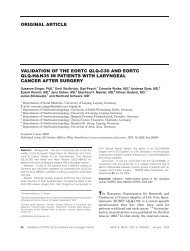
ORIGINAL ARTICLE<br />
FEASIBILITY OF TRANSORAL ROBOTIC-ASSISTED<br />
SUPRAGLOTTIC LARYNGECTOMY<br />
Eran E. Alon, MD, Jan L. Kasperbauer, MD, Kerry D. Olsen, MD, Eric J. Moore, MD<br />
Department <strong>of</strong> Otorhinolaryngology, Mayo Clinic, Rochester, Minnesota. E-mail: moore.eric@mayo.edu<br />
Accepted 13 December 2010<br />
Published online 15 April 2011 in Wiley Online Library (wileyonlinelibrary.com). DOI: 10.1002/hed.21719<br />
Abstract: Background. We aimed to describe our experience<br />
with <strong>transoral</strong> robotic-assisted <strong>supraglottic</strong> <strong>laryngectomy</strong>.<br />
Methods. We retrospectively reviewed the records <strong>of</strong><br />
patients who underwent <strong>transoral</strong> robotic-assisted <strong>supraglottic</strong><br />
<strong>laryngectomy</strong> at our institution between May 2007 and November<br />
2008.<br />
Results. During the study period, 7 patients with malignancy<br />
underwent the procedure (4 men, 3 women; average<br />
age, 61 years). All tumors were completely resected, with clear<br />
margins on pathologic analysis. Six patients underwent neck<br />
dissection at the time <strong>of</strong> <strong>transoral</strong> surgery. One intraoperative<br />
complication occurred: thermal injury to the anterior cervical<br />
skin. Four patients underwent primary tracheotomy tube placement,<br />
3 <strong>of</strong> whom were decannulated without difficulty. Two<br />
patients required long-term gastrostomy tubes while receiving<br />
adjuvant radiotherapy.<br />
Conclusions. Supraglottic malignancies can be successfully<br />
resected <strong>transoral</strong>ly with robotic assistance. The potential<br />
benefits <strong>of</strong> this method are improved access and visualization.<br />
Further long-term follow-up is needed to compare oncologic<br />
and functional outcomes with those <strong>of</strong> current approaches.<br />
VC 2011 Wiley Periodicals, Inc. Head Neck 34: 225–229, 2012<br />
Keywords: <strong>supraglottic</strong> <strong>laryngectomy</strong>; <strong>transoral</strong> robotic surgery<br />
Transoral laser microsurgery was initially described<br />
by Vaughan et al 1,2 and further popularized by<br />
Steiner 3 for resection <strong>of</strong> malignant tumors <strong>of</strong> the<br />
upper aerodigestive tract. Numerous studies have<br />
since been published describing <strong>transoral</strong> resection <strong>of</strong><br />
malignancies in the <strong>supraglottic</strong> larynx. 4–12 Recently,<br />
Ambrosch 13 reviewed the literature on laser microsurgery<br />
for the treatment <strong>of</strong> laryngeal cancer. The 5-year<br />
survival rates for early <strong>supraglottic</strong> carcinomas (T1<br />
and T2) achieved with laser microsurgery were comparable<br />
to the rates reported for open <strong>supraglottic</strong> <strong>laryngectomy</strong>.<br />
Radiotherapy for T1 and T2 cancers was<br />
shown to have comparable tumor control rates; how-<br />
Correspondence to: E. J. Moore<br />
The Mayo Clinic does not endorse the products mentioned in this<br />
article.<br />
This work was presented at Combined Otolaryngology Spring Meeting<br />
in Phoenix, Arizona, May 28–31, 2009.<br />
VC 2011 Wiley Periodicals, Inc.<br />
ever, for patients with local recurrence, 14% to 31% <strong>of</strong><br />
patients required total <strong>laryngectomy</strong>. Only a few<br />
reports in the literature describe <strong>transoral</strong> treatment<br />
outcomes for moderately advanced (T3) cancers.<br />
These studies show significantly better control and<br />
organ preservation with laser microsurgery or open<br />
approaches than those with radiotherapy. 13 Furthermore,<br />
treatment by <strong>transoral</strong> approaches is associated<br />
with better swallowing outcomes, shorter durations <strong>of</strong><br />
feeding tube placement, and decreased need for tracheotomy<br />
tube compared with open approaches. 13<br />
Transoral robotic surgery was introduced by Weinstein<br />
et al 14 in 2005. Previous work from the University<br />
<strong>of</strong> Pennsylvania has shown the feasibility and<br />
safety <strong>of</strong> this approach to the larynx. 15,16 Furthermore,<br />
Solares and Strome 17 demonstrated on a cadaveric<br />
model the feasibility <strong>of</strong> the daVinci Surgical<br />
System (Intuitive Surgical Inc, Sunnyvale, CA) for<br />
accessing the <strong>supraglottic</strong> larynx. The advantages<br />
cited for using the daVinci robotic system in <strong>transoral</strong><br />
surgery include movable, high-definition, 3-dimensional<br />
imaging, not necessarily requiring line <strong>of</strong> sight;<br />
natural hand movement translated to the robotic<br />
instrument; and a shorter learning curve compared<br />
with that <strong>of</strong> traditional <strong>transoral</strong> techniques. 18<br />
At our institution, more than 100 <strong>transoral</strong><br />
approaches to tumors in the oropharynx, tongue base,<br />
and larynx have been performed. Here, we describe<br />
our experience with <strong>transoral</strong> robotic-assisted resection<br />
<strong>of</strong> <strong>supraglottic</strong> malignancies.<br />
MATERIALS AND METHODS<br />
This study was approved by the Mayo Clinic Institutional<br />
Review Board. We retrospectively searched our<br />
patient database for records <strong>of</strong> patients who underwent<br />
<strong>transoral</strong> robotic-assisted resection <strong>of</strong> <strong>supraglottic</strong><br />
malignancies between May 2007 and November<br />
2008. Information obtained from the medical records<br />
included surgical procedure, tumor type and stage,<br />
postoperative recovery, and any complications.<br />
Operative Technique. The patients were placed in a<br />
supine position and intubated with a laser-protected<br />
endotracheal tube. Paralysis was induced to assist in<br />
Robotic Transoral Supraglottic Laryngectomy HEAD & NECK—DOI 10.1002/hed February 2012 225
FIGURE 1. Feyh-Kastenbauer oral retractor. [Color figure can be<br />
viewed in the online issue, which is available at wileyonlinelibrary.<br />
com.]<br />
<strong>transoral</strong> exposure. A Feyh-Kastenbauer oral retractor<br />
(Gyrus ACMI, Bartlett, TN) was used for <strong>transoral</strong> exposure<br />
(see Figure 1). This retractor includes several<br />
adjustable blades; for <strong>supraglottic</strong> exposure, the laryngeal<br />
blade (Wollenberg Laryngeal Blade; Gyrus ACMI)<br />
(see Figure 2) was found to be most beneficial because<br />
it reached deep into the vallecula. The laryngoscope<br />
holder and chest support were used to suspend the<br />
retractor. The retractor allowed the surgeon to advance<br />
the blade and to pivot the angle <strong>of</strong> the blade to obtain<br />
maximal exposure. Slim cheek retractors can be<br />
mounted to help secure the endotracheal tube to the<br />
side, and suction may be attached to the frame <strong>of</strong> the<br />
retractor, although we did not find this efficacious.<br />
Once we achieved adequate exposure, the daVinci<br />
Surgical System was brought into the surgical field,<br />
alongside the operating table. Three robotic arms<br />
were used: 1 arm held the camera at midline; the<br />
other 2 arms held the various 5-mm instruments. In<br />
most cases, the Schertel grasper (Intuitive Surgical)<br />
(see Figure 3) was found to be most useful in handling<br />
the tissue, and a spatula tip cautery (Intuitive<br />
Surgical) was placed in the other arm (see Figure 4).<br />
A30 upward-facing, binocular, rigid endoscope was<br />
FIGURE 3. Operative photograph. Operative exposure with<br />
assistant at head <strong>of</strong> bed and robot in position. [Color figure can be<br />
viewed in the online issue, which is available at wileyonlinelibrary.<br />
com.]<br />
used in all cases. The 30 lens allowed for placement<br />
<strong>of</strong> the camera in the oropharynx, keeping it out <strong>of</strong> the<br />
way <strong>of</strong> the other instruments. The grasping instrument<br />
and cautery were brought in at an angle and<br />
the trocars supporting these instruments were positioned<br />
at cheek level to maximize the range <strong>of</strong> motion<br />
and to avoid collision <strong>of</strong> the instruments. The primary<br />
surgeon operated the daVinci system from the console.<br />
An assistant was positioned at the head <strong>of</strong> the<br />
patient with 2 suction instruments, assisting in<br />
smoke evacuation, retraction, and hemostasis.<br />
The approach to the tumors depended on their<br />
location and extent. If the tumors were small enough<br />
to be visualized in their entirety, an attempt was<br />
made to resect them en bloc. For large, deep, infiltrating<br />
tumors, dissection was carried through the<br />
tumors to establish depth and orientation, and the<br />
tumors were resected piecemeal. Margins were submitted<br />
separately for frozen section pathologic analysis<br />
in all cases. All frozen section margins were<br />
obtained using the robotic system. If the epiglottis<br />
was resected, the branch <strong>of</strong> the superior laryngeal artery<br />
passing in the pharyngoepiglottic fold was identified<br />
and ligated with a surgical clip (see Figure 5). A<br />
suction cautery was also available for hemostasis.<br />
FIGURE 2. Wollenberg laryngeal blade retractor. [Color figure<br />
can be viewed in the online issue, which is available at<br />
wileyonlinelibrary.com.]<br />
FIGURE 4. Monopolar spatula cautery. (Adapted from Intuitive<br />
Surgical, Inc. EndoWrist Instrument and Accessory Catalog<br />
September 2010; p. 12 [Internet]. Sunnyvale (CA). Available at:<br />
http://www.intuitivesurgical.com/products/endowrist_instruments/<br />
871145_L_I-and-A_Catalog_2010_Public_small.pdf. Used with<br />
permission.)<br />
226 Robotic Transoral Supraglottic Laryngectomy HEAD & NECK—DOI 10.1002/hed February 2012
FIGURE 5. Supraglottic resection with clips on a branch <strong>of</strong> the<br />
left superior laryngeal artery. [Color figure can be viewed in the<br />
online issue, which is available at wileyonlinelibrary.com.]<br />
RESULTS<br />
Patients. Search <strong>of</strong> the database identified 7<br />
patients who underwent <strong>transoral</strong> robotic-assisted<br />
resection <strong>of</strong> <strong>supraglottic</strong> malignancies during the<br />
study period. The cohort included 4 men and 3<br />
women, with an average age <strong>of</strong> 61 years (range, 45–<br />
72 years) (Table 1). In all patients, biopsy <strong>of</strong> the tumor<br />
indicated a diagnosis <strong>of</strong> squamous cell carcinoma.<br />
On the basis <strong>of</strong> the 2003 American Joint Committee<br />
on Cancer (AJCC) TNM classification system, 2<br />
patients had T1 tumors, 4 patients had T2 tumors,<br />
and 1 patient had a T3 tumor. One patient had stage<br />
I disease, 2 patients had stage II disease, 2 patients<br />
had stage III disease, and 2 patients had stage IVA<br />
disease. Once hemostasis and clear margins were<br />
achieved, all patients underwent neck dissection at<br />
the time <strong>of</strong> their initial surgery, except 1 patient who<br />
was undergoing surgery for a previously treated recurrence.<br />
Tracheotomy tubes were placed in 4 <strong>of</strong> the 7<br />
patients at the time <strong>of</strong> surgery because <strong>of</strong> concomitant<br />
bilateral neck dissections and immediate concern for<br />
airway obstruction. Nasogastric feeding tubes were<br />
placed in 5 patients.<br />
Surgical Outcome. Adequate exposure was achieved<br />
in all cases, with visualization <strong>of</strong> the tumor and the<br />
ability to manipulate the robotic arms to allow resection<br />
(see Figure 3). In fact, as compared with our experience<br />
with <strong>transoral</strong> laser microsurgery, <strong>transoral</strong><br />
robotic surgery allowed for (1) a wider field <strong>of</strong> visualization,<br />
without the need to readjust the laryngoscope;<br />
(2) access to larger and bulkier tumors, which<br />
in other circumstances might have been resected with<br />
a traditional open approach; and (3) the ability to use<br />
2 surgeons, thereby allowing for a more efficient<br />
resection. Furthermore, <strong>transoral</strong> robotic surgery<br />
allowed us to gain access to various areas that would<br />
have been hidden with traditional line-<strong>of</strong>-sight microlaryngoscopy<br />
and bronchoscopy, especially in the anterior<br />
structures <strong>of</strong> the supraglottis. The extent <strong>of</strong><br />
infrahyoid versus suprahyoid involvement and anterior<br />
vs posterior involvement was not a limiting factor<br />
in the resections. No procedures were converted to<br />
open traditional partial <strong>laryngectomy</strong>. Negative surgical<br />
margins were achieved in all cases. The average<br />
blood loss for the entire procedure (<strong>transoral</strong> surgery<br />
and neck dissections) was 82 mL (range, 30–125 mL),<br />
and the average hospital stay was 5 days (range, 4–8<br />
days).<br />
One immediate intraoperative complication was<br />
identified at the end <strong>of</strong> the procedure. The patient<br />
had a T3 tumor extending into the pre-epiglottic<br />
space and adjacent to the thyroid cartilage. For<br />
improved exposure, the patient’s larynx was taped<br />
down to the operative table. This resulted in a thermal<br />
injury due to the proximity <strong>of</strong> the electrocautery<br />
to the skin. The skin involved was excised at the time<br />
<strong>of</strong> the neck dissection without difficulty. The same<br />
patient also had a late complication <strong>of</strong> <strong>supraglottic</strong><br />
stenosis, most likely attributable to the extent <strong>of</strong> her<br />
surgery and disease. She subsequently required a<br />
supraglottoplasty with stenting <strong>of</strong> her airway and is<br />
currently decannulated.<br />
Three <strong>of</strong> the 4 patients with tracheotomy tubes<br />
were decannulated between postoperative days 4 and<br />
45. One patient with <strong>supraglottic</strong> stenosis required<br />
replacement <strong>of</strong> a tracheotomy tube, and 1 patient who<br />
Table 1. Patient clinical data.<br />
Patient/age, y<br />
TNM<br />
classification Procedure RT Trach † NG tube ‡ Outcome<br />
1/72 T2N1M0 TORS, Rt ND N — N ALF<br />
2/51 T1N0M0 TORS, Rt ND N — 56 days ALF<br />
3/45 T3N0M0 TORS, Trach Bilat ND N 4 days 38 days ALF<br />
4/57 T2N0M0 TORS, Trach Bilat ND N 45 days 45 days ALF<br />
5/67 T2N2bM0 TORS, Trach Y Trach dependent G tube during RT Death, unknown<br />
cause<br />
6/67 T1N1M0 TORS N — N ALF<br />
7/71 T2N2cM0 TORS, Trach Bilat ND Y 21 days G tube during RT ALF<br />
Abbreviations: ALF, alive at last follow-up; Bilat, bilateral; G tube, gastrostomy tube; N, none; ND, neck dissection; NG, nasogastric feeding tube; Rt, right; RT, radiotherapy;<br />
TORS, <strong>transoral</strong> robotic surgery; Trach, tracheotomy tube; Y, yes.<br />
† Duration <strong>of</strong> tracheotomy tube use.<br />
‡ Duration <strong>of</strong> nasogastric feeding tube use.<br />
Robotic Transoral Supraglottic Laryngectomy HEAD & NECK—DOI 10.1002/hed February 2012 227
equired postoperative radiotherapy was kept with a<br />
tracheotomy tube for airway protection and pulmonary<br />
hygiene ascribed to aspiration. Three <strong>of</strong> the 5<br />
patients who required nasogastric feeding tubes had<br />
the tubes removed between postoperative days 38 and<br />
56, and the other 2 patients, who received adjuvant<br />
radiotherapy, required a gastrostomy tube. Only 2<br />
patients required adjuvant radiotherapy for extensive<br />
nodal disease. Although the follow-up period was<br />
brief, no recurrence has been identified in any patient<br />
since the surgery.<br />
One patient in our cohort died <strong>of</strong> unknown causes<br />
6 months after his surgery. He had completed adjuvant<br />
radiotherapy and was dependent on tracheotomy<br />
and gastrostomy tubes, without clinical evidence <strong>of</strong><br />
recurrent disease at the time <strong>of</strong> death.<br />
DISCUSSION<br />
Early reports on the use <strong>of</strong> <strong>transoral</strong> endoscopic<br />
approaches limited the role <strong>of</strong> these approaches to biopsy<br />
<strong>of</strong> suspicious lesions. 13 With the pioneering work<br />
<strong>of</strong> Steiner and others, <strong>transoral</strong> endoscopic laser surgery<br />
has evolved to be a sound oncologic approach to<br />
early <strong>supraglottic</strong> cancers and, in certain circumstances,<br />
to advanced cancers. 18 The main advantages <strong>of</strong><br />
this approach compared with the traditional open<br />
approaches are preservation <strong>of</strong> organ structure and<br />
function. Fewer patients require tracheotomy tubes,<br />
rehabilitation <strong>of</strong> swallowing and vocal function is<br />
quicker, and the incidence <strong>of</strong> aspiration pneumonia is<br />
lower. 19 Furthermore, this approach allows for additional<br />
treatment, and the <strong>transoral</strong> approach can be<br />
converted to an open approach at any time. 20<br />
There is general consensus that success is dependent<br />
on an experienced surgeon with sound knowledge<br />
<strong>of</strong> the surgical technique, the understanding <strong>of</strong> piecemeal<br />
resection techniques, and endoscopic understanding<br />
<strong>of</strong> the anatomy. 20 The experience <strong>of</strong> the<br />
surgeon may be reflected in the number <strong>of</strong> complications<br />
encountered. 21 The traditional endoscopic <strong>transoral</strong><br />
approach has several technical limitations. It<br />
uses a microscope that is limited by ‘‘line <strong>of</strong> sight,’’<br />
which may limit the surgeon’s view <strong>of</strong> the surgical<br />
field and require frequent repositioning <strong>of</strong> the microscope<br />
and/or the laryngoscope. Furthermore, this<br />
approach allows for only 1 surgeon to operate at a<br />
given time.<br />
Robotic surgery has gained wide acceptance in the<br />
urologic field and has since been implemented in cardiac,<br />
gynecologic, and general surgery. In endoscopic<br />
<strong>transoral</strong> approaches, the robotic system allows for 2<br />
surgeons to participate in the procedure; the primary<br />
surgeon operates the arms from a remote console,<br />
while the assistant at the head <strong>of</strong> the patient can<br />
assist with suctioning, retraction, and hemostasis.<br />
The primary surgeon maintains a 3-dimensional view<br />
and can reposition the camera at will. Another<br />
advantage <strong>of</strong> the robotic system is the elimination <strong>of</strong><br />
the ‘‘fulcrum effect’’; the surgeon no longer uses long<br />
instruments but controls the distal end <strong>of</strong> the robotic<br />
instruments with 7 degrees <strong>of</strong> freedom, with scaling<br />
<strong>of</strong> movement, increased precision, and tremor<br />
filtration.<br />
The current robotic system, however, does have<br />
some limitations. The robotic system lacks haptic<br />
feedback; the surgeon cannot sense the pressure<br />
placed on the tissue or the firmness <strong>of</strong> the tissue.<br />
This creates a limitation when attempting to assess<br />
extent and depth <strong>of</strong> disease. The current system is<br />
also restricted by the size <strong>of</strong> the instruments.<br />
Although we were able to expose all the tumors and<br />
resect them with negative margins, the 5-mm robotic<br />
arms were cumbersome in a narrow surgical field.<br />
In our patient group, 1 intraoperative complication<br />
occurred: a monopolar cautery burn. Most likely,<br />
the use <strong>of</strong> a cautery instrument in this area is suboptimal<br />
because <strong>of</strong> the dissemination <strong>of</strong> heat and the<br />
char that is created on the margin edges <strong>of</strong> the specimen,<br />
which can make it difficult to evaluate by frozen<br />
section pathologic analysis. The use <strong>of</strong> lasers, such as<br />
thulium lasers, integrated into the robotic system<br />
may solve this problem.<br />
Further tailoring <strong>of</strong> the robotic systems to our surgical<br />
needs may <strong>of</strong>fer patients safe and oncologically<br />
sound <strong>transoral</strong> procedures with a shorter hospital<br />
stay, quicker return to an oral diet, and limited need<br />
for a tracheotomy tube.<br />
CONCLUSIONS<br />
Supraglottic malignancy can be successfully resected<br />
<strong>transoral</strong>ly with robotic assistance. As experience<br />
grows, many malignancies that have been traditionally<br />
approached with open partial <strong>laryngectomy</strong> may<br />
be candidates for a more minimally invasive<br />
approach. The potential benefits <strong>of</strong> this method are<br />
improved access and visualization. Further long-term<br />
follow-up is needed to compare oncologic and functional<br />
outcomes with those <strong>of</strong> current approaches.<br />
REFERENCES<br />
1. Vaughan CW. Transoral laryngeal surgery using the CO 2 laser:<br />
laboratory experiments and clinical experience. Laryngoscope<br />
1978;88:1399–1420.<br />
2. Vaughan CW, Strong MS, Jako GJ. Laryngeal carcinoma: <strong>transoral</strong><br />
treatment utilizing the CO 2 laser. Am J Surg 1978;136:<br />
490–493.<br />
3. Steiner W. Experience in endoscopic laser surgery <strong>of</strong> malignant<br />
tumours <strong>of</strong> the upper aero-digestive tract. Adv Otorhinolaryngol<br />
1988;39:135–144.<br />
4. Ambrosch P, Kron M, Steiner W. Carbon dioxide laser microsurgery<br />
for early <strong>supraglottic</strong> carcinoma. Ann Otol Rhinol Laryngol<br />
1998;107:680–688.<br />
5. Davis RK, Hayes JK. Management <strong>of</strong> <strong>supraglottic</strong> cancer:<br />
selected endoscopic laser resection and postoperative irradiation.<br />
Adv Otorhinolaryngol 1995;49:231–236.<br />
6. Davis RK, Shapshay SM, Strong MS, Hyams VJ. Transoral partial<br />
<strong>supraglottic</strong> resection using the CO 2 laser. Laryngoscope<br />
1983;93:429–432.<br />
7. Eckel HE. Endoscopic laser resection <strong>of</strong> <strong>supraglottic</strong> carcinoma.<br />
Otolaryngol Head Neck Surg 1997;117:681–687.<br />
228 Robotic Transoral Supraglottic Laryngectomy HEAD & NECK—DOI 10.1002/hed February 2012
8. Iro H, Waldfahrer F, Altendorf-H<strong>of</strong>mann A, Weidenbecher M,<br />
Sauer R, Steiner W. Transoral laser surgery <strong>of</strong> <strong>supraglottic</strong> cancer:<br />
follow-up <strong>of</strong> 141 patients. Arch Otolaryngol Head Neck Surg<br />
1998;124:1245–1250.<br />
9. Rudert HH, Werner JA, H<strong>of</strong>t S. Transoral carbon dioxide laser<br />
resection <strong>of</strong> <strong>supraglottic</strong> carcinoma. Ann Otol Rhinol Laryngol<br />
1999;108:819–827.<br />
10. Steiner W. Results <strong>of</strong> curative laser microsurgery <strong>of</strong> laryngeal<br />
carcinomas. Am J Otolaryngol 1993;14:116–121.<br />
11. Zeitels SM, Vaughan CW, Domanowski GF. Endoscopic management<br />
<strong>of</strong> early <strong>supraglottic</strong> cancer. Ann Otol Rhinol Laryngol<br />
1990;99:951–956.<br />
12. Grant DG, Salassa JR, Hinni ML, Pearson BW, Hayden RE, Perry<br />
WC. Transoral laser microsurgery for carcinoma <strong>of</strong> the <strong>supraglottic</strong><br />
larynx. Otolaryngol Head Neck Surg 2007;136:900–906.<br />
13. Ambrosch P. The role <strong>of</strong> laser microsurgery in the treatment <strong>of</strong><br />
laryngeal cancer. Curr Opin Otolaryngol Head Neck Surg 2007;<br />
15:82–88.<br />
14. Weinstein GS, O’Malley BW Jr, Hockstein NG. Transoral robotic<br />
surgery: <strong>supraglottic</strong> <strong>laryngectomy</strong> in a canine model. Laryngoscope<br />
2005;115:1315–1319.<br />
15. Hockstein NG, Nolan JP, O’Malley BW Jr, Woo YJ. Robotic<br />
microlaryngeal surgery: a technical feasibility study using the<br />
daVinci surgical robot and an airway mannequin. Laryngoscope<br />
2005;115:780–785.<br />
16. Hockstein NG, O’Malley BW Jr, Weinstein GS. Assessment <strong>of</strong><br />
intraoperative safety in <strong>transoral</strong> robotic surgery. Laryngoscope<br />
2006;116:165–168.<br />
17. Solares CA, Strome M. Transoral robot-assisted CO 2 laser <strong>supraglottic</strong><br />
<strong>laryngectomy</strong>: experimental and clinical data. Laryngoscope<br />
2007;117:817–820.<br />
18. Weinstein GS, O’Malley BW Jr, Snyder W, Hockstein NG.<br />
Transoral robotic surgery: <strong>supraglottic</strong> partial <strong>laryngectomy</strong>.<br />
Ann Otol Rhinol Laryngol 2007;116:19–23.<br />
19. Kollisch M, Werner JA, Lippert BM, Rudert H. Functional<br />
results following partial <strong>supraglottic</strong> resection: comparison <strong>of</strong><br />
conventional surgery vs. <strong>transoral</strong> laser microsurgery. Adv Otorhinolaryngol<br />
1995;49:237–240.<br />
20. Jackel MC, Martin A, Steiner W. Twenty-five years experience<br />
with laser surgery for head and neck tumors: report <strong>of</strong> an international<br />
symposium, Gottingen, Germany, 2005. Eur Arch Otorhinolaryngol<br />
2007;264:577–585.<br />
21. Vilaseca-Gonzalez I, Bernal-Sprekelsen M, Blanch-Alejandro JL,<br />
Moragas-Lluis M. Complications in <strong>transoral</strong> CO 2 laser surgery<br />
for carcinoma <strong>of</strong> the larynx and hypopharynx. Head Neck 2003;<br />
25:382–388.<br />
Robotic Transoral Supraglottic Laryngectomy HEAD & NECK—DOI 10.1002/hed February 2012 229