REVERSE OPG - A REVIVAL. C. Pravda, D. Koteeswaran. - Aosr.co.in
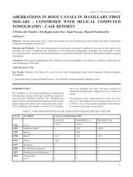
REVERSE OPG - A REVIVAL. C. Pravda, D. Koteeswaran. - Aosr.co.in
REVERSE OPG - A REVIVAL. C. Pravda, D. Koteeswaran. - Aosr.co.in

You also want an ePaper? Increase the reach of your titles
YUMPU automatically turns print PDFs into web optimized ePapers that Google loves.
Reverse <strong>OPG</strong><br />
Patient position<strong>in</strong>g is critical and needs to be accurate<br />
such that <strong>co</strong>ndylar region is close to the lateral centre<br />
of rotation. The speed is reduced so that the image<br />
layer moves closer to the centre of rotation and<br />
be<strong>co</strong>mes th<strong>in</strong>ner result<strong>in</strong>g <strong>in</strong> a clearer and less distorted<br />
view. This view is used especially when the mouth<br />
open<strong>in</strong>g is limited.<br />
Errors <strong>in</strong> the technique mostly occur with improper<br />
position<strong>in</strong>g of the patient.<br />
Advantages<br />
- Shows the <strong>co</strong>ndyle and its neighbor<strong>in</strong>g structures<br />
with the ascend<strong>in</strong>g ramus that is usually overlapped<br />
by the soft tissue shadows <strong>in</strong> the <strong>OPG</strong><br />
- The technique is simple to perform. Retakes are<br />
possible. And quality <strong>co</strong>ntrol is easier to ma<strong>in</strong>ta<strong>in</strong>.<br />
- Requires m<strong>in</strong>imum <strong>co</strong>operation from the patient<br />
- It practically elim<strong>in</strong>ates problem <strong>in</strong> patient with<br />
trismus or un<strong>co</strong>operative patient<br />
Limitations<br />
- Reverse <strong>OPG</strong> is not without problems. Position<strong>in</strong>g<br />
of the patient is more critical than for the standard<br />
view.<br />
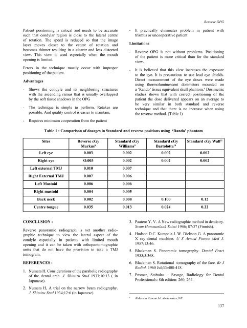
- It is believed that this view <strong>in</strong>creases the exposure<br />
to the eye. It is precautious to use lead eye shields.<br />
Direct measurement of the eye doses were made<br />
us<strong>in</strong>g thermolum<strong>in</strong>escent dosimeters mounted on<br />
a ‘Rando’ tissue equivalent skull phantom. † Dosimetric<br />
studies shows that with <strong>co</strong>rrect position<strong>in</strong>g of the<br />
patient the dose delivered appears on an average to<br />
be very similar <strong>in</strong> both standard and reverse<br />
technique and that there is no <strong>in</strong>crease when us<strong>in</strong>g<br />
the reverse method. (Table 1)<br />
Table 1 : Comparison of dosages <strong>in</strong> Standard and reverse positions us<strong>in</strong>g ‘Rando’ phantom<br />
Sites<br />
Reverse cGy<br />
Markus 8<br />
Standard cGy<br />
Williams 9<br />
Standard cGy<br />
Bartolotta 10 Standard cGy Wall 11<br />
Left eye 0.003 0.002 0.002 0.002<br />
Right eye O.003 0.002 0.002 0.002<br />
Left external TMJ 0.010 0.007<br />
Right External TMJ 0.007 0.006<br />
Left Mastoid 0.006 0.006<br />
Right mastoid 0.004 0.005<br />
Back neck 0.002 0.008 0.100 0.12<br />
Centre tongue 0.035 0.013 0.024 0.22<br />
CONCLUSION :<br />
Reverse panoramic radiograph is yet another radiographic<br />
technique to view the lateral aspect of the<br />
<strong>co</strong>ndyle especially <strong>in</strong> patients with limited mouth<br />
open<strong>in</strong>g and it can be taken with orthopantomographic<br />
units that do not have the provision to take a TMJ<br />
tomogram.<br />
REFERENCES :<br />
1. Numata H. Considerations of the parabolic radiography<br />
of the dental arch. J. Shimizu Stud 1933;10:13 ( <strong>in</strong><br />
Japanese).<br />
2. Numata H, A trial on the narrow beam radiography.<br />
J. Shimizu Stud 1934;12:6 (<strong>in</strong> Japanese).<br />
3. Paatero Y. V. A New radiographic method <strong>in</strong> dentistry.<br />
Svom Hammaslaak Toimi 1946; 87:37 (F<strong>in</strong>nish).<br />
4. Hudson D.C. Kumpala J. W. Dickson G. A panoramic<br />
X ray dental mach<strong>in</strong>e. U S Armed Forces Med J.<br />
1957;13:46.<br />
5. Blackman S. Panoramic tomography. Dental Pract<br />
1955;5:368.<br />
6. Blackman S. Rotational tomography of the face. Br J<br />
Radiol. 1960 Jul;33:408-418.<br />
7. Fromer, Stabulas – Savage, Radiology for Dental<br />
Professionals: 8th edition: 260, 264.<br />
†<br />
Alderson Research Laboratories, NY.<br />
137