BILE DUCT SYSTEM MALFORMATION ... - Journal of IMAB
BILE DUCT SYSTEM MALFORMATION ... - Journal of IMAB
BILE DUCT SYSTEM MALFORMATION ... - Journal of IMAB

You also want an ePaper? Increase the reach of your titles
YUMPU automatically turns print PDFs into web optimized ePapers that Google loves.
<strong>Journal</strong> <strong>of</strong> <strong>IMAB</strong> - Annual Proceeding (Scientific Papers) 2009, book 1<br />
<strong>BILE</strong> <strong>DUCT</strong> <strong>SYSTEM</strong> <strong>MALFORMATION</strong> -<br />
EMBRYOLOGICAL AND PATHOLOGICAL<br />
ASSOCIATION. TREATMENT /REVIEW ARTICLE/<br />
Ludmil M. Veltchev 1 , Manol A. Kalniev 2 , Todor A. Todorov 3 ,<br />
1) Fellow, Master’s Program in Hepatobiliary Pancreatic Surgery, Henri Bismuth<br />
Hepatobiliary Institute, 12-14, avenue Paul Vaillant-Couturier, 94804 Villejuif<br />
Cedex<br />
2) Department <strong>of</strong> Anatomy, Cytology and Histology, University <strong>of</strong> Medicine,<br />
S<strong>of</strong>ia, Bulgaria<br />
3) Department <strong>of</strong> Pathology, University <strong>of</strong> Medicine, S<strong>of</strong>ia, Bulgaria<br />
ABSTRACT<br />
Cystic diseases <strong>of</strong> the liver which are in most cases<br />
hereditary, are related to an embryonic disorder know as<br />
ductal plate malformation. These diseases correspond to<br />
partial or total arrest <strong>of</strong> remodeling <strong>of</strong> the ductal plate,<br />
leading to more or less complete persistence <strong>of</strong> the excess<br />
<strong>of</strong> embryonic biliary structures. The ductal plate<br />
malformation may concern different segments <strong>of</strong> the<br />
intrahepatic biliary tree (segmental bile ducts, interlobular<br />
bile ducts and the smallest bile duct ramifications) leading<br />
to various pathoclinical entities Congenital cystic lesions<br />
<strong>of</strong> bile ducts may affect intra or extrahepatic bile ducts.<br />
Intrahepatic lesions include five entities: congenital hepatic<br />
fibrosis, Caroli’s syndrome, von Meyenburg complexes,<br />
simple cyst <strong>of</strong> the liver and polycystic liver disease.<br />
Congenital hepatic fibrosis and von Meyenburg complexes<br />
are secondary to ductal plate malformation affecting the<br />
smallest intrahepatic bile ducts.<br />
Choledocal cysts, Caroli’s disease and Caroli’s<br />
syndrome belong to the some family <strong>of</strong> congenital<br />
malformations <strong>of</strong> the large bile ducts (1). The former affects<br />
the extrahepatic bile duct (including occasionally the left and<br />
right branch <strong>of</strong> the hepatic duct) while the latter affects<br />
segmental intrahepatic bile ducts. Both are extremely rare<br />
(in the order <strong>of</strong> 1:10.000 or 100.000 and 1:1.000.000 births<br />
respectively.<br />
Key words: Caroli’s disease, biliary dilation,<br />
complications<br />
INTRO<strong>DUCT</strong>ION<br />
The malformation responsible for Caroli’s disease is<br />
an anomalous rearramgement <strong>of</strong> the ductal plate, part <strong>of</strong><br />
hilum plate (2). It is consisted <strong>of</strong> bile ducts and vessels<br />
surrounded by a shealth that is continuous with Glisson’s<br />
capsule intrahepatecaly and hepatoduodenal ligament<br />
extrahepatically. Insaid it contain lymphatic, venous and<br />
arterial network.<br />
Portal vein usually is dislocated posterior to the<br />
common hepatic bile duct and hepatic artery and by the<br />
description <strong>of</strong> Cauinaud it is covered by separate sheath <strong>of</strong><br />
loose connective tissues that permit easy dissection from<br />
other components.<br />
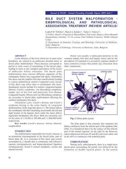
Fig. 1. Hilum plate system<br />
The hilar plate is that structure that separates the<br />
biliary confluence from the inferior part <strong>of</strong> the quadrate lobe<br />
(S4a). It is boundered above by the surface <strong>of</strong> S4a-inferior<br />
part <strong>of</strong> the medial segment, on the right by the Rounvier<br />
sulcus (depression between S5 and S6) and cystic plate, and<br />
in the left it is continuous with umbilical plate.<br />
EMBRYOGENESIS<br />
During early embryogenesis, there is a single-layer<br />
ductal plate surrounding the portal vein followed by the<br />
formation <strong>of</strong> double layered plates. In the normal<br />
66
developmental, extensive resorption <strong>of</strong> the primitive bile<br />
ducts leads to the final stage, in which a network <strong>of</strong> fine<br />
bile ducts surrounds the portal vein .Insufficient resorption<br />
<strong>of</strong> ductal plate can lead to large dilated segments <strong>of</strong> the<br />
primitive bile duct surrounding the central portal vein (bile<br />
ducts that originally encircle the portal vein fail to involve<br />
properly, giving rise to a cystic dilation). This malformation<br />
may occur at the level <strong>of</strong> the large segmental ducts (giving<br />
rise to Caroli’s disease with enlarged segmental ducts,<br />
sometimes localized) <strong>of</strong> intermediate size bile ducts (giving<br />
rise to Caroli’s syndrome with combines enlarged segmental<br />
bile ducts that are more diffuse and congenital hepatic<br />
fibrosis) These events can also occur at the level <strong>of</strong> the<br />
interlobular bile ducts (giving rise to Von Meyerburg<br />
complexes or to polycystic liver disease (3).<br />
The malformation responsible for (most) choledocal<br />
cyst is an anomalous junction <strong>of</strong> the biliary and pancreatic<br />
ducts. In the normal situation, both ducts join into a<br />
common channel, the length <strong>of</strong> which is shorter than the<br />
length <strong>of</strong> Oddi’s sphincter. Hence, reflux from pancreatic<br />
fluid into common bile duct (or <strong>of</strong> bile duct into the<br />
pancreatic duct) is prevented. Should an anomalous<br />
proliferation <strong>of</strong> the biliary epithelium occur during fetal life,<br />
this common duct will become longer than the length <strong>of</strong> the<br />
sphincter (which remains constant) and reflux will occur. The<br />
reflux <strong>of</strong> pancreatic fluid into common bile duct is thought<br />
to be responsible for bile duct dilation and inflammation <strong>of</strong><br />
the epithelium.<br />
This common channel which can be identified by<br />
cholangio-pancreato MRI, endoscopic ultrasound or<br />
retrograde cholangiography, is present in 95% or more <strong>of</strong><br />
choledocal cysts.<br />
However, a common channel will not necessarily<br />
produce a choledocal cyst;beside , functional pancreatobiliary<br />
reflux may occur despite a normal common channel(<br />
this entity is called occult pancreatic-biliary reflux) (4).<br />
The common consequences <strong>of</strong> Coroli’s disease /<br />
syndrome and choledocal cysts are bile stasis, pigmental<br />
stone formation (as well as protein plugs in the case <strong>of</strong><br />
choledocal cyst) and malignant transformation. As a rule,<br />
the disease may remain silent for decades or give rise to<br />
biliary pain (simple obstruction) or pancreatitis (should a<br />
stone or a protein plug migrate). However, symptoms<br />
become much more severe once bile becomes infected,<br />
either spontaneously, or as a result <strong>of</strong> endoscopic<br />
manoeuvres. Cholangitis at that stage becomes the leading<br />
symptom and endoscopic (or percutaneous) invasive<br />
approaches should be avoided in asymptomatic or<br />
symptomatic patients.<br />
MALIGNIZATION<br />
The risk <strong>of</strong> malignant transformation (a likely<br />
dysplasia, adenoma, adenocarcinoma sequence) is related<br />
to stasis and chronic inflammation. This risk is correlated<br />
with age (is exceptional in children) and septic contamination<br />
<strong>of</strong> bile (5, 6). For choledocal cysts, the incidence is<br />
estimated to be 8% before age <strong>of</strong> 40 and 25% thereafter and<br />
is clearly increased when a prior cysto-digestive<br />
anastomosis has been performed (this treatment is<br />
nowadays contraindicated) (7). For Caroli disease/<br />
syndrome, the reported incidence ranges between 10 and<br />
25 %( 8).Of not, pancreatic fluid reflux ( for choledocal cysts)<br />
may in itself result in malignant transformation, nor only in<br />
the choledocal cyst bur also anywhere else in the bile duct<br />
and in particular the gallbladder where stasis naturally<br />
occurs. Hence, gallbladder malignancy may occur not only<br />
in patients with a choledocal cysts with anomalous<br />
pancreatico-biliary junction, but also in patients with an<br />
anomalous pancreatico-biliary junction without a choledocal<br />
cyst( and recognizing this condition is an indicatin for<br />
prophylactic cholecystectomy).<br />
TREATMENT<br />
The risk <strong>of</strong> malignant transformation is an indication<br />
for treatment <strong>of</strong> choledocal cyst that should include<br />
resection <strong>of</strong> all the cystic dilation, cholecystectomy and<br />
Roux-en-Y bilio-digestive anastomosis. This apparently<br />
straightforward procedure in fact turns out to be associated<br />
with high morbidity rate, inparticulare from the intra- and<br />
retropancreatic dissection <strong>of</strong> the cystic dilatation(5).<br />
Treatment <strong>of</strong> Caroli’s disease (without congenital<br />
hepatic fibrosis) may occasionally rely on partial liver<br />
resection if the involvement is localased.Caroli’s syndrome<br />
(diffuse involvement with congenital hepatic fibrosis) can<br />
only be cured by liver transplantation.<br />
The most common indication for transplantation in<br />
these series was recurrent cholangitis. Most patients had<br />
associated polycystic kidney disease or hepatic fibrosis. As<br />
the risk <strong>of</strong> this treatment is not very different from the risk<br />
<strong>of</strong> malignant transformation, there is no consensus that<br />
prophylactic transplantation should be indicated in<br />
symptomatic patients.<br />
CONCLUSION:<br />
Cystic diseases <strong>of</strong> the system are congenital<br />
disorders, related to the embryological developmental and<br />
basis structure called hilar plate. Clinical presentation<br />
includes dilatation <strong>of</strong> intra and extra hepatic bile ducts,<br />
cholangitis, bile duct stone formation, dilatation and<br />
jaundice. The malignant transformation is the most<br />
dangerous complications. Used imaging techniques such as<br />
ultrasound, CT, MRI cholangiography diagnosis can be<br />
verified. If monolobar liver dilatation is diagnosed, liver<br />
resection is method <strong>of</strong> choice. For complicated and bilobar<br />
liver involvement liver transplantation is indicated.<br />
Extra hepatic dilatation presented by choledocal cysts<br />
necessitates resection and biliodigestive anastomosis.<br />
67
REFERENCES:<br />
1. Todani T, Watanabe Y, Narusue M,<br />
Tabuchi k, Okajima K (1977). “Congenital<br />
bile duct cysts: Classification, operative<br />
procedures, and review <strong>of</strong> thirty-seven<br />
cases including cancer arising from<br />
choledochal cyst”. Am. J. Surg. 134 (2):<br />
263–9 PMID 889044.<br />
2. Desmet VJ. Congenital diseases <strong>of</strong><br />
intrahepatic bile ducts: variations on the<br />
theme “ductal plate malformation”.<br />
Hepatology. 1992 Oct; 16 (4):1069-83<br />
3. Vijayan V, Tan CE. Development<br />
<strong>of</strong> the human intrahepatic biliary system.<br />
Ann Acad Med Singapore. 1999 Jan; 28(1):<br />
105-8.<br />
4. Sugiyama, Y. Atomi 1 Pancreatic juice<br />
can reflux into the bile duct in patients<br />
without anomalous pancreaticobiliary<br />
junction. J Gastroenterol 2004; 39:1021–<br />
1022.<br />
5. Kasper HU, Stippel DL, Töx U,<br />
Drebber U, Dienes HP. Primary cholangiocarcinoma<br />
in a case <strong>of</strong> Caroli’s disease: case<br />
report and literature. Pathologe. 2006<br />
Jul;27(4):300-4.<br />
6. Etienne JC, Bouillot JL, Alexandre<br />
JH.Cholangiocarcinoma associated with<br />
Caroli’s disease. Apropos <strong>of</strong> a case. Review<br />
<strong>of</strong> the literature]J Chir (Paris). 1987 Mar;<br />
124(3):161-4. PMID: 3034936<br />
7. Zemskov VS, Bobrov OE, Shelemba<br />
MV. Surgical treatment <strong>of</strong> biliary cyst. Klin<br />
Khir. 1992; ( 11):1-3. PMID: 1296051<br />
8. Sans M, Rimola A, Navasa M,<br />
Grande L, Garcia-Valdecasas JC, Andreu<br />
H, et al. Liver transplantation in patients<br />
with Caroli’s disease and recurrent<br />
cholangitis. Transpl Int1997; 10:241-244.<br />
68<br />
Corresponding author:<br />
Ludmil Marinov Veltchev, MD PhD<br />
Mobile: +359 876 259 685<br />
E-mail: drlmarinov@yahoo.com