The Anatomy of the Medial Part of the Knee
The Anatomy of the Medial Part of the Knee
The Anatomy of the Medial Part of the Knee

You also want an ePaper? Increase the reach of your titles
YUMPU automatically turns print PDFs into web optimized ePapers that Google loves.
2002<br />
THE JOURNAL OF BONE & JOINT SURGERY · JBJS.ORG<br />
VOLUME 89-A · NUMBER 9 · SEPTEMBER 2007<br />
T HE ANATOMY OF THE MEDIAL PART OF THE KNEE<br />
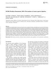
Fig. 1<br />
Photograph <strong>of</strong> a femur from a bone box specimen (distal-medial view, right knee) with pointers<br />
demonstrating <strong>the</strong> relationships between <strong>the</strong> medial epicondyle (ME), <strong>the</strong> adductor tubercle (AT),<br />
and <strong>the</strong> gastrocnemius tubercle (GT). <strong>The</strong> dots are placed at <strong>the</strong> highest point <strong>of</strong> each structure.<br />
<strong>the</strong> adductor tubercle and was close to a small depression,<br />
which corresponded to <strong>the</strong> location <strong>of</strong> <strong>the</strong> attachment <strong>of</strong> <strong>the</strong><br />
medial gastrocnemius tendon (Figs. 1 and 2).<br />
Quantitative analysis <strong>of</strong> <strong>the</strong>se osseous landmarks in <strong>the</strong><br />
dissected knees revealed that <strong>the</strong> adductor tubercle was 12.6<br />
mm (range, 9.0 to 15.2 mm) proximal and 8.3 mm (range,<br />
5.9 to 11.6 mm) posterior to <strong>the</strong> medial epicondyle. <strong>The</strong> gastrocnemius<br />
tubercle was 9.4 mm (range, 7.1 to 11.8 mm) distal<br />
and 8.7 mm (range, 6.8 to 12.5 mm) posterior to <strong>the</strong><br />
adductor tubercle and 6.0 mm (range, 4.4 to 8.9 mm) proximal<br />
and 13.7 mm (range, 10.8 to 15.8 mm) posterior to <strong>the</strong><br />
medial epicondyle.<br />
Superficial <strong>Medial</strong> Collateral Ligament<br />
(Tibial Collateral Ligament)<br />
<strong>The</strong> superficial medial collateral ligament was <strong>the</strong> largest<br />
structure over <strong>the</strong> medial aspect <strong>of</strong> <strong>the</strong> knee. It had one femoral<br />
and two tibial attachments. <strong>The</strong> quantitative relationships<br />
and attachment areas <strong>of</strong> <strong>the</strong> superficial medial collateral ligament<br />
are listed in tables in <strong>the</strong> Appendix.<br />
<strong>The</strong> femoral attachment <strong>of</strong> <strong>the</strong> superficial medial collateral<br />
ligament was round to slightly oval in shape and was<br />
located in a depression that was an average <strong>of</strong> 3.2 mm (range,<br />
1.6 to 5.2 mm) proximal and 4.8 mm (range, 2.5 to 6.3 mm)<br />
posterior to <strong>the</strong> medial epicondyle (Figs. 2 and 3). <strong>The</strong>re was<br />
no firm attachment between <strong>the</strong> superficial medial collateral<br />
ligament and <strong>the</strong> underlying deep medial collateral ligament,<br />
and no definable bursae were identified between <strong>the</strong>se two<br />
structures.<br />
As <strong>the</strong> superficial medial collateral ligament coursed<br />
distally, it had two separate tibial attachments (Figs. 2 and 4).<br />
Between <strong>the</strong>se two distinct tibial attachments, <strong>the</strong> superficial<br />
medial collateral ligament was separated from <strong>the</strong> tibia by <strong>the</strong><br />
inferior medial genicular artery and vein, along with its corresponding<br />
nerve branch from <strong>the</strong> tibial nerve, and some fine<br />
fascial and adipose tissues. <strong>The</strong> proximal attachment <strong>of</strong> <strong>the</strong> superficial<br />
medial collateral ligament was primarily to s<strong>of</strong>t tissues<br />
ra<strong>the</strong>r than directly to bone. <strong>The</strong> majority <strong>of</strong> <strong>the</strong> s<strong>of</strong>t<br />
tissue deep to <strong>the</strong> proximal tibial attachment <strong>of</strong> <strong>the</strong> superficial<br />
medial collateral ligament was <strong>the</strong> anterior arm <strong>of</strong> <strong>the</strong> semimembranosus<br />
tendon, which itself attached directly to bone.<br />
<strong>The</strong> distal tibial attachment was broad-based and was located<br />
just anterior to <strong>the</strong> posteromedial crest <strong>of</strong> <strong>the</strong> tibia. <strong>The</strong> majority<br />
<strong>of</strong> <strong>the</strong> distal attachment was located within <strong>the</strong> pes<br />
anserine bursa and formed a large portion <strong>of</strong> <strong>the</strong> posterior<br />
floor <strong>of</strong> this bursa. <strong>The</strong> posterior aspect <strong>of</strong> <strong>the</strong> tibial portion <strong>of</strong><br />
<strong>the</strong> superficial medial collateral ligament blended with <strong>the</strong> distal<br />
tibial expansion <strong>of</strong>f <strong>the</strong> semimembranosus tendon 24 along<br />
its distal aspect.<br />
Deep <strong>Medial</strong> Collateral Ligament<br />
(Mid-Third <strong>Medial</strong> Capsular Ligament)<br />
<strong>The</strong> deep medial collateral ligament was a thickening <strong>of</strong> <strong>the</strong><br />
medial joint capsule that was most distinct along its anterior<br />
border, where it roughly paralleled <strong>the</strong> anterior aspect <strong>of</strong> <strong>the</strong><br />
superficial medial collateral ligament. It was most easily identified<br />
along its anterior femoral course, where <strong>the</strong> joint capsule<br />
that coursed toward <strong>the</strong> medial part <strong>of</strong> <strong>the</strong> patella was