The Anatomy of the Medial Part of the Knee
The Anatomy of the Medial Part of the Knee
The Anatomy of the Medial Part of the Knee

Create successful ePaper yourself
Turn your PDF publications into a flip-book with our unique Google optimized e-Paper software.
This is an enhanced PDF from <strong>The</strong> Journal <strong>of</strong> Bone and Joint Surgery<br />
<strong>The</strong> PDF <strong>of</strong> <strong>the</strong> article you requested follows this cover page.<br />
<strong>The</strong> <strong>Anatomy</strong> <strong>of</strong> <strong>the</strong> <strong>Medial</strong> <strong>Part</strong> <strong>of</strong> <strong>the</strong> <strong>Knee</strong><br />
Robert F. LaPrade, Anders Hauge Engebretsen, Thuan V. Ly, Steinar Johansen, Fred A. Wentorf and Lars<br />
Engebretsen<br />
J Bone Joint Surg Am. 2007;89:2000-2010. doi:10.2106/JBJS.F.01176<br />
This information is current as <strong>of</strong> September 26, 2007<br />
Supplementary material<br />
Reprints and Permissions<br />
Publisher Information<br />
Commentary and Perspective, data tables, additional images, video clips and/or<br />
translated abstracts are available for this article. This information can be accessed<br />
at http://www.ejbjs.org/cgi/content/full/89/9/2000/DC1<br />
Click here to order reprints or request permission to use material from this<br />
article, or locate <strong>the</strong> article citation on jbjs.org and click on <strong>the</strong> [Reprints and<br />
Permissions] link.<br />
<strong>The</strong> Journal <strong>of</strong> Bone and Joint Surgery<br />
20 Pickering Street, Needham, MA 02492-3157<br />
www.jbjs.org
2000<br />
COPYRIGHT © 2007 BY THE JOURNAL OF BONE AND JOINT SURGERY, INCORPORATED<br />
<strong>The</strong> <strong>Anatomy</strong> <strong>of</strong> <strong>the</strong> <strong>Medial</strong> <strong>Part</strong> <strong>of</strong> <strong>the</strong> <strong>Knee</strong><br />
By Robert F. LaPrade, MD, PhD, Anders Hauge Engebretsen, Medical Student,<br />
Thuan V. Ly, MD, Steinar Johansen, MD, Fred A. Wentorf, MS, and Lars Engebretsen, MD, PhD<br />
Investigation performed at <strong>the</strong> University <strong>of</strong> Minnesota, Minneapolis, Minnesota<br />
Background: While <strong>the</strong> anatomy <strong>of</strong> <strong>the</strong> medial part <strong>of</strong> <strong>the</strong> knee has been described qualitatively, quantitative descriptions<br />
<strong>of</strong> <strong>the</strong> attachment sites <strong>of</strong> <strong>the</strong> main medial knee structures have not been reported. <strong>The</strong> purpose <strong>of</strong> <strong>the</strong><br />
present study was to verify <strong>the</strong> qualitative anatomy <strong>of</strong> medial knee structures and to perform a quantitative evaluation<br />
<strong>of</strong> <strong>the</strong>ir anatomic attachment sites as well as <strong>the</strong>ir relationships to pertinent osseous landmarks.<br />
Methods: Dissections were performed and measurements were made for eight nonpaired fresh-frozen cadaveric<br />
knees with use <strong>of</strong> an electromagnetic three-dimensional tracking sensor system.<br />
Results: In addition to <strong>the</strong> medial epicondyle and <strong>the</strong> adductor tubercle, a third osseous prominence, <strong>the</strong> gastrocnemius<br />
tubercle, which corresponded to <strong>the</strong> attachment site <strong>of</strong> <strong>the</strong> medial gastrocnemius tendon, was identified. <strong>The</strong><br />
average length <strong>of</strong> <strong>the</strong> superficial medial (tibial) collateral ligament was 94.8 mm. <strong>The</strong> superficial medial collateral ligament<br />
femoral attachment was 3.2 mm proximal and 4.8 mm posterior to <strong>the</strong> medial epicondyle. <strong>The</strong> superficial medial<br />
collateral ligament had two separate attachments on <strong>the</strong> tibia. <strong>The</strong> distal attachment <strong>of</strong> <strong>the</strong> superficial medial<br />
collateral ligament on <strong>the</strong> tibia was 61.2 mm distal to <strong>the</strong> knee joint. <strong>The</strong> deep medial collateral ligament consisted<br />
<strong>of</strong> menisc<strong>of</strong>emoral and meniscotibial portions. <strong>The</strong> posterior oblique ligament femoral attachment was 7.7 mm distal<br />
and 6.4 mm posterior to <strong>the</strong> adductor tubercle and 1.4 mm distal and 2.9 mm anterior to <strong>the</strong> gastrocnemius tubercle.<br />
<strong>The</strong> medial patell<strong>of</strong>emoral ligament attachment on <strong>the</strong> femur was 1.9 mm anterior and 3.8 mm distal to <strong>the</strong> adductor<br />
tubercle.<br />
Conclusions: <strong>The</strong> medial knee ligament structures have a consistent attachment pattern.<br />
Clinical Relevance: Identification <strong>of</strong> <strong>the</strong> gastrocnemius tubercle and <strong>the</strong> quantitative relationships presented here<br />
will be useful in <strong>the</strong> study <strong>of</strong> anatomic repairs and reconstructions <strong>of</strong> complex ligamentous injuries that involve <strong>the</strong><br />
medial knee structures.<br />
While <strong>the</strong> medial collateral ligament is <strong>the</strong> most frequently<br />
injured ligament in <strong>the</strong> knee 1-4 , and while a<br />
better understanding <strong>of</strong> its functional anatomy,<br />
biomechanics, and healing has been obtained over <strong>the</strong> past<br />
twenty years 5-9 , we have found that its anatomy has only been<br />
described qualitatively, and <strong>the</strong>re is controversy about descriptions<br />
<strong>of</strong> some aspects <strong>of</strong> its anatomy that have been contradictory<br />
or incomplete 2,6,10-15 . <strong>The</strong> medial ligament complex <strong>of</strong><br />
<strong>the</strong> knee includes one large ligament and a series <strong>of</strong> capsular<br />
thickenings and tendinous attachments. <strong>The</strong> superficial medial<br />
collateral ligament is commonly called <strong>the</strong> tibial collateral<br />
ligament, whereas <strong>the</strong> deep medial collateral ligament is also<br />
called <strong>the</strong> mid-third medial capsular ligament 10,16 . <strong>The</strong> capsular<br />
attachments from <strong>the</strong> main common tendon <strong>of</strong> <strong>the</strong><br />
semimembranosus have been called <strong>the</strong> posterior oblique<br />
ligament 5,17-20 . However, <strong>the</strong>re appears to be controversy about<br />
whe<strong>the</strong>r <strong>the</strong> posterior oblique ligament is a distinct structure<br />
or if it is a portion <strong>of</strong> <strong>the</strong> superficial medial collateral ligament,<br />
termed <strong>the</strong> oblique fibers <strong>of</strong> <strong>the</strong> superficial medial collateral<br />
ligament 2,10,13-17 .<br />
An extensive literature search revealed that, while <strong>the</strong>re<br />
are many qualitative descriptions <strong>of</strong> <strong>the</strong> anatomy <strong>of</strong> <strong>the</strong> medial<br />
part <strong>of</strong> <strong>the</strong> knee 2,5,6,10,13-15,21 , <strong>the</strong>re are no specific quantitative<br />
descriptions <strong>of</strong> <strong>the</strong> medial knee structures. Many <strong>of</strong> <strong>the</strong>se<br />
Disclosure: In support <strong>of</strong> <strong>the</strong>ir research for or preparation <strong>of</strong> this work, one or more <strong>of</strong> <strong>the</strong> authors received, in any one year, outside funding or<br />
grants in excess <strong>of</strong> $10,000 from Health East, Norway, and <strong>the</strong> Norwegian Research Council (grant #42692) and <strong>the</strong> Sports Medicine Research<br />
Fund <strong>of</strong> <strong>the</strong> Minnesota Medical Foundation. Nei<strong>the</strong>r <strong>the</strong>y nor a member <strong>of</strong> <strong>the</strong>ir immediate families received payments or o<strong>the</strong>r benefits or a commitment<br />
or agreement to provide such benefits from a commercial entity. No commercial entity paid or directed, or agreed to pay or direct, any benefits<br />
to any research fund, foundation, division, center, clinical practice, or o<strong>the</strong>r charitable or nonpr<strong>of</strong>it organization with which <strong>the</strong> authors, or a member<br />
<strong>of</strong> <strong>the</strong>ir immediate families, are affiliated or associated.<br />
J Bone Joint Surg Am. 2007;89:2000-10 • doi:10.2106/JBJS.F.01176
2001<br />
THE JOURNAL OF BONE & JOINT SURGERY · JBJS.ORG<br />
VOLUME 89-A · NUMBER 9 · SEPTEMBER 2007<br />
T HE ANATOMY OF THE MEDIAL PART OF THE KNEE<br />
complex structures have been illustrated ei<strong>the</strong>r with oversimplifications<br />
<strong>of</strong> <strong>the</strong>ir attachments to both bone and o<strong>the</strong>r structures<br />
or with liberal interpretations <strong>of</strong> <strong>the</strong>ir courses by <strong>the</strong><br />
illustrators, which makes it difficult to compare <strong>the</strong> attachments<br />
and courses <strong>of</strong> many separate structures among<br />
studies 2,5,6,10,13-15,21 . <strong>The</strong> purpose <strong>of</strong> <strong>the</strong> present study was to verify<br />
<strong>the</strong> qualitative anatomy <strong>of</strong> medial knee structures and to<br />
perform a quantitative evaluation <strong>of</strong> <strong>the</strong>ir anatomic attachment<br />
sites as well as <strong>the</strong>ir relationships to pertinent osseous<br />
landmarks.<br />
Materials and Methods<br />
Gross <strong>Anatomy</strong> Dissections<br />
wenty femora from <strong>the</strong> bone box specimens <strong>of</strong> <strong>the</strong> De-<br />
<strong>of</strong> <strong>Anatomy</strong> at <strong>the</strong> University <strong>of</strong> Minnesota were<br />
Tpartment<br />
qualitatively analyzed to examine <strong>the</strong> osseous prominences <strong>of</strong><br />
<strong>the</strong> medial side <strong>of</strong> <strong>the</strong> knee. <strong>The</strong> locations <strong>of</strong> <strong>the</strong>se osseous<br />
prominences were <strong>the</strong>n used to help to identify and analyze<br />
<strong>the</strong> osseous prominences seen during <strong>the</strong> fresh-frozen knee<br />
dissections.<br />
Dissections were performed on eight nonpaired freshfrozen<br />
cadaveric knees that had no sign <strong>of</strong> previous injury,<br />
knee abnormality, or disease. <strong>The</strong> mean age <strong>of</strong> <strong>the</strong> donors<br />
had been fifty-nine years (range, forty-four to seventy-two<br />
years) at <strong>the</strong> time <strong>of</strong> death. Each cadaveric knee was stored<br />
frozen at −20°C and was allowed to thaw overnight prior to<br />
dissection.<br />
Anatomic Measurements<br />
<strong>The</strong> Polhemus FASTRAK electromagnetic three-dimensional<br />
tracking sensor system (Polhemus, Colchester, Vermont) was<br />
used to quantitatively identify <strong>the</strong> insertion sites <strong>of</strong> <strong>the</strong> measured<br />
structures and related osseous landmarks 22,23 . This device<br />
is a six-degrees-<strong>of</strong>-freedom measuring device that tracks<br />
<strong>the</strong> position and orientation <strong>of</strong> a receiver relative to a transmitter<br />
with use <strong>of</strong> low-frequency magnetic fields. <strong>The</strong> transmitter<br />
device produces a pulsed magnetic field. In turn, <strong>the</strong><br />
receiver device contains a small solenoid that senses <strong>the</strong> magnetic<br />
field. <strong>The</strong> magnetic field produced by <strong>the</strong> transmitter<br />
device has different effects depending on <strong>the</strong> receiver position<br />
in <strong>the</strong> magnetic field, and <strong>the</strong> position and orientation with<br />
respect to <strong>the</strong> axes <strong>of</strong> <strong>the</strong> transmitter can <strong>the</strong>n be calculated<br />
instantaneously (MotionMonitor; Innovative Sports Training,<br />
Chicago, Illinois). <strong>The</strong> transmitter-to-receiver separation<br />
range in <strong>the</strong> present study was 300 to 480 mm, which<br />
was within <strong>the</strong> previously reported optimal range <strong>of</strong> 100 to<br />
700 mm for <strong>the</strong>se testing conditions to minimize positional<br />
error 22 . <strong>The</strong> knee was placed into a device that fixed <strong>the</strong> specimen<br />
relative to <strong>the</strong> transmitter device. A probe was connected<br />
to <strong>the</strong> electromagnetic tracking system and acted as <strong>the</strong> receiver<br />
device to measure <strong>the</strong> three-dimensional coordinate<br />
location <strong>of</strong> <strong>the</strong> structure or structures <strong>of</strong> interest. Distances <strong>of</strong><br />
interest were calculated with use <strong>of</strong> three-dimensional data<br />
points. <strong>The</strong> accuracy <strong>of</strong> this measurement system has been reported<br />
to be within 0.3° and 0.3 mm 23 .<br />
After placement <strong>of</strong> <strong>the</strong> knee into <strong>the</strong> holding device, meticulous<br />
sharp dissection <strong>of</strong> <strong>the</strong> structures <strong>of</strong> <strong>the</strong> medial and<br />
posteromedial aspects <strong>of</strong> <strong>the</strong> knee was performed with use <strong>of</strong><br />
ei<strong>the</strong>r a knife blade or a fine-pointed hemostat. After <strong>the</strong> initial<br />
measurements <strong>of</strong> each specific structure were made by<br />
placing <strong>the</strong> Polhemus measuring probe against <strong>the</strong> edge <strong>of</strong> <strong>the</strong><br />
structure and recording its three-dimensional coordinate location,<br />
<strong>the</strong> attachment sites were dissected down to bone and<br />
outlined and <strong>the</strong> perimeters <strong>of</strong> <strong>the</strong> attachment sites were identified<br />
with <strong>the</strong> measuring probe.<br />
<strong>The</strong> perimeters <strong>of</strong> <strong>the</strong> tibial attachment sites <strong>of</strong> <strong>the</strong> medial<br />
structures were identified first. All measurements were<br />
made by <strong>the</strong> same individual (R.F.L.) to avoid interobserver<br />
error. Each attachment site was recorded by tracing its outline<br />
with <strong>the</strong> measuring probe immediately after it was<br />
sharply dissected <strong>of</strong>f bone. Measurements were made along<br />
<strong>the</strong> periphery <strong>of</strong> each attachment site. Joint line measurements<br />
were made to <strong>the</strong> edge <strong>of</strong> <strong>the</strong> articular cartilage surfaces<br />
<strong>of</strong> <strong>the</strong> medial femoral condyle for structures attached to<br />
<strong>the</strong> femur and to <strong>the</strong> medial tibial plateau for structures attached<br />
to <strong>the</strong> tibia.<br />
Once all <strong>of</strong> <strong>the</strong> desired structures and osseous landmarks<br />
were identified, <strong>the</strong> outlines <strong>of</strong> both <strong>the</strong> distal part <strong>of</strong><br />
<strong>the</strong> femur and <strong>the</strong> proximal part <strong>of</strong> <strong>the</strong> tibia were collected to<br />
establish a three-dimensional axis on which to map <strong>the</strong> locations<br />
<strong>of</strong> <strong>the</strong> structures. <strong>The</strong> coordinates <strong>of</strong> each identified<br />
point were used to calculate <strong>the</strong> areas <strong>of</strong> <strong>the</strong> insertion sites, <strong>the</strong><br />
centroid <strong>of</strong> each insertion, and <strong>the</strong> distances between <strong>the</strong> centroids.<br />
<strong>The</strong> distances between structures were <strong>the</strong>n broken<br />
down into anterior-posterior, medial-lateral, and proximaldistal<br />
components. <strong>The</strong> distances measured with this system<br />
were straight-line distances and did not take into account osseous<br />
prominences or depressions. For this reason, small variations<br />
in measured distances occurred between <strong>the</strong> osseous<br />
landmarks and <strong>the</strong> separate anatomic structures.<br />
Results<br />
easurements are reported to <strong>the</strong> midpoint <strong>of</strong> a struc-<br />
attachment site and osseous landmarks. All dis-<br />
Mture’s<br />
tances and areas are reported as averages for each structure<br />
(see Appendix). Attachment areas for identified structures are<br />
listed in a table <strong>the</strong> Appendix. Straight-line distances between<br />
<strong>the</strong> centers <strong>of</strong> structures are reported in tables in <strong>the</strong> Appendix,<br />
whereas proximal-distal and anterior-posterior attachment<br />
relationships are described in this section.<br />
<strong>Medial</strong> Femoral Osseous Landmarks<br />
Qualitative analysis <strong>of</strong> <strong>the</strong> femora from <strong>the</strong> bone box specimens<br />
revealed that <strong>the</strong> medial epicondyle was <strong>the</strong> most anterior<br />
and distal osseous prominence over <strong>the</strong> medial aspect <strong>of</strong><br />
<strong>the</strong> medial femoral condyle. <strong>The</strong> adductor tubercle was located<br />
at <strong>the</strong> distal edge <strong>of</strong> a thin ridge <strong>of</strong> bone, called <strong>the</strong> medial<br />
supracondylar line, along <strong>the</strong> medial aspect <strong>of</strong> <strong>the</strong> distal<br />
part <strong>of</strong> <strong>the</strong> femur. <strong>The</strong> adductor tubercle was located proximal<br />
and posterior to <strong>the</strong> medial epicondyle. A third osseous prominence,<br />
which we have called <strong>the</strong> gastrocnemius tubercle, was<br />
identified; this structure was slightly distal and posterior to
2002<br />
THE JOURNAL OF BONE & JOINT SURGERY · JBJS.ORG<br />
VOLUME 89-A · NUMBER 9 · SEPTEMBER 2007<br />
T HE ANATOMY OF THE MEDIAL PART OF THE KNEE<br />
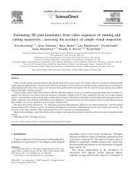
Fig. 1<br />
Photograph <strong>of</strong> a femur from a bone box specimen (distal-medial view, right knee) with pointers<br />
demonstrating <strong>the</strong> relationships between <strong>the</strong> medial epicondyle (ME), <strong>the</strong> adductor tubercle (AT),<br />
and <strong>the</strong> gastrocnemius tubercle (GT). <strong>The</strong> dots are placed at <strong>the</strong> highest point <strong>of</strong> each structure.<br />
<strong>the</strong> adductor tubercle and was close to a small depression,<br />
which corresponded to <strong>the</strong> location <strong>of</strong> <strong>the</strong> attachment <strong>of</strong> <strong>the</strong><br />
medial gastrocnemius tendon (Figs. 1 and 2).<br />
Quantitative analysis <strong>of</strong> <strong>the</strong>se osseous landmarks in <strong>the</strong><br />
dissected knees revealed that <strong>the</strong> adductor tubercle was 12.6<br />
mm (range, 9.0 to 15.2 mm) proximal and 8.3 mm (range,<br />
5.9 to 11.6 mm) posterior to <strong>the</strong> medial epicondyle. <strong>The</strong> gastrocnemius<br />
tubercle was 9.4 mm (range, 7.1 to 11.8 mm) distal<br />
and 8.7 mm (range, 6.8 to 12.5 mm) posterior to <strong>the</strong><br />
adductor tubercle and 6.0 mm (range, 4.4 to 8.9 mm) proximal<br />
and 13.7 mm (range, 10.8 to 15.8 mm) posterior to <strong>the</strong><br />
medial epicondyle.<br />
Superficial <strong>Medial</strong> Collateral Ligament<br />
(Tibial Collateral Ligament)<br />
<strong>The</strong> superficial medial collateral ligament was <strong>the</strong> largest<br />
structure over <strong>the</strong> medial aspect <strong>of</strong> <strong>the</strong> knee. It had one femoral<br />
and two tibial attachments. <strong>The</strong> quantitative relationships<br />
and attachment areas <strong>of</strong> <strong>the</strong> superficial medial collateral ligament<br />
are listed in tables in <strong>the</strong> Appendix.<br />
<strong>The</strong> femoral attachment <strong>of</strong> <strong>the</strong> superficial medial collateral<br />
ligament was round to slightly oval in shape and was<br />
located in a depression that was an average <strong>of</strong> 3.2 mm (range,<br />
1.6 to 5.2 mm) proximal and 4.8 mm (range, 2.5 to 6.3 mm)<br />
posterior to <strong>the</strong> medial epicondyle (Figs. 2 and 3). <strong>The</strong>re was<br />
no firm attachment between <strong>the</strong> superficial medial collateral<br />
ligament and <strong>the</strong> underlying deep medial collateral ligament,<br />
and no definable bursae were identified between <strong>the</strong>se two<br />
structures.<br />
As <strong>the</strong> superficial medial collateral ligament coursed<br />
distally, it had two separate tibial attachments (Figs. 2 and 4).<br />
Between <strong>the</strong>se two distinct tibial attachments, <strong>the</strong> superficial<br />
medial collateral ligament was separated from <strong>the</strong> tibia by <strong>the</strong><br />
inferior medial genicular artery and vein, along with its corresponding<br />
nerve branch from <strong>the</strong> tibial nerve, and some fine<br />
fascial and adipose tissues. <strong>The</strong> proximal attachment <strong>of</strong> <strong>the</strong> superficial<br />
medial collateral ligament was primarily to s<strong>of</strong>t tissues<br />
ra<strong>the</strong>r than directly to bone. <strong>The</strong> majority <strong>of</strong> <strong>the</strong> s<strong>of</strong>t<br />
tissue deep to <strong>the</strong> proximal tibial attachment <strong>of</strong> <strong>the</strong> superficial<br />
medial collateral ligament was <strong>the</strong> anterior arm <strong>of</strong> <strong>the</strong> semimembranosus<br />
tendon, which itself attached directly to bone.<br />
<strong>The</strong> distal tibial attachment was broad-based and was located<br />
just anterior to <strong>the</strong> posteromedial crest <strong>of</strong> <strong>the</strong> tibia. <strong>The</strong> majority<br />
<strong>of</strong> <strong>the</strong> distal attachment was located within <strong>the</strong> pes<br />
anserine bursa and formed a large portion <strong>of</strong> <strong>the</strong> posterior<br />
floor <strong>of</strong> this bursa. <strong>The</strong> posterior aspect <strong>of</strong> <strong>the</strong> tibial portion <strong>of</strong><br />
<strong>the</strong> superficial medial collateral ligament blended with <strong>the</strong> distal<br />
tibial expansion <strong>of</strong>f <strong>the</strong> semimembranosus tendon 24 along<br />
its distal aspect.<br />
Deep <strong>Medial</strong> Collateral Ligament<br />
(Mid-Third <strong>Medial</strong> Capsular Ligament)<br />
<strong>The</strong> deep medial collateral ligament was a thickening <strong>of</strong> <strong>the</strong><br />
medial joint capsule that was most distinct along its anterior<br />
border, where it roughly paralleled <strong>the</strong> anterior aspect <strong>of</strong> <strong>the</strong><br />
superficial medial collateral ligament. It was most easily identified<br />
along its anterior femoral course, where <strong>the</strong> joint capsule<br />
that coursed toward <strong>the</strong> medial part <strong>of</strong> <strong>the</strong> patella was
2003<br />
THE JOURNAL OF BONE & JOINT SURGERY · JBJS.ORG<br />
VOLUME 89-A · NUMBER 9 · SEPTEMBER 2007<br />
T HE ANATOMY OF THE MEDIAL PART OF THE KNEE<br />
<strong>the</strong> superficial, central (tibial), and <strong>the</strong> capsular arms 2,17 (Fig.<br />
6). <strong>The</strong> distances from <strong>the</strong> femoral attachment <strong>of</strong> <strong>the</strong> posterior<br />
oblique ligament to o<strong>the</strong>r specific osseous landmarks are listed<br />
in a table in <strong>the</strong> Appendix. On <strong>the</strong> average, <strong>the</strong> posterior oblique<br />
ligament attached on <strong>the</strong> femur 7.7 mm (range, 6.1 to<br />
9.8 mm) distal and 6.4 mm (range, 4.5 to 10.6 mm) posterior<br />
to <strong>the</strong> adductor tubercle and 1.4 mm (range, 0.8 to 2.1 mm)<br />
distal and 2.9 mm (range, 2.1 to 4.1 mm) anterior to <strong>the</strong> third<br />
osseous prominence over <strong>the</strong> medial part <strong>of</strong> <strong>the</strong> knee, <strong>the</strong> gastrocnemius<br />
tubercle.<br />
<strong>The</strong> superficial arm <strong>of</strong> <strong>the</strong> posterior oblique ligament<br />
consisted <strong>of</strong> a thin fascial expansion. Proximally it coursed<br />
medial to <strong>the</strong> anterior arm <strong>of</strong> <strong>the</strong> semimembranosus, and distally<br />
it followed <strong>the</strong> posterior border <strong>of</strong> <strong>the</strong> superficial medial<br />
collateral ligament (Fig. 6). Proximally it blended into <strong>the</strong> central<br />
arm <strong>of</strong> <strong>the</strong> posterior oblique ligament, whereas distally it<br />
was parallel to <strong>the</strong> posterior border <strong>of</strong> <strong>the</strong> superficial medial<br />
collateral ligament until it blended into <strong>the</strong> distal tibial expansion<br />
<strong>of</strong> <strong>the</strong> semimembranosus and its tibial attachment 24 .<br />
<strong>The</strong> central arm was <strong>the</strong> largest and thickest portion <strong>of</strong><br />
<strong>the</strong> posterior oblique ligament (Figs. 7-A and 7-B). It coursed<br />
from <strong>the</strong> distal aspect <strong>of</strong> <strong>the</strong> main semimembranosus tendon<br />
and was a thick fascial reinforcement <strong>of</strong> both <strong>the</strong> menisc<strong>of</strong>em-<br />
Fig. 2<br />
Illustration <strong>of</strong> <strong>the</strong> femoral osseous landmarks and attachment sites <strong>of</strong><br />
<strong>the</strong> main medial knee structures. AT = adductor tubercle, GT = gastrocnemius<br />
tubercle, ME = medial epicondyle, AMT = adductor magnus tendon,<br />
MGT = medial gastrocnemius tendon, sMCL = superficial medial<br />
collateral ligament, MPFL = medial patell<strong>of</strong>emoral ligament, and POL =<br />
posterior oblique ligament.<br />
visibly thinner and had a different fiber orientation. <strong>The</strong> posterior<br />
border <strong>of</strong> <strong>the</strong> deep medial collateral ligament blended<br />
with and became inseparable from <strong>the</strong> central arm <strong>of</strong> <strong>the</strong> posterior<br />
oblique ligament, just posterior to <strong>the</strong> posterior edge <strong>of</strong><br />
<strong>the</strong> superficial medial collateral ligament.<br />
<strong>The</strong> deep medial collateral ligament consisted <strong>of</strong> distinct<br />
menisc<strong>of</strong>emoral and meniscotibial ligament components (Fig.<br />
5). <strong>The</strong> menisc<strong>of</strong>emoral ligament was consistently longer in<br />
<strong>the</strong> proximal-to-distal direction than <strong>the</strong> meniscotibial portion<br />
(see Appendix). <strong>The</strong> meniscotibial ligament portion <strong>of</strong><br />
<strong>the</strong> deep medial collateral ligament was a consistently shorter<br />
and thicker structure and attached just distal to <strong>the</strong> edge <strong>of</strong> <strong>the</strong><br />
articular cartilage <strong>of</strong> <strong>the</strong> medial tibial plateau (see Appendix).<br />
Posterior Oblique Ligament<br />
<strong>The</strong> posterior oblique ligament consisted <strong>of</strong> three fascial attachments<br />
that coursed <strong>of</strong>f <strong>the</strong> distal aspect <strong>of</strong> <strong>the</strong> semimembranosus<br />
tendon at <strong>the</strong> knee and have been previously termed<br />
Fig. 3<br />
Illustration <strong>of</strong> <strong>the</strong> main medial knee structures (right knee). VMO = vastus<br />
medialis obliquus muscle, MPFL = medial patell<strong>of</strong>emoral ligament,<br />
POL = posterior oblique ligament, sMCL = superficial medial collateral<br />
ligament, SM = semimembranosus muscle, MGT = medial gastrocnemius<br />
tendon, and AMT = adductor magnus tendon.
2004<br />
THE JOURNAL OF BONE & JOINT SURGERY · JBJS.ORG<br />
VOLUME 89-A · NUMBER 9 · SEPTEMBER 2007<br />
T HE ANATOMY OF THE MEDIAL PART OF THE KNEE<br />
oral and meniscotibial portions <strong>of</strong> <strong>the</strong> posteromedial capsule,<br />
and it also had a stout attachment to <strong>the</strong> medial meniscus. Anteriorly,<br />
it merged with <strong>the</strong> posterior fibers <strong>of</strong> <strong>the</strong> superficial<br />
medial collateral ligament. <strong>The</strong> central arm <strong>of</strong> <strong>the</strong> posterior<br />
oblique ligament could be differentiated from <strong>the</strong> superficial<br />
medial collateral ligament by <strong>the</strong> proximal course <strong>of</strong> its fanlike<br />
fibers, which ran more posteriorly toward its femoral<br />
attachment than did <strong>the</strong> fibers <strong>of</strong> <strong>the</strong> superficial medial collateral<br />
ligament, which coursed more anteriorly toward its<br />
femoral attachment (Fig. 6). Its distal attachment was primarily<br />
to <strong>the</strong> posteromedial aspect <strong>of</strong> <strong>the</strong> medial meniscus, <strong>the</strong><br />
meniscotibial portion <strong>of</strong> <strong>the</strong> posteromedial capsule, and <strong>the</strong><br />
posteromedial part <strong>of</strong> <strong>the</strong> tibia.<br />
<strong>The</strong> capsular arm <strong>of</strong> <strong>the</strong> posterior oblique ligament consisted<br />
<strong>of</strong> a thin proximal fascial expansion <strong>of</strong>f <strong>the</strong> anterior aspect<br />
<strong>of</strong> <strong>the</strong> distal part <strong>of</strong> <strong>the</strong> semimembranosus tendon (Fig.<br />
6). It was located posterior and lateral to <strong>the</strong> menisc<strong>of</strong>emoral<br />
capsular attachments <strong>of</strong> <strong>the</strong> central arm and had no fibers that<br />
coursed toward <strong>the</strong> tibia. <strong>The</strong> capsular arm primarily blended<br />
with <strong>the</strong> menisc<strong>of</strong>emoral portion <strong>of</strong> <strong>the</strong> posteromedial joint<br />
capsule and <strong>the</strong> medial aspect <strong>of</strong> <strong>the</strong> oblique popliteal ligament,<br />
and it also attached to <strong>the</strong> s<strong>of</strong>t tissues over <strong>the</strong> medial<br />
gastrocnemius tendon, <strong>the</strong> adductor magnus tendon expansion<br />
to <strong>the</strong> medial gastrocnemius, and <strong>the</strong> adductor magnus<br />
tendon femoral attachment. Qualitatively, it was less stout<br />
overall than <strong>the</strong> central arm, and it did not have any osseous<br />
attachment.<br />
Fig. 4<br />
Illustration <strong>of</strong> <strong>the</strong> superficial medial collateral ligament (sMCL) (medial<br />
aspect, left knee). <strong>The</strong> proximal forceps are under <strong>the</strong> anterior edge <strong>of</strong><br />
<strong>the</strong> femoral portion, and <strong>the</strong> distal hemostat is between <strong>the</strong> proximal<br />
and distal tibial attachments. SM = semimembranosus, and POL =<br />
posterior oblique ligament.<br />
<strong>Medial</strong> Patell<strong>of</strong>emoral Ligament<br />
<strong>The</strong> medial patell<strong>of</strong>emoral ligament was located anterior to,<br />
and in a distinct extra-articular layer from, <strong>the</strong> medial joint<br />
capsule. <strong>The</strong> distal border <strong>of</strong> <strong>the</strong> vastus medialis obliquus<br />
muscle attached along <strong>the</strong> majority <strong>of</strong> <strong>the</strong> proximal edge <strong>of</strong> <strong>the</strong><br />
medial patell<strong>of</strong>emoral ligament (Fig. 8). It was from this proximal<br />
margin that <strong>the</strong> medial patell<strong>of</strong>emoral ligament was consistently<br />
identified. Distally, it could be distinguished as a<br />
distinct thickening within <strong>the</strong> fascial layer, which coursed between<br />
<strong>the</strong> proximal-medial edge <strong>of</strong> <strong>the</strong> patella and its femoral<br />
attachment. <strong>The</strong> medial patell<strong>of</strong>emoral ligament had a broadbased<br />
attachment to <strong>the</strong> superomedial aspect <strong>of</strong> <strong>the</strong> medial<br />
border <strong>of</strong> <strong>the</strong> patella. On <strong>the</strong> average, <strong>the</strong> midpoint <strong>of</strong> <strong>the</strong> medial<br />
patell<strong>of</strong>emoral ligament patellar attachment was located<br />
41.4% <strong>of</strong> <strong>the</strong> length from <strong>the</strong> proximal tip <strong>of</strong> <strong>the</strong> patella along<br />
<strong>the</strong> total patellar length (proximal to distal). <strong>The</strong> average overall<br />
length <strong>of</strong> <strong>the</strong> patella in <strong>the</strong>se knees was 48.4 mm (range,<br />
38.1 to 55.8 mm). <strong>The</strong> ligament <strong>the</strong>n coursed medially toward<br />
<strong>the</strong> femoral attachments <strong>of</strong> <strong>the</strong> adductor magnus tendon and<br />
superficial medial collateral ligament and attached primarily<br />
to s<strong>of</strong>t tissues between <strong>the</strong>se two structures (Fig. 2). <strong>The</strong> me-<br />
Fig. 5<br />
Photograph <strong>of</strong> <strong>the</strong> menisc<strong>of</strong>emoral (MF) and meniscotibial (MT) portions<br />
<strong>of</strong> <strong>the</strong> deep medial collateral ligament (medial aspect, left knee)<br />
with <strong>the</strong> posterior oblique ligament and remaining medial capsule removed.<br />
<strong>The</strong> asterisk indicates <strong>the</strong> femoral attachment site <strong>of</strong> superficial<br />
medial collateral ligament. MM = posterior aspect <strong>of</strong> medial<br />
meniscus, MFC = posterior aspect <strong>of</strong> medial femoral condyle, and<br />
MTP = posterior aspect <strong>of</strong> medial tibial plateau.
2005<br />
THE JOURNAL OF BONE & JOINT SURGERY · JBJS.ORG<br />
VOLUME 89-A · NUMBER 9 · SEPTEMBER 2007<br />
T HE ANATOMY OF THE MEDIAL PART OF THE KNEE<br />
Fig. 6<br />
Illustration <strong>of</strong> <strong>the</strong> three arms <strong>of</strong> <strong>the</strong> posterior oblique ligament (posteromedial<br />
aspect, right knee). sMCL = superficial medial collateral ligament,<br />
SM = semimembranosus muscle, MGT = medial gastrocnemius<br />
tendon, and OPL = oblique popliteal ligament.<br />
dial patell<strong>of</strong>emoral ligament attachment on <strong>the</strong> femur was an<br />
average <strong>of</strong> 10.6 mm (range, 8.0 to 13.4 mm) proximal and 8.8<br />
mm (range, 6.7 to 10.3 mm) posterior to <strong>the</strong> medial epicondyle<br />
and 1.9 mm (range, 1.3 to 3.2 mm) anterior and 3.8<br />
mm (range, 2.1 to 6.3 mm) distal to <strong>the</strong> adductor tubercle.<br />
<strong>The</strong> average length <strong>of</strong> <strong>the</strong> medial patell<strong>of</strong>emoral ligament was<br />
65.2 mm (range, 56.8 to 77.8 mm) between its patellar and<br />
femoral attachment sites.<br />
Adductor Magnus Tendon<br />
<strong>The</strong> adductor magnus tendon attached in an osseous depression<br />
an average <strong>of</strong> 3.0 mm (range, 1.8 to 4.6 mm) posterior<br />
and 2.7 mm (range, 1.6 to 4.3 mm) proximal to <strong>the</strong> adductor<br />
tubercle and did not attach directly to <strong>the</strong> adductor tubercle<br />
(Fig. 2). <strong>The</strong> distal-medial aspect <strong>of</strong> <strong>the</strong> adductor magnus tendon<br />
had a thick fascial expansion, which fanned out posteromedially<br />
and attached to <strong>the</strong> medial gastrocnemius tendon,<br />
<strong>the</strong> capsular arm <strong>of</strong> <strong>the</strong> posterior oblique ligament, and <strong>the</strong><br />
posteromedial capsule (Fig. 9). At its attachment site to <strong>the</strong><br />
medial gastrocnemius tendon, <strong>the</strong> fascial expansion averaged<br />
15.7 mm (range, 11.2 to 21.3 mm) in width.<br />
Fig. 7-A<br />
Fig. 7-B<br />
Photograph (Fig. 7-A) and illustration (Fig. 7-B) demonstrating <strong>the</strong> central arm (CA) <strong>of</strong> <strong>the</strong> posterior oblique ligament (medial aspect, right knee). <strong>The</strong><br />
asterisk indicates <strong>the</strong> femoral attachment <strong>of</strong> <strong>the</strong> superficial medial collateral ligament (sMCL) (removed). <strong>The</strong> tip <strong>of</strong> <strong>the</strong> forceps is at <strong>the</strong> medial gastrocnemius<br />
attachment (removed). MGT = medial gastrocnemius tendon, SM = semimembranosus tendon, MFC = anterior aspect <strong>of</strong> medial femoral<br />
condyle, ME = medial epicondyle, POL = posterior oblique ligament, and VMO = vastus medialis obliquus muscle.
2006<br />
THE JOURNAL OF BONE & JOINT SURGERY · JBJS.ORG<br />
VOLUME 89-A · NUMBER 9 · SEPTEMBER 2007<br />
T HE ANATOMY OF THE MEDIAL PART OF THE KNEE<br />
<strong>The</strong> distal-lateral aspect <strong>of</strong> <strong>the</strong> adductor magnus tendon<br />
had a very thick tendinous sheath that attached to <strong>the</strong><br />
medial supracondylar line. <strong>The</strong> vastus medialis obliquus muscle<br />
had its medial attachment both along this thick tendinous<br />
sheath and also along <strong>the</strong> lateral aspect <strong>of</strong> <strong>the</strong> adductor magnus<br />
tendon.<br />
<strong>Medial</strong> Gastrocnemius Tendon<br />
<strong>The</strong> medial gastrocnemius tendon was formed at <strong>the</strong> medial<br />
edge <strong>of</strong> <strong>the</strong> medial gastrocnemius muscle belly (Fig. 10). It attached<br />
an average <strong>of</strong> 2.6 mm (range, 1.4 to 4.4 mm) proximal<br />
and 3.1 mm (range, 2.6 to 3.6 mm) posterior in a depression<br />
adjacent to a third osseous prominence over <strong>the</strong> medial aspect<br />
<strong>of</strong> <strong>the</strong> medial femoral condyle, <strong>the</strong> gastrocnemius tubercle,<br />
and <strong>the</strong> tendon attachment was an average <strong>of</strong> 5.3 mm (range,<br />
4.0 to 7.2 mm) distal and 8.1 mm (range, 6.1 to 10.3 mm) posterior<br />
to <strong>the</strong> adductor tubercle (Figs. 2 and 10) (see Appendix).<br />
As noted previously, <strong>the</strong> medial gastrocnemius tendon<br />
had a thick fascial attachment along its lateral aspect to <strong>the</strong> adductor<br />
magnus tendon and a thin fascial attachment along its<br />
medial and posterior aspect to <strong>the</strong> capsular arm <strong>of</strong> <strong>the</strong> posterior<br />
oblique ligament.<br />
Pes Anserine Tendon Attachments<br />
<strong>The</strong> pes anserine tibial attachment consisted <strong>of</strong> <strong>the</strong> sartorius,<br />
gracilis, and <strong>the</strong> semitendinosus tendinous attachments on<br />
<strong>the</strong> anteromedial aspect <strong>of</strong> <strong>the</strong> proximal part <strong>of</strong> <strong>the</strong> tibia. <strong>The</strong><br />
sartorius tendon fascia was intimately attached to <strong>the</strong> superficial<br />
fascial layer, whereas <strong>the</strong> gracilis and semitendinosus<br />
tendons were located on <strong>the</strong> posterior (deep) surface <strong>of</strong> <strong>the</strong><br />
superficial fascial layer over <strong>the</strong> medial aspect <strong>of</strong> <strong>the</strong> knee.<br />
Once <strong>the</strong> pes anserine tendons were reflected laterally, <strong>the</strong>ir<br />
distinct individual attachment sites were easily identified as<br />
each individual tendon attached in an almost linear fashion<br />
at <strong>the</strong> lateral edge <strong>of</strong> <strong>the</strong> pes anserine bursa, which was<br />
present in all knees (Fig. 11). <strong>The</strong> sartorius tendon attached<br />
more proximally, followed by <strong>the</strong> gracilis tendon and <strong>the</strong><br />
semitendinosus tendon. <strong>The</strong> average tendon widths were 8.0<br />
mm (range, 5.7 to 9.3 mm) for <strong>the</strong> sartorius, 8.4 mm (range,<br />
6.2 to 11.4 mm) for <strong>the</strong> gracilis, and 11.3 mm (range, 7.5 to<br />
15.8 mm) for <strong>the</strong> semitendinosus at <strong>the</strong>ir tibial attachment<br />
sites. <strong>The</strong> midpoint <strong>of</strong> <strong>the</strong> lateral attachment <strong>of</strong> <strong>the</strong> gracilis<br />
on <strong>the</strong> tibia averaged 8.2 mm (range, 2.8 to 11.3 mm) proximal<br />
and 13.4 mm (range, 10.3 to 15.5 mm) anterior to <strong>the</strong><br />
distal osseous attachment <strong>of</strong> <strong>the</strong> superficial medial collateral<br />
ligament.<br />
Semimembranosus Tendon Tibial Attachments<br />
<strong>The</strong> semimembranosus tendinous attachments on <strong>the</strong> medial<br />
and posteromedial parts <strong>of</strong> <strong>the</strong> tibia consisted <strong>of</strong> <strong>the</strong> anterior<br />
and direct arms (see Appendix). <strong>The</strong> anterior arm attached<br />
deep to <strong>the</strong> proximal tibial attachment <strong>of</strong> <strong>the</strong> superficial medial<br />
collateral ligament in an oval-shaped pattern, and its attachment<br />
was distal to <strong>the</strong> tibial joint line. <strong>The</strong> direct arm<br />
attached to <strong>the</strong> proximal aspect <strong>of</strong> <strong>the</strong> posteromedial part <strong>of</strong><br />
<strong>the</strong> tibia in a small groove just proximal to <strong>the</strong> tuberculum<br />
Fig. 8<br />
Photograph <strong>of</strong> <strong>the</strong> isolated medial patell<strong>of</strong>emoral ligament (MPFL) with<br />
<strong>the</strong> posterior vastus medialis obliquus (VMO) fibers elevated <strong>of</strong>f <strong>the</strong> ligament<br />
(medial view, right knee). <strong>The</strong> needle driver is under <strong>the</strong> medial<br />
patell<strong>of</strong>emoral ligament, and <strong>the</strong> pointer is at <strong>the</strong> distal edge <strong>of</strong> <strong>the</strong><br />
medial patell<strong>of</strong>emoral ligament. <strong>The</strong> deeper medial capsule has been<br />
removed. AMT = adductor magnus tendon, p = patella, and SM = semimembranosus<br />
tendon.<br />
Fig. 9<br />
Photograph <strong>of</strong> <strong>the</strong> femoral attachment <strong>of</strong> <strong>the</strong> adductor magnus tendon<br />
(AMT) and its fascial expansion (arrows) to <strong>the</strong> medial gastrocnemius<br />
tendon (MGT) and posteromedial capsule (PMC) (medial view, right<br />
knee). <strong>The</strong> forceps is holding <strong>the</strong> anterior edge <strong>of</strong> superficial medial<br />
collateral ligament (sMCL). SM = semimembranosus tendon.
2007<br />
THE JOURNAL OF BONE & JOINT SURGERY · JBJS.ORG<br />
VOLUME 89-A · NUMBER 9 · SEPTEMBER 2007<br />
T HE ANATOMY OF THE MEDIAL PART OF THE KNEE<br />
tendinis prominence 25 . <strong>The</strong> attachment was posterior to <strong>the</strong><br />
medial tibial crest and distal to <strong>the</strong> posteromedial aspect <strong>of</strong> <strong>the</strong><br />
joint line. <strong>The</strong> semimembranosus bursa had its distal border<br />
along <strong>the</strong> proximal edge <strong>of</strong> <strong>the</strong> tibial attachment <strong>of</strong> <strong>the</strong> direct<br />
arm. <strong>The</strong> semimembranosus bursa continued medial to <strong>the</strong><br />
anterior arm, until <strong>the</strong> anterior arm attached to bone along<br />
<strong>the</strong> posteromedial part <strong>of</strong> <strong>the</strong> tibia.<br />
Fig. 10<br />
Illustration <strong>of</strong> <strong>the</strong> femoral attachment sites <strong>of</strong> <strong>the</strong> medial gastrocnemius<br />
and adductor magnus tendons and <strong>the</strong>ir relationship to <strong>the</strong> adductor<br />
and gastrocnemius tubercles (medial view, right knee). Both<br />
tendons are detached from <strong>the</strong>ir osseous attachments. AT = adductor<br />
tubercle, GT = gastrocnemius tubercle, MCL = medial collateral ligament,<br />
ME = medial epicondyle, MFC = medial femoral condyle, MPFL =<br />
medial patell<strong>of</strong>emoral ligament, POL = posterior oblique ligament, and<br />
VMO = vastus medialis obliquus muscle.<br />
Fig. 11<br />
Illustration <strong>of</strong> <strong>the</strong> lateral edge <strong>of</strong> <strong>the</strong> pes anserine bursa, demonstrating<br />
<strong>the</strong> distinct attachment sites <strong>of</strong> <strong>the</strong> sartorius, gracilis, and semitendinosus<br />
tendons (medial view, left knee). <strong>The</strong> hemostat is under <strong>the</strong><br />
gap between <strong>the</strong> proximal and distal tibial attachments <strong>of</strong> <strong>the</strong> superficial<br />
medial collateral ligament (sMCL).<br />
Discussion<br />
hile <strong>the</strong> qualitative anatomy <strong>of</strong> <strong>the</strong> medial side <strong>of</strong> <strong>the</strong><br />
W knee has been described previously 2,10,11,13-15,17,20,26 , no<br />
comprehensive detailed quantitative anatomy descriptions<br />
have been published, to our knowledge. We found that many<br />
previous descriptions <strong>of</strong> <strong>the</strong> qualitative attachment sites <strong>of</strong> <strong>the</strong><br />
medial part <strong>of</strong> <strong>the</strong> knee were inaccurate once <strong>the</strong> individual<br />
structures were isolated and measured, especially for <strong>the</strong><br />
femoral attachment sites <strong>of</strong> <strong>the</strong> superficial medial collateral<br />
ligament, <strong>the</strong> posterior oblique ligament, and <strong>the</strong> medial patell<strong>of</strong>emoral<br />
ligament.<br />
We believe that it is important to report on and understand<br />
<strong>the</strong> course <strong>of</strong> <strong>the</strong> individual structures and <strong>the</strong> attachment<br />
sites ra<strong>the</strong>r than to attempt to describe <strong>the</strong>m in layers 10 .<br />
<strong>The</strong> layered description is not useful for surgical approaches<br />
because <strong>the</strong> area where <strong>the</strong> medial sided structures are actually<br />
separated into three layers is quite small 10 . In fact, <strong>the</strong> authors<br />
who introduced this layered description reported that <strong>the</strong> only<br />
distinct location where <strong>the</strong>re were three tissue planes was directly<br />
over <strong>the</strong> superficial medial collateral ligament and occasionally<br />
over <strong>the</strong> medial patell<strong>of</strong>emoral ligament 10 . In <strong>the</strong><br />
majority <strong>of</strong> locations, <strong>the</strong>re are only individual structures deep<br />
to <strong>the</strong> superficial crural fascial layer and <strong>the</strong>re is not an intervening<br />
middle layer. Thus, we recommend that consideration<br />
be made to minimizing <strong>the</strong> use <strong>of</strong> <strong>the</strong> three-layered anatomy<br />
description <strong>of</strong> medial-sided knee structures and that clinical<br />
and magnetic resonance imaging descriptions <strong>of</strong> medial knee<br />
anatomy refer to individual structures.<br />
Femoral Osseous Prominences<br />
In all femora that were analyzed for <strong>the</strong> present study, <strong>the</strong>re<br />
were always three separate osseous prominences over <strong>the</strong><br />
medial aspect <strong>of</strong> <strong>the</strong> knee. Until this study, we were unaware<br />
<strong>of</strong> <strong>the</strong> presence <strong>of</strong> a third femoral osseous prominence. In<br />
some knees, this prominence was <strong>the</strong> largest <strong>of</strong> <strong>the</strong> three. It<br />
was located slightly distal and posterior to <strong>the</strong> adductor tubercle.<br />
As <strong>the</strong> medial gastrocnemius tendon attached close to<br />
this third osseous prominence, we propose that it be named<br />
<strong>the</strong> gastrocnemius tubercle. We also found that <strong>the</strong> posterior<br />
oblique ligament attachment was adjacent to <strong>the</strong> gastrocnemius<br />
tubercle, which means that its attachment was closer to<br />
<strong>the</strong> gastrocnemius tubercle than to <strong>the</strong> adductor tubercle.<br />
We believe that a great deal <strong>of</strong> <strong>the</strong> confusion in <strong>the</strong> previously<br />
published literature with regard to <strong>the</strong> location <strong>of</strong> <strong>the</strong><br />
femoral attachments <strong>of</strong> <strong>the</strong> medial patell<strong>of</strong>emoral ligament<br />
and <strong>the</strong> posterior oblique ligament has been due to <strong>the</strong> lack<br />
<strong>of</strong> recognition <strong>of</strong> <strong>the</strong> gastrocnemius tubercle. In addition, we<br />
believe that it is important for clinicians to recognize <strong>the</strong>
2008<br />
THE JOURNAL OF BONE & JOINT SURGERY · JBJS.ORG<br />
VOLUME 89-A · NUMBER 9 · SEPTEMBER 2007<br />
T HE ANATOMY OF THE MEDIAL PART OF THE KNEE<br />
presence <strong>of</strong> this gastrocnemius tubercle as it could be incorrectly<br />
identified as <strong>the</strong> adductor tubercle by palpation, resulting<br />
in non-anatomic repairs or reconstructions <strong>of</strong> medial<br />
knee injuries.<br />
Superficial <strong>Medial</strong> Collateral Ligament<br />
(Tibial Collateral Ligament)<br />
<strong>The</strong> superficial medial collateral ligament is <strong>the</strong> largest structure<br />
<strong>of</strong> <strong>the</strong> medial part <strong>of</strong> <strong>the</strong> knee and has been qualitatively<br />
well described in <strong>the</strong> literature 10,13-15,20 . Our measurements agree<br />
with those <strong>of</strong> previous investigators, who have described it to<br />
be between 10 and 12 cm in overall length 10,20,26 .<br />
We found <strong>the</strong> superficial medial collateral ligament to<br />
have one proximal femoral attachment, which was not directly<br />
to <strong>the</strong> medial epicondyle but was centered in a small depression<br />
slightly proximal and posterior to <strong>the</strong> center <strong>of</strong> <strong>the</strong> medial<br />
epicondyle. Palmer’s description 27<br />
<strong>of</strong> <strong>the</strong> femoral attachment<br />
<strong>of</strong> <strong>the</strong> superficial medial collateral ligament, although vague,<br />
seems to be <strong>the</strong> closest one to our findings; he noted that it attached<br />
in an approximately 2-cm oval pattern “in <strong>the</strong> neighborhood<br />
<strong>of</strong> <strong>the</strong> area over which <strong>the</strong> condylar axis shifts”. While<br />
o<strong>the</strong>r authors have reported that <strong>the</strong> superficial medial collateral<br />
ligament attached directly to <strong>the</strong> medial epicondyle 10,13-<br />
15,17,18,20,25,26,28,29<br />
, we did not find any instances in which it attached<br />
directly <strong>the</strong>re.<br />
We found <strong>the</strong> superficial medial collateral ligament<br />
had two distinct tibial attachments. <strong>The</strong> proximal tibial attachment<br />
was primarily to s<strong>of</strong>t tissues directly over <strong>the</strong> anterior<br />
arm <strong>of</strong> <strong>the</strong> semimembranosus, whereas <strong>the</strong> distal tibial<br />
attachment was directly to bone. Brantigan and Voshell 14,15<br />
also previously reported that <strong>the</strong> superficial medial collateral<br />
ligament attached inferiorly to two points on <strong>the</strong> tibia, and<br />
o<strong>the</strong>r investigators 14,27 have reported that <strong>the</strong> distal aspect <strong>of</strong><br />
<strong>the</strong> superficial medial collateral ligament attached approximately<br />
6 cm distal to <strong>the</strong> joint line, which is in agreement<br />
with our findings.<br />
Deep <strong>Medial</strong> Collateral Ligament<br />
(Mid-Third <strong>Medial</strong> Capsular Ligament)<br />
We found <strong>the</strong> deep medial collateral ligament to consist <strong>of</strong> a<br />
thickening <strong>of</strong> <strong>the</strong> medial joint capsule, deep and firmly<br />
adherent to, but separable from, <strong>the</strong> superficial medial collateral<br />
ligament, with distinct menisc<strong>of</strong>emoral and meniscotibial<br />
components. <strong>The</strong> menisc<strong>of</strong>emoral ligament portion<br />
attached distal and deep to <strong>the</strong> femoral attachment <strong>of</strong> <strong>the</strong><br />
superficial medial collateral ligament. <strong>The</strong> meniscotibial<br />
portion, which was much shorter and thicker than <strong>the</strong> menisc<strong>of</strong>emoral<br />
ligament portion, attached just distal to <strong>the</strong><br />
edge <strong>of</strong> <strong>the</strong> articular cartilage <strong>of</strong> <strong>the</strong> medial tibial plateau.<br />
O<strong>the</strong>rs have also reported that <strong>the</strong> deep medial collateral ligament<br />
was composed <strong>of</strong> menisc<strong>of</strong>emoral and meniscotibial<br />
portions 26,30 .<br />
Posterior Oblique Ligament<br />
<strong>The</strong> attachment sites and course <strong>of</strong> <strong>the</strong> posterior oblique ligament<br />
have been a source <strong>of</strong> confusion in <strong>the</strong> literature<br />
17,18,20,25 . We found <strong>the</strong> three components <strong>of</strong> <strong>the</strong> posterior<br />
oblique ligament, previously described by Hughston and<br />
Eilers 17 as <strong>the</strong> superficial, central, and capsular arms, to be<br />
readily identified. While we found that all three structures<br />
were continuous with each o<strong>the</strong>r, defined attachment patterns<br />
were consistently identified and outlined. <strong>The</strong> two primarily<br />
anterior arms, <strong>the</strong> central and superficial arms,<br />
blended into each o<strong>the</strong>r to form a common femoral attachment,<br />
which was proximal and posterior to <strong>the</strong> femoral attachment<br />
<strong>of</strong> <strong>the</strong> superficial medial collateral ligament. <strong>The</strong><br />
femoral attachment <strong>of</strong> <strong>the</strong> posterior oblique ligament was<br />
not to <strong>the</strong> adductor tubercle 17,18 or medial epicondyle 25 , as described<br />
previously; ra<strong>the</strong>r, <strong>the</strong> ligament attached 7.7 mm distal<br />
and 6.4 mm posterior to <strong>the</strong> adductor tubercle and 1.4<br />
mm distal and 2.9 mm anterior to <strong>the</strong> gastrocnemius tubercle.<br />
Thus, in effect, <strong>the</strong> femoral attachment <strong>of</strong> <strong>the</strong> posterior<br />
oblique ligament is closer to <strong>the</strong> gastrocnemius tubercle than<br />
to <strong>the</strong> adductor tubercle.<br />
We also found that <strong>the</strong> central arm <strong>of</strong> <strong>the</strong> posterior oblique<br />
ligament forms <strong>the</strong> main portion <strong>of</strong> this structure.<br />
While <strong>the</strong> central arm has been also referred to as <strong>the</strong> tibial<br />
arm in <strong>the</strong> literature 2,4,16,17 , we have chosen to call this portion<br />
<strong>of</strong> <strong>the</strong> posterior oblique ligament <strong>the</strong> central arm according<br />
to <strong>the</strong> original description by Hughston and Eilers 17 . This is<br />
because this structure is centrally located, with its main<br />
structure and static function 5,9 being more intertwined with<br />
its proximal femoral course ra<strong>the</strong>r than its more distally<br />
based tibial course. <strong>The</strong> o<strong>the</strong>r two components are thin<br />
structures. <strong>The</strong> superficial layer is a thin structure that runs<br />
parallel to <strong>the</strong> posterior aspect <strong>of</strong> <strong>the</strong> superficial medial collateral<br />
ligament, which blends distally with <strong>the</strong> distal tibial<br />
expansion <strong>of</strong> <strong>the</strong> semimembranosus 24 , while <strong>the</strong> capsular<br />
arm is also thin and attaches primarily to <strong>the</strong> posteromedial<br />
joint capsule. Thus, it appears that <strong>the</strong> main structure that<br />
would need to be repaired or reconstructed in this anatomic<br />
area following a posteromedial knee injury would be <strong>the</strong><br />
central arm <strong>of</strong> <strong>the</strong> posterior oblique ligament. In fact, we<br />
found that <strong>the</strong> central arm was <strong>the</strong> portion <strong>of</strong> <strong>the</strong> posterior<br />
oblique ligament that merged with and reinforced <strong>the</strong> posteromedial<br />
capsule, was adherent to <strong>the</strong> medial meniscus,<br />
and formed <strong>the</strong> main portion <strong>of</strong> <strong>the</strong> femoral attachment <strong>of</strong><br />
<strong>the</strong> posterior oblique ligament.<br />
In some <strong>of</strong> <strong>the</strong> earlier literature on medial knee anatomy<br />
10,13-15 , <strong>the</strong> superficial medial collateral ligament was reported<br />
to have an oblique posterior portion, which is now<br />
recognized as <strong>the</strong> posterior oblique ligament. All <strong>of</strong> those<br />
previous descriptions 10,13,14,15<br />
fit with our description <strong>of</strong> <strong>the</strong><br />
main portion <strong>of</strong> <strong>the</strong> central arm <strong>of</strong> <strong>the</strong> posterior oblique<br />
ligament.<br />
<strong>Medial</strong> Patell<strong>of</strong>emoral Ligament<br />
We found that <strong>the</strong> medial patell<strong>of</strong>emoral ligament was a distinct<br />
structure that was located anterior to <strong>the</strong> deeper medial<br />
joint capsule and was distinctly extracapsular from <strong>the</strong> underlying<br />
medial joint capsule in all cases. We found that its<br />
attachment width along <strong>the</strong> superomedial border <strong>of</strong> <strong>the</strong>
2009<br />
THE JOURNAL OF BONE & JOINT SURGERY · JBJS.ORG<br />
VOLUME 89-A · NUMBER 9 · SEPTEMBER 2007<br />
T HE ANATOMY OF THE MEDIAL PART OF THE KNEE<br />
patella was similar to <strong>the</strong> attachment width described by<br />
Steensen et al. 31 . It <strong>the</strong>n coursed distal-medial to <strong>the</strong> adductor<br />
tubercle to its femoral attachment. <strong>The</strong> location <strong>of</strong> its<br />
femoral attachment has been variably described to be at<br />
ei<strong>the</strong>r <strong>the</strong> medial epicondyle 25,32 , at <strong>the</strong> anterior aspect <strong>of</strong> <strong>the</strong><br />
medial epicondyle 31,33 , or just distal to <strong>the</strong> adductor tubercle<br />
34 . As noted previously, we found its femoral attachment<br />
to be located closer to <strong>the</strong> adductor tubercle than to <strong>the</strong> medial<br />
epicondyle, which agrees with <strong>the</strong> description provided<br />
by Tuxoe et al. 34 .<br />
Adductor Magnus Tendon<br />
In <strong>the</strong> present study, we found that <strong>the</strong> adductor magnus<br />
tendon attached in a small depression slightly posterior and<br />
proximal to <strong>the</strong> adductor tubercle and not directly to <strong>the</strong> tip<br />
<strong>of</strong> <strong>the</strong> tubercle as described previously 13,25,29 . It also had a<br />
thick fascial attachment, which extended posteriorly from<br />
<strong>the</strong> distal aspect <strong>of</strong> <strong>the</strong> tendon to attach to <strong>the</strong> proximal<br />
aspect <strong>of</strong> <strong>the</strong> medial gastrocnemius tendon and posteromedial<br />
joint capsule. To our knowledge, this fascial attachment<br />
between <strong>the</strong> adductor magnus and medial gastrocnemius<br />
tendons has not been specifically described previously.<br />
<strong>Medial</strong> Gastrocnemius Tendon<br />
We found that <strong>the</strong> medial gastrocnemius tendon attached in<br />
a small depression that was proximal and adjacent to a third<br />
osseous prominence, which we have called <strong>the</strong> gastrocnemius<br />
tubercle, located over <strong>the</strong> posteromedial edge <strong>of</strong> <strong>the</strong><br />
medial femoral condyle. Our findings differed somewhat<br />
from those reported by Standring 25 , who did not note <strong>the</strong><br />
presence <strong>of</strong> <strong>the</strong> gastrocnemius tubercle and who reported<br />
that <strong>the</strong> medial gastrocnemius tendon attached in a depression<br />
at <strong>the</strong> upper and posterior aspect <strong>of</strong> <strong>the</strong> medial femoral<br />
condyle, just behind <strong>the</strong> adductor tubercle. While we did<br />
find that <strong>the</strong> medial gastrocnemius tendon attached in a<br />
small osseous depression in this region, it was actually posterior<br />
to <strong>the</strong> gastrocnemius tubercle and not <strong>the</strong> adductor<br />
tubercle.<br />
Overview<br />
n <strong>the</strong> present study, we quantitatively determined <strong>the</strong> anatomic<br />
attachment sites <strong>of</strong> <strong>the</strong> medial knee structures and<br />
I<br />
<strong>the</strong>ir relationships to pertinent osseous landmarks. In addition,<br />
a third osseous prominence over <strong>the</strong> medial part <strong>of</strong> <strong>the</strong><br />
knee, <strong>the</strong> gastrocnemius tubercle, was identified. With <strong>the</strong><br />
improved knowledge <strong>of</strong> <strong>the</strong> attachment anatomy and courses<br />
<strong>of</strong> structures <strong>of</strong> <strong>the</strong> medial part <strong>of</strong> <strong>the</strong> knee, knee surgeons<br />
and radiologists should be able to improve <strong>the</strong>ir interpretation<br />
<strong>of</strong> injuries to <strong>the</strong> s<strong>of</strong>t-tissue structures <strong>of</strong> this area. In<br />
addition, this detailed knowledge <strong>of</strong> <strong>the</strong> quantitative attachment<br />
sites <strong>of</strong> <strong>the</strong>se medial knee structures will prove to be<br />
useful in <strong>the</strong> evaluation <strong>of</strong> techniques and outcomes studies<br />
<strong>of</strong> anatomic repairs and reconstructions <strong>of</strong> posttraumatic<br />
ligamentous injuries that involve <strong>the</strong> medial and posteromedial<br />
knee structures.<br />
Appendix<br />
Tables showing details <strong>of</strong> <strong>the</strong> measurements made in this<br />
study are available with <strong>the</strong> electronic versions <strong>of</strong> this article,<br />
on our web site at jbjs.org (go to <strong>the</strong> article citation and<br />
click on “Supplementary Material”) and on our quarterly CD-<br />
ROM (call our subscription department, at 781-449-9780, to<br />
order <strong>the</strong> CD-ROM). •<br />
NOTE: <strong>The</strong> authors thank Melissa Kath, BA, and John Redmond, BA, for <strong>the</strong>ir work on <strong>the</strong> laboratory<br />
portion <strong>of</strong> this project.<br />
Robert F. LaPrade, MD, PhD<br />
Thuan V. Ly, MD<br />
Fred A. Wentorf, MS<br />
Department <strong>of</strong> Orthopaedic Surgery, University <strong>of</strong> Minnesota, 2450<br />
Riverside Avenue, R200, Minneapolis, MN 55454. E-mail address for R.F.<br />
LaPrade: lapra001@umn.edu<br />
Anders Hauge Engebretsen, Medical Student<br />
Steinar Johansen, MD<br />
Lars Engebretsen, MD, PhD<br />
Department <strong>of</strong> Orthopaedic Surgery, Ulleval University Hospital, University<br />
<strong>of</strong> Oslo, N-0407 Oslo, Norway<br />
References<br />
1. Grood ES, Noyes FR, Butler DL, Suntay WJ. Ligamentous and capsular restraints<br />
preventing straight medial and lateral laxity in intact human cadaver<br />
knees. J Bone Joint Surg Am. 1981;63:1257-69.<br />
2. Hughston JC. <strong>The</strong> importance <strong>of</strong> <strong>the</strong> posterior oblique ligament in repairs <strong>of</strong><br />
acute tears <strong>of</strong> <strong>the</strong> medial ligaments in knees with and without an associated rupture<br />
<strong>of</strong> <strong>the</strong> anterior cruciate ligament. J Bone Joint Surg Am. 1994;76:1328-44.<br />
3. Kannus P. Long-term results <strong>of</strong> conservatively treated medial collateral ligament<br />
injuries <strong>of</strong> <strong>the</strong> knee joint. Clin Orthop Relat Res. 1988;226:103-12.<br />
4. LaPrade RF. <strong>The</strong> medial collateral ligament complex and <strong>the</strong> posterolateral aspect<br />
<strong>of</strong> <strong>the</strong> knee. In: Arendt EA, editor. Orthopaedic knowledge update. Sports<br />
medicine 2. Rosemont, IL: American Academy <strong>of</strong> Orthopaedic Surgeons; 1999.<br />
p327-40.<br />
5. Fischer RA, Arms SW, Johnson RJ, Pope MH. <strong>The</strong> functional relationship <strong>of</strong> <strong>the</strong><br />
posterior oblique ligament to <strong>the</strong> medial collateral ligament <strong>of</strong> <strong>the</strong> human knee.<br />
Am J Sports Med. 1985;13:390-7.<br />
6. Haimes JL, Wroble RR, Grood ES, Noyes FR. Role <strong>of</strong> <strong>the</strong> medial structures in<br />
<strong>the</strong> intact and anterior cruciate ligament-deficient knee. Limits <strong>of</strong> motion in <strong>the</strong><br />
human knee. Am J Sports Med. 1994;22:402-9.<br />
7. Inoue M, McGurk-Burleson E, Hollis JM, Woo SL. Treatment <strong>of</strong> <strong>the</strong> medial<br />
collateral ligament injury. I: <strong>The</strong> importance <strong>of</strong> anterior cruciate ligament on <strong>the</strong><br />
varus-valgus knee laxity. Am J Sports Med. 1987;15:15-21.<br />
8. Quapp KM, Weiss JA. Material characterization <strong>of</strong> human medial collateral ligament.<br />
J Biomech Eng. 1998;120:757-63.<br />
9. Warren LF, Marshall JL, Girgis F. <strong>The</strong> prime static stabilizer <strong>of</strong> <strong>the</strong> medial side<br />
<strong>of</strong> <strong>the</strong> knee. J Bone Joint Surg Am. 1974;56:665-74.<br />
10. Warren LF, Marshall JL. <strong>The</strong> supporting structures and layers on <strong>the</strong> medial<br />
side <strong>of</strong> <strong>the</strong> knee: an anatomical analysis. J Bone Joint Surg Am. 1979;<br />
61:56-62.<br />
11. Ivey M, Prud’homme J. Anatomic variations <strong>of</strong> <strong>the</strong> pes anserinus: a cadaver<br />
study. Orthopedics. 1993;16:601-6.<br />
12. Kaplan EB. Factors responsible for <strong>the</strong> stability <strong>of</strong> <strong>the</strong> knee joint. Bull Hosp<br />
Joint Dis. 1957;18:51-9.<br />
13. Last RJ. Some anatomical details <strong>of</strong> <strong>the</strong> knee joint. J Bone Joint Surg Br.<br />
1948;30:683-9.<br />
14. Brantigan OC, Voshell AF. <strong>The</strong> mechanics <strong>of</strong> <strong>the</strong> ligaments and menisci <strong>of</strong><br />
<strong>the</strong> knee joint. J Bone Joint Surg Am. 1941;23:44-66.<br />
15. Brantigan OC, Voshell AF. <strong>The</strong> tibial collateral ligament: its function, its
2010<br />
THE JOURNAL OF BONE & JOINT SURGERY · JBJS.ORG<br />
VOLUME 89-A · NUMBER 9 · SEPTEMBER 2007<br />
T HE ANATOMY OF THE MEDIAL PART OF THE KNEE<br />
bursae, and its relation to <strong>the</strong> medial meniscus. J Bone Joint Surg Am. 1943;<br />
25:121-31.<br />
16. Hughston JC, Andrews JR, Cross MJ, Moschi A. Classification <strong>of</strong> knee ligament<br />
instabilities. <strong>Part</strong> I. <strong>The</strong> medial compartment and cruciate ligaments. J<br />
Bone Joint Surg Am. 1976;58:159-72.<br />
17. Hughston JC, Eilers AF. <strong>The</strong> role <strong>of</strong> <strong>the</strong> posterior oblique ligament in repairs<br />
<strong>of</strong> acute medial (collateral) ligament tears <strong>of</strong> <strong>the</strong> knee. J Bone Joint Surg Am.<br />
1973;55:923-40.<br />
18. Sims WF, Jacobson KE. <strong>The</strong> posteromedial corner <strong>of</strong> <strong>the</strong> knee: medial-sided<br />
injury patterns revisited. Am J Sports Med. 2004;32:337-45.<br />
19. Poliacu Prose L, Lohman AH, Huson A. <strong>The</strong> collateral ligaments <strong>of</strong> <strong>the</strong> knee<br />
joint in <strong>the</strong> cat and man. Morphological and functional study <strong>of</strong> <strong>the</strong> internal arrangement<br />
<strong>of</strong> fibers. Acta Anat (Basel). 1988;133:70-8.<br />
20. Loredo R, Hodler J, Pedowitz R, Yeh LR, Trudell D, Resnick D. Posteromedial<br />
corner <strong>of</strong> <strong>the</strong> knee: MR imaging with gross anatomic correlation. Skeletal Radiol.<br />
1999;28:305-11.<br />
21. Sullivan D, Levy IM, Sheskier S, Torzilli PA, Warren RF. <strong>Medial</strong> restraints to<br />
anterior-posterior motion <strong>of</strong> <strong>the</strong> knee. J Bone Joint Surg Am. 1984;66:930-6.<br />
22. An KN, Jacobsen C, Berglund LJ, Chao EY. Application <strong>of</strong> a magnetic tracking<br />
device to kinesiologic studies. J Biomech. 1988;21:613-20.<br />
23. McKellop H, H<strong>of</strong>fman R, Sarmiento A, Ebramzadeh E. Control <strong>of</strong> motion <strong>of</strong><br />
tibial fractures with use <strong>of</strong> a functional brace or an external fixator. A study <strong>of</strong><br />
cadavera with use <strong>of</strong> a magnetic motion sensor. J Bone Joint Surg Am. 1993;<br />
75:1019-25.<br />
24. LaPrade RF, Morgan PM, Wentorf FA, Johansen S, Engebretsen L. <strong>The</strong> anatomy<br />
<strong>of</strong> <strong>the</strong> posterior aspect <strong>of</strong> <strong>the</strong> knee: an anatomic study. J Bone Joint Surg<br />
Am. 2007;89:758-64.<br />
25. <strong>Knee</strong>. In: Standring S, editor. Gray’s anatomy: <strong>the</strong> anatomical basis<br />
<strong>of</strong> clinical practice. 39th ed. New York: Churchill Livingstone; 2005.<br />
p 1471-88.<br />
26. De Maeseneer M, Van Roy F, Lenchik L, Barbaix E, De Ridder F, Osteaux M.<br />
Three layers <strong>of</strong> <strong>the</strong> medial capsular and supporting structures <strong>of</strong> <strong>the</strong> knee: MR<br />
imaging-anatomic correlation. Radiographics. 2000;20:S83-9.<br />
27. Palmer I. On <strong>the</strong> injuries to <strong>the</strong> ligaments <strong>of</strong> <strong>the</strong> knee joint. A clinical study.<br />
Acta Chir Scand. 1938;81 Suppl 53:3-282.<br />
28. Leg/knee. In: Thompson JC, editor. Netter’s concise atlas <strong>of</strong> orthopaedic<br />
anatomy. Teterboro, NJ: Icon Learning Systems; 2002. p 199-242.<br />
29. Moore KL, Dalley AF. Clinically oriented anatomy. New York: Williams and<br />
Wilkins; 1999. Lower limb; p 503-663.<br />
30. Slocum DB, Larson RL. Rotatory instability <strong>of</strong> <strong>the</strong> knee. Its pathogenesis<br />
and a clinical test to demonstrate its presence. J Bone Joint Surg Am. 1968;<br />
50:211-25.<br />
31. Steensen RN, Dopirak RM, McDonald WG 3rd. <strong>The</strong> anatomy and isometry <strong>of</strong><br />
<strong>the</strong> medial patell<strong>of</strong>emoral ligament: implications for reconstruction. Am J Sports<br />
Med. 2004;32:1509-13.<br />
32. Amis AA, Firer P, Mountney J, Senavongse W, Thomas NP. <strong>Anatomy</strong> and biomechanics<br />
<strong>of</strong> <strong>the</strong> medial patell<strong>of</strong>emoral ligament. <strong>Knee</strong>. 2003;10:215-20. Erratum<br />
in: <strong>Knee</strong>. 2004;11:73.<br />
33. Feller JA, Feagin JA Jr, Garrett WE Jr. <strong>The</strong> medial patell<strong>of</strong>emoral ligament<br />
revisited: an anatomical study. <strong>Knee</strong> Surg Sports Traumatol Arthrosc. 1993;<br />
1:184-6.<br />
34. Tuxoe JL, Teir M, Winge S, Nielsen PL. <strong>The</strong> medial patell<strong>of</strong>emoral ligament:<br />
a dissection study. <strong>Knee</strong> Surg Sports Traumatol Arthrosc. 2002;<br />
10:138-40.