The proteome of rat olfactory sensory cilia
The proteome of rat olfactory sensory cilia
The proteome of rat olfactory sensory cilia

Create successful ePaper yourself
Turn your PDF publications into a flip-book with our unique Google optimized e-Paper software.
322 DOI 10.1002/pmic.200800149<br />
Proteomics 2009, 9, 322–334<br />
RESEARCH ARTICLE<br />
<strong>The</strong> <strong>proteome</strong> <strong>of</strong> <strong>rat</strong> <strong>olfactory</strong> <strong>sensory</strong> <strong>cilia</strong><br />
Ulrich Mayer 1 , Alexander Küller 2 *, Philipp C. Daiber 1 , Inge Neudorf 1 , Uwe Warnken 3 ,<br />
Martina Schnölzer 3 , Stephan Frings 1 and Frank Möhrlen 1<br />
1 Department <strong>of</strong> Molecular Physiology, Institute <strong>of</strong> Zoology, University <strong>of</strong> Heidelberg, Heidelberg, Germany<br />
2 Department <strong>of</strong> Applied Physical Chemistry, Institute <strong>of</strong> Physical Chemistry, University <strong>of</strong> Heidelberg,<br />
Heidelberg, Germany<br />
3 Functional Proteome Analysis, German Cancer Research Center (DKFZ), Heidelberg, Germany<br />
Olfactory <strong>sensory</strong> neurons expose to the inhaled air chemo<strong>sensory</strong> <strong>cilia</strong> which bind odorants and<br />
ope<strong>rat</strong>e as transduction organelles. Odorant receptors in the <strong>cilia</strong>ry membrane activate a transduction<br />
cascade which uses cAMP and Ca 21 for <strong>sensory</strong> signaling in the <strong>cilia</strong>ry lumen. Although<br />
the canonical transduction pathway is well established, molecular components for more complex<br />
aspects <strong>of</strong> <strong>sensory</strong> transduction, like adaptation, regulation, and termination <strong>of</strong> the receptor response<br />
have not been systematically identified. Moreover, open questions in <strong>olfactory</strong> physiology<br />
include how the <strong>cilia</strong> exchange solutes with the surrounding mucus, assemble their highly<br />
polarized set <strong>of</strong> proteins, and cope with noxious substances in the ambient air. A specific <strong>cilia</strong>ry<br />
<strong>proteome</strong> would promote research efforts in all <strong>of</strong> these fields. We have improved a method to<br />
detach <strong>cilia</strong> from <strong>rat</strong> <strong>olfactory</strong> <strong>sensory</strong> neurons and have isolated a prepa<strong>rat</strong>ion specifically enriched<br />
in <strong>cilia</strong>ry membrane proteins. Using LC-ESI-MS/MS analysis, we identified 377 proteins<br />
which constitute the <strong>olfactory</strong> <strong>cilia</strong> <strong>proteome</strong>. <strong>The</strong>se proteins represent a comprehensive data set<br />
for <strong>olfactory</strong> research since more than 80% can be attributed to the characteristic functions <strong>of</strong><br />
<strong>olfactory</strong> <strong>sensory</strong> neurons and their <strong>cilia</strong>: signal processing, protein targeting, neurogenesis,<br />
solute transport, and cytoprotection. Organellar proteomics thus yielded decisive information<br />
about the diverse physiological functions <strong>of</strong> a <strong>sensory</strong> organelle.<br />
Received: 14 February, 2008<br />
Revised: 16 July, 2008<br />
Accepted: 1 August, 2008<br />
Keywords:<br />
Mass spectrometry / Olfactory receptor neurons / Proteomic analysis / Sensory <strong>cilia</strong> /<br />
Signal transduction<br />
1 Introduction<br />
Correspondence: Dr. Frank Möhrlen, Department <strong>of</strong> Molecular<br />
Physiology, University <strong>of</strong> Heidelberg, Im Neuenheimer Feld 230,<br />
69120 Heidelberg, Germany<br />
E-mail: moehrlen@uni-hd.de<br />
Fax: 149-6221-54-5627<br />
Abbreviations: AC III, adenylyl cylase type III; CNG, cyclic nucleotide<br />
gated; FA, formic acid; OR, odorant receptor; ORN, <strong>olfactory</strong><br />
receptor neuron; SEM, scanning electron microscopy<br />
In the mammalian nose, odorants are detected by primary<br />
afferent <strong>olfactory</strong> receptor neurons (ORNs). <strong>The</strong>se cells<br />
project a single dendrite to the surface <strong>of</strong> the <strong>olfactory</strong> neuroepithelium<br />
where a tuft <strong>of</strong> <strong>sensory</strong> <strong>cilia</strong> protrudes from<br />
the dendritic ending into a mucus layer, the interface between<br />
air and tissue. Odorants dissolve in this mucus and<br />
make contact with the <strong>cilia</strong>ry membrane where they bind to<br />
receptor proteins and trigger a metabotropic transduction<br />
cascade. <strong>The</strong> canonical transduction pathway (reviews: [1,<br />
2]) begins with adenylyl cylase type III (AC III) which is<br />
activated by odorant receptors (ORs) via a stimulatory GTPbinding<br />
protein (G olf ) and synthesizes cAMP as second<br />
messenger. cAMP opens Ca 21 -permeable transduction<br />
channels which assemble in the <strong>cilia</strong>ry membrane from the<br />
* Current address: CeloNova BioSciences Germany GmbH, Im<br />
Neuenheimer Feld 515, 69120 Heidelberg, Germany.<br />
© 2009 WILEY-VCH Verlag GmbH & Co. KGaA, Weinheim www.proteomics-journal.com
Proteomics 2009, 9, 322–334 323<br />
three subunits CNGA2, CNGA4, and CNGB1b. <strong>The</strong> Ca 21<br />
influx through these cyclic nucleotide-gated (CNG) channels<br />
leads to opening <strong>of</strong> <strong>cilia</strong>ry Ca 21 -activated chloride<br />
channels which amplify the receptor potential and cause<br />
electrical excitation. While major components <strong>of</strong> this transduction<br />
chain are known, there are many gaps in our<br />
knowledge which hinder progress in <strong>olfactory</strong> research<br />
considerably. For example, the main charge carrier <strong>of</strong> the<br />
<strong>olfactory</strong> receptor current is chloride, but the molecular<br />
identity <strong>of</strong> the relevant chloride channel has not yet been<br />
established with certainty [3–5]. <strong>The</strong> homeostasis <strong>of</strong> chloride<br />
is <strong>of</strong> central importance for ORN function, but it is not clear<br />
which chloride transporters are responsible [6–9]. Various<br />
molecular candidates have been suggested to contribute to<br />
ORN adaptation, but a definite role was not proven for any<br />
<strong>of</strong> them [10–14]. In addition to these <strong>sensory</strong> tasks, a continuous<br />
process <strong>of</strong> adult neurogenesis upholds the total<br />
number <strong>of</strong> ORNs, which live for only a few weeks, and<br />
maintains the ordered connectivity to the <strong>olfactory</strong> bulb <strong>of</strong><br />
the brain [15, 16]. Moreover, the assembly <strong>of</strong> the <strong>cilia</strong> and<br />
the targeting <strong>of</strong> <strong>cilia</strong>ry membrane proteins is an emerging<br />
topic in <strong>olfactory</strong> research [17, 18]. And finally, it has<br />
become clear in recent years that several distinct types <strong>of</strong><br />
receptor neurons coexist in the <strong>olfactory</strong> epithelium, each<br />
serving a specific <strong>sensory</strong> purpose and ope<strong>rat</strong>ing with a<br />
specific set <strong>of</strong> signal processing proteins [19–21]. In view <strong>of</strong><br />
the increasingly complex protein networks that constitute<br />
our concepts <strong>of</strong> olfaction, a survey <strong>of</strong> protein expression in<br />
ORN <strong>cilia</strong> would be helpful as reference for future studies.<br />
Such a survey can be obtained from a proteomic analysis <strong>of</strong><br />
isolated <strong>cilia</strong> <strong>of</strong> sufficient purity.<br />
In a recent study, we have analyzed the purity <strong>of</strong> the<br />
<strong>cilia</strong> prepa<strong>rat</strong>ion routinely used in <strong>olfactory</strong> biochemistry<br />
[22]. For this prepa<strong>rat</strong>ion, ORN <strong>cilia</strong> are sepa<strong>rat</strong>ed from<br />
<strong>olfactory</strong> epithelium by a calcium shock and harvested by<br />
ultracentrifugation. We characterized 268 proteins in this<br />
prepa<strong>rat</strong>ion and allocated these proteins to the different<br />
cell types and subcellular compartments <strong>of</strong> the <strong>olfactory</strong><br />
epithelium. <strong>The</strong> concent<strong>rat</strong>ion <strong>of</strong> <strong>cilia</strong>ry marker proteins<br />
revealed that the calcium-shock prepa<strong>rat</strong>ion was highly<br />
enriched in <strong>cilia</strong>ry membranes. However, almost half <strong>of</strong> the<br />
protein set did not originate from <strong>cilia</strong>ry membranes but<br />
represented contaminations from non<strong>cilia</strong>ry structures.<br />
<strong>The</strong>refore, the conventional <strong>cilia</strong> prepa<strong>rat</strong>ion is not the appropriate<br />
material for the <strong>cilia</strong>ry protein survey intended<br />
here.<br />
In the present study, we improved the method <strong>of</strong> <strong>cilia</strong><br />
isolation and documented the quality <strong>of</strong> de<strong>cilia</strong>tion by scanning<br />
electron microscopy (SEM) and immunohistochemistry.<br />
An advanced LC protocol prior to MS was then used to<br />
characterize the <strong>cilia</strong> <strong>proteome</strong> <strong>of</strong> <strong>rat</strong> ORNs. <strong>The</strong> result is a<br />
comprehensive data set <strong>of</strong> 377 proteins most likely originating<br />
from <strong>olfactory</strong> <strong>cilia</strong>. Functionally, these proteins could be<br />
attributed to <strong>olfactory</strong> signal processing, neurogenesis, <strong>cilia</strong>ry<br />
protein targeting, transport processes, and cytoprotection.<br />
2 Materials and methods<br />
2.1 Animals<br />
Prepa<strong>rat</strong>ions <strong>of</strong> <strong>rat</strong> <strong>olfactory</strong> epithelium and <strong>olfactory</strong> <strong>cilia</strong><br />
were obtained from 3 to 6-month-old Wistar <strong>rat</strong>s. Animals<br />
were killed by CO 2 inhalation and decapitation. All experiments<br />
were performed in accordance with the Animal Protection<br />
Law and the guidelines and permissions <strong>of</strong> the University<br />
<strong>of</strong> Heidelberg. For immunohistochemical treatment,<br />
the rostral part <strong>of</strong> the skull, containing the nasal cavity, was<br />
dissected. For membrane prepa<strong>rat</strong>ions, the head capsule was<br />
opened by a sagittal section to remove the <strong>olfactory</strong> epithelium<br />
from the dorsal posterior part <strong>of</strong> the nasal septum.<br />
2.2 Immunohistochemistry and microscopy<br />
Immunohistochemistry <strong>of</strong> cryosections from <strong>rat</strong> <strong>olfactory</strong><br />
epithelium was performed as previously described [23]. <strong>The</strong><br />
following primary antibodies and dilutions were used: rabbit<br />
anti-AC III 1:500 (Santa Cruz Biotech, #sc-588), goat-anti<br />
ezrin 1:50 (Santa Cruz Biotech, #sc-6409), mouse monoclonal<br />
anti-SLC4A1 1:50 (Abcam, BIII 136, #ab11012), rabbit<br />
anti-Flotilin-1 1:250 (Santa Cruz Biotech, H-104, #sc-25506),<br />
rabbit antiplexin B2 1:50 (Santa Cruz Biotech, H-90, #sc-<br />
67034), and rabbit anti-CLIC6 1:500 (AVIVA Systems Biology,<br />
#ARP35388). Visualization <strong>of</strong> primary antibodies was<br />
performed using donkey antirabbit Alexa488 (Molecular<br />
Probes, A-21206, 1:500), donkey antigoat Alexa568 (Molecular<br />
Probes, A-11057, 1:500), and goat antimouse Alexa 568<br />
(Molecular Probes, A-11004, 1:500) secondary antibodies. A<br />
1:300 dilution <strong>of</strong> a 90 mM DAPI solution (Molecular Probes,<br />
C-7509) was used to stain nuclei. Appropriate controls were<br />
performed for all antibodies used. Sections were analyzed<br />
using a Nikon Eclipse E600 FN epifluorescence microscope<br />
with a PCO SensiCam 12 Bit Cooled Imaging digital camera.<br />
2.3 SEM<br />
Isolated <strong>olfactory</strong> tissue was washed for 5 min in solution A<br />
(140 mM NaCl, 3 mM KCl, 2 mM MgSO 4 , 7.5 mM D-glucose,<br />
20 mM HEPES, pH 7.4) or B (140 mM NaCl, 2 mM MgSO 4 ,<br />
7.5 mM D-glucose, 20 mM HEPES, pH 7.4) each containing<br />
5 mM EGTA. After treatment with solutions A, C (20 mM<br />
CaCl 2 in A), or D (20 mM CaCl 2 , 30 mM KCl in B), the tissue<br />
was placed in fixative for 2 h (2.5% w/v glutaraldehyde in<br />
0.1 M sodium cacodylate buffer, pH 7.4). Tissue was postfixed<br />
for 2 h in 1% v/v osmium tetroxide/1.5% v/v potassium<br />
hexacyan<strong>of</strong>er<strong>rat</strong>. Following fixation and washing in 0.05 M<br />
maleate buffer, pH 2.5, the specimen was dehyd<strong>rat</strong>ed in a<br />
graded series <strong>of</strong> aceton, critical point dried (BAL-TEC CPD<br />
030, critical point dryer) with several wash-and-rinse cycles <strong>of</strong><br />
CO 2 , sputter-coated with gold (BAL-TEC, MED 020 coating<br />
system, 15 nm, 5610 22 mbar argon), and examined in a<br />
Zeiss LEO 1530 scanning electron microscope at an accele<strong>rat</strong>ing<br />
voltage <strong>of</strong> 3 kVand a base pressure <strong>of</strong> ,5610 26 mbar.<br />
© 2009 WILEY-VCH Verlag GmbH & Co. KGaA, Weinheim www.proteomics-journal.com
324 U. Mayer et al. Proteomics 2009, 9, 322–334<br />
2.4 Isolation <strong>of</strong> <strong>olfactory</strong> <strong>cilia</strong><br />
Isolation <strong>of</strong> <strong>olfactory</strong> <strong>cilia</strong> was performed both using (i) the<br />
established “calcium-shock” method as described in ref.<br />
[22] and (ii) a new protocol combining a Ca 21 /K 1 -shock<br />
with NaBr treatment, subsequently designated as “Ca 21 /<br />
K 1 -shock” method. <strong>The</strong> latter protocol starts with washing<br />
the <strong>olfactory</strong> epithelia <strong>of</strong> seven <strong>rat</strong>s in ice-cold solution B<br />
containing 5 mM EGTA and 1% protease-inhibitor-mix<br />
(50 mM PMSF, 10 mM iodoacetamide, 100 mM pepstatin<br />
A, 10 mM o-phenanthroline, solubilized in DMSO). Afterwards,<br />
<strong>olfactory</strong> <strong>cilia</strong> were detached by gentle agitation in<br />
de<strong>cilia</strong>tion solution (solution B containing 20 mM CaCl 2 ,<br />
30 mM KCl, and 1% protease-inhibitor-mix) for 20 min at<br />
47C. Sequential centrifugation at 8506g and 65006g for<br />
10 min at 47C was performed to remove pieces <strong>of</strong> tissue.<br />
<strong>The</strong> supernatant containing detached <strong>cilia</strong> was collected<br />
and loaded on top <strong>of</strong> a 45% w/v sucrose solution in de<strong>cilia</strong>tion<br />
solution, and was concent<strong>rat</strong>ed at 100 0006g for<br />
60 min at 47C using a Beckman L-70 ultracentrifuge. Cilia<br />
were visible as a pallid white layer at the sucrose–supernatant<br />
interface, and were diluted in ten-fold volume <strong>of</strong><br />
de<strong>cilia</strong>tion solution and centrifuged at 100 0006g for<br />
60 min at 47C. <strong>The</strong> resulting <strong>cilia</strong> pellet was sequentially<br />
resuspended in 2 and 1 M NaBr solution and each centrifuged<br />
at 100 0006g for 60 min at 47C. Finally, the <strong>cilia</strong><br />
were resuspended in washing solution (10 mM Tris, 3 mM<br />
MgCl 2 , 2 mM EGTA, pH 7.4) and centrifuged at<br />
100 0006g for 60 min at 47C. <strong>The</strong> protein yield was measured<br />
using the Bradford assay [24] and resulted in 10 mg<br />
protein per seven <strong>rat</strong>s.<br />
2.5 Whole tissue membrane protein purification<br />
Whole membrane protein extracts <strong>of</strong> the <strong>olfactory</strong> epithelium<br />
were prepared as described in ref. [22].<br />
2.6 Immunoblot<br />
Six micrograms <strong>of</strong> proteins from <strong>cilia</strong> detached by calciumshock<br />
or Ca 21 /K 1 -shock and proteins from whole membrane<br />
extracts were loaded on 10% 1-D SDS gels [25], electroblotted<br />
to PVDF membranes (Machery & Nagel; Germany) and<br />
incubated with appropriate antibodies as described in ref.<br />
[22]. <strong>The</strong> following antibodies and dilutions were used: rabbit<br />
anti-AC III 1:200 (Santa Cruz Biotech, #sc-588); mouse anti-<br />
CNGA4 1:20 (mAB7B11, monoclonal, directed against c-terminal<br />
residues 392–575; developed by [12]; obtained from<br />
the Developmental Studies Hybridoma Bank developed<br />
under the auspices <strong>of</strong> the NICHD and maintained by <strong>The</strong><br />
University <strong>of</strong> Iowa, Department <strong>of</strong> Biological Sciences, Iowa<br />
City, IA 52242); goat antiezrin 1:200 (Santa Cruz Biotech,<br />
#sc-6409). <strong>The</strong> ECL plus enhanced chemoluminescence system<br />
(Amersham Biosciences, UK) was used to monitor<br />
bound antibodies.<br />
2.7 MS<br />
Proteins <strong>of</strong> <strong>cilia</strong> derived from the Ca 21 /K 1 -shock method<br />
were sepa<strong>rat</strong>ed on a denaturing 7–12.5% gradient SDS gel<br />
according to [25] and stained with colloidal Coomassie CBB<br />
G250 using Roti © -Blue (Roth, Karlsruhe, Germany). <strong>The</strong><br />
resulting gel was cut into 29 pieces and digested with trypsin<br />
(Promega, Madison, WI). <strong>The</strong> supernatant from the tryptic<br />
digestion and the extracts from step 1 (acetonitrile (MeCN)/<br />
H 2 O/formic acid (FA), 50.0/49.9/0.1 v/v/v), 2 (MeCN 100%),<br />
3(H 2 O/FA, 99.9/0.1 v/v), and 4 (MeCN/H 2 O/FA, 50.0/49.9/<br />
0.1 v/v/v) were combined, evapo<strong>rat</strong>ed, and dissolved in H 2 O/<br />
FA, 99.9/0.1 v/v.<br />
Nanoscale LC-MS analysis <strong>of</strong> the extract aliquots was<br />
performed using the CapLC capillary LC system (Waters,<br />
Eschborn, Germany) which was coupled to a hybrid quadrupole<br />
orthogonal accele<strong>rat</strong>ion TOF tandem mass spectrometer<br />
(Q-TOF; Micromass, Manchester, UK). <strong>The</strong> LC-ESI-MS/<br />
MS device was adjusted with a PicoTip Emitter (New<br />
Objective, Woburn, MA, USA) fitted on a Z-spray (Micromass)<br />
interface. For peptide trapping, a Symmetry 300<br />
NanoEase C18 column (Waters) was used. Chromatographic<br />
sepa<strong>rat</strong>ions were performed on an RP capillary column<br />
(Atlantis C18, 3 mm particle size, 75 mm inner diameter,<br />
15 cm length (Waters) with a flow <strong>rat</strong>e <strong>of</strong> 200 nL/min.<br />
For all gel slices (Fig. 2B), the chromatography was carried<br />
out using a long linear gradient from solvent A (99.9%<br />
v/v H 2 O, 0.1% v/v FA) to 55% solvent B (5% v/v H 2 O, 94.9%<br />
v/v MeCN, 0.1% v/v FA) in 2.3 h and from 55 to 100% solvent<br />
B in 3.8 h. A nanoelectrospray ion source was used for<br />
ionization <strong>of</strong> eluted peptides. <strong>The</strong> cone voltage was set to<br />
80 V and the capillary voltage was set to 2400 V. Data acquisition<br />
was controlled by MassLynx 4.0 s<strong>of</strong>tware (Waters).<br />
Low-energy CID was performed with argon as a collision gas<br />
(pressure in the collision cell was set to 5610 25 mbar), and<br />
the collision energy was optimized for all precursor ions dependent<br />
on their charge state and molecular weight in the<br />
range <strong>of</strong> 25–40 eV. Mass Lynx raw data files were processed<br />
with Protein Lynx Global Server 2.2 s<strong>of</strong>tware (Waters). Deisotoping<br />
was performed using the MaxEnt3 algorithm.<br />
2.8 Protein Identification<br />
Processed data were searched against the <strong>rat</strong> protein subdatabase<br />
(37 521 protein sequences) <strong>of</strong> the National Center<br />
for Biotechnology Information (NCBI) nonredundant<br />
(120 506; 4 196 452 protein sequences) database using the<br />
MASCOT algorithm version v2.1 (Matrix Science, London,<br />
UK). <strong>The</strong> mass tolerance was set to 0.1 Da for fragment ions<br />
and 200 ppm for precursor ions. No fragment ions score cut<strong>of</strong>f<br />
was applied. <strong>The</strong> following search parameters were<br />
selected: variable modification due to methionine oxidation,<br />
fixed cysteine modification with the carbamidomethyl-side<br />
chain, one missed cleavage site in the case <strong>of</strong> incomplete<br />
trypsin hydrolysis. MASCOT result files were analyzed using<br />
the s<strong>of</strong>tware MASCOT file analyzer (MFA), available at<br />
© 2009 WILEY-VCH Verlag GmbH & Co. KGaA, Weinheim www.proteomics-journal.com
Proteomics 2009, 9, 322–334 325<br />
http://www.zoo.uni-heidelberg.de/mfa/PIC. <strong>The</strong> following<br />
settings were applied: annotation threshold60%, minimum<br />
sum score 50, minimum number <strong>of</strong> peptides 2.<br />
Furthermore, protein hits were taken as identified if a minimum<br />
<strong>of</strong> one peptide had an individual ion score exceeding<br />
the MASCOT identity threshold. Under the applied search<br />
parameters a sum MASCOT score <strong>of</strong> 50 typically refers to a<br />
match probability <strong>of</strong> p0.0005, where p is the probability that<br />
the observed match is a random event. Redundancy <strong>of</strong> proteins<br />
that appeared in the database under different names<br />
and accession numbers was eliminated.<br />
2.9 Bioinformatics<br />
For fast information procurement on each annotated protein,<br />
we used Protein Information Crawler (PIC; [26]). For<br />
further annotation <strong>of</strong> protein sequences, Basic Local Alignment<br />
Search Tool (BLAST) searches [27, 28] at NCBI and<br />
sequence based Harvester searches [29] were used. Prediction<br />
<strong>of</strong> transmembrane helices was performed by using<br />
TMHMM Server v. 2.0 [30] and SOSUI [31]. For subcellular<br />
localization, the Subcellular Localization Database Locate<br />
was used [32]. All analyses were complemented by evaluating<br />
peer-reviewed publications.<br />
3 Results<br />
3.1 Prepa<strong>rat</strong>ion <strong>of</strong> <strong>cilia</strong>ry proteins<br />
Our <strong>rat</strong>ionale for improving the purity <strong>of</strong> the <strong>olfactory</strong> <strong>cilia</strong><br />
prepa<strong>rat</strong>ion was to achieve selective isolation <strong>of</strong> <strong>cilia</strong> while<br />
leaving the epithelial tissue unaffected. We monitored the<br />
efficiency <strong>of</strong> the de<strong>cilia</strong>tion protocols using SEM. Figure 1A<br />
shows that the surface <strong>of</strong> untreated tissue is covered with the<br />
tubular <strong>cilia</strong> structures as well as with numerous small glo-<br />
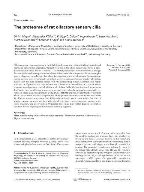
Figure 1. Quality <strong>of</strong> de<strong>cilia</strong>tion by calcium shock and Ca 21 /K 1 -shock. (A) SEM image <strong>of</strong> control <strong>olfactory</strong> neuroepithelium with <strong>sensory</strong> <strong>cilia</strong><br />
(tubular structures) and mucus globules (surface view). <strong>The</strong> inset gives a closer look on the <strong>cilia</strong>ry structures. Both scale bars indicate 2 mm.<br />
(B) Coronal cryosection showing the polarized expression <strong>of</strong> AC III (green), used as a <strong>cilia</strong>ry marker protein in this study. (C) Colocalization<br />
<strong>of</strong> AC III with the microvilli marker protein ezrin (red) at the apical rim <strong>of</strong> the epithelium. (D) <strong>The</strong> conventional “calcium-shock” de<strong>cilia</strong>tion<br />
method reduces the <strong>cilia</strong> density, but the remaining muco<strong>cilia</strong>ry matrix still covers the surface (see inset). (E) <strong>The</strong> AC III signal is partly<br />
removed but much <strong>of</strong> the <strong>cilia</strong>ry matrix remains in place. (F) Both AC III and ezrin signals are reduced, illust<strong>rat</strong>ing that the calcium shock<br />
detaches both <strong>cilia</strong> and microvilli. (G) Treatment with the Ca 21 /K 1 -shock leaves the apical surface practically free <strong>of</strong> <strong>cilia</strong>. Dendritic endings<br />
can be seen (inset) with stumps <strong>of</strong> broken-<strong>of</strong>f <strong>cilia</strong>. (H) <strong>The</strong> punctate pattern <strong>of</strong> the AC III immunosignal originates from the remaining<br />
dendritic endings. (I) While de<strong>cilia</strong>tion has virtually abolished the AC III signal, the remaining ezrin signal documents the differential effect<br />
<strong>of</strong> the Ca 21 /K 1 -shock on <strong>cilia</strong> and microvilli. Blue structures in B, E, H are DAPI-stained nuclei. Scale bars indicate 2 mm in the SEM images<br />
(A, D, G; also insets) and 25 mm in the fluorescence images.<br />
© 2009 WILEY-VCH Verlag GmbH & Co. KGaA, Weinheim www.proteomics-journal.com
326 U. Mayer et al. Proteomics 2009, 9, 322–334<br />
bules scattered across the apical surface. <strong>The</strong>se globules<br />
result from the fixation <strong>of</strong> the <strong>olfactory</strong> mucus, as a wash<br />
with the mucolytic agent DTT (3 mM) partly removed these<br />
structures (not shown). <strong>The</strong> apical endings <strong>of</strong> the ORN dendrites<br />
from which the <strong>cilia</strong> emanate are not visible in the<br />
dense <strong>cilia</strong>ry matrix (inset to Fig. 1A). This matrix gives an<br />
intense immunosignal for AC III in coronal cryosections <strong>of</strong><br />
the <strong>olfactory</strong> epithelium (Fig. 1B), reflecting the polarized<br />
expression <strong>of</strong> AC III in the <strong>cilia</strong>ry membrane. Examining the<br />
tissue after the calcium-shock treatment revealed that the<br />
<strong>cilia</strong>ry matrix was only partially removed. <strong>The</strong> apical surface<br />
was still densely <strong>cilia</strong>ted (Fig. 1D), and dendritic endings<br />
were mostly concealed underneath the <strong>cilia</strong> (inset to Fig. 1D).<br />
Accordingly, cryosections from treated tissue show an almost<br />
continuous AC III immunosignal which was, however,<br />
markedly reduced when compared to the control tissue<br />
(Fig. 1E). This result shows that much <strong>of</strong> the <strong>cilia</strong>ry material<br />
is not detached by the calcium-shock method.<br />
To increase the yield <strong>of</strong> <strong>cilia</strong>ry material, we adapted a<br />
de<strong>cilia</strong>tion procedure that was originally developed for the<br />
detergent-free isolation <strong>of</strong> <strong>cilia</strong> from Paramecium [33]. Basically,<br />
this method consists <strong>of</strong> keeping the cells in a low-salt,<br />
Ca 21 -free buffer, before adding CaCl 2 and KCl (10 and<br />
30 mM, respectively) to trigger the de<strong>cilia</strong>tion process.<br />
Importantly, this procedure did not induce cell lysis or blistering<br />
in Paramecium, both <strong>of</strong> which would introduce non<strong>cilia</strong>ry<br />
proteins into the prepa<strong>rat</strong>ion. With <strong>olfactory</strong> epithelium,<br />
we found a combined Ca 21 /K 1 -shock with 20 mM CaCl 2<br />
and 30 mM KCl most effective for de<strong>cilia</strong>tion (see Section 2).<br />
Tissue treated with this protocol showed a smooth apical<br />
surface, almost completely devoid <strong>of</strong> <strong>cilia</strong>, as well as good<br />
tissue integrity (Fig. 1G). Many dendritic endings were now<br />
visible at the apical surface, bearing the stumps <strong>of</strong> <strong>cilia</strong> which<br />
usually broke <strong>of</strong>f near the <strong>cilia</strong>ry base at the dendritic knobs<br />
(inset to Fig. 1G). In the immunohistochemical analysis, this<br />
tissue showed a residual, punctate staining pattern at the<br />
apical rim, consistent with a signal originating from the<br />
de<strong>cilia</strong>ted dendritic knobs. AC III is expressed both in dendritic<br />
knobs and proximal <strong>cilia</strong> segments, although at lower<br />
density than in the distal parts <strong>of</strong> the <strong>cilia</strong> [34]. Thus, the<br />
Ca 21 /K 1 -shock produces a better gain <strong>of</strong> <strong>cilia</strong>ry material than<br />
the conventional calcium-shock method.<br />
A higher gain <strong>of</strong> <strong>cilia</strong>ry material can result in an<br />
increased purity <strong>of</strong> the <strong>cilia</strong> prepa<strong>rat</strong>ion, provided that the<br />
Ca 21 /K 1 -shock protocol does not also lead to an increase <strong>of</strong><br />
non<strong>cilia</strong>ry material. To monitor the sepa<strong>rat</strong>ion <strong>of</strong> <strong>cilia</strong> and<br />
non<strong>cilia</strong>ry material, we used ezrin, a protein which is located<br />
in immediate proximity to the <strong>cilia</strong> (Fig. 1C). Ezrin is not<br />
expressed in <strong>cilia</strong> but in the microvilli <strong>of</strong> the epithelial supporting<br />
cells. <strong>The</strong>se cells form a single layer at the top <strong>of</strong> the<br />
<strong>olfactory</strong> epithelium where their microvilli are interspersed<br />
with the ORN <strong>cilia</strong>. As such, ezrin is a suitable marker protein<br />
for non<strong>cilia</strong>ry apical membranes. Immunosignals <strong>of</strong> AC<br />
III and ezrin completely match in untreated tissue (Fig. 1C),<br />
as expected from earlier studies [35, 36]. Following the Ca 21 /<br />
K 1 -shock, the AC III signal is reduced to a punctate pattern<br />
while the ezrin signal remains strong (Fig. 1I). Thus, the<br />
Ca 21 /K 1 -shock exerts a differential effect on <strong>cilia</strong> and microvilli.<br />
It detaches <strong>cilia</strong> while leaving the microvilli largely<br />
intact. In contrast, tissue treated by the conventional calcium-shock<br />
treatment (Fig. 1F) appears more damaged with<br />
sections <strong>of</strong> the microvilli layer missing. <strong>The</strong>se data show that<br />
the Ca 21 /K 1 -shock method is better suited to sepa<strong>rat</strong>e <strong>cilia</strong>ry<br />
from non<strong>cilia</strong>ry material.<br />
To assess the purity <strong>of</strong> the material, we determined the<br />
degree <strong>of</strong> enrichment <strong>of</strong> two <strong>cilia</strong>ry-marker proteins, AC III<br />
and CNGA4, in the <strong>cilia</strong> prepa<strong>rat</strong>ion. Membrane proteins<br />
were isolated from (i) total <strong>olfactory</strong> epithelium, (ii) the <strong>cilia</strong><br />
prepa<strong>rat</strong>ion obtained by calcium-shock, and (iii) the <strong>cilia</strong><br />
prepa<strong>rat</strong>ion obtained by the Ca 21 /K 1 -shock. <strong>The</strong> same<br />
amount <strong>of</strong> total protein from each <strong>of</strong> these prepa<strong>rat</strong>ions<br />
(6 mg; verified by densitometric analysis <strong>of</strong> gel stainings) was<br />
sepa<strong>rat</strong>ed by SDS-PAGE, and the amounts <strong>of</strong> AC III and<br />
CNGA4 proteins were compared on Western blots. Figure 2A<br />
shows a representative result for 12 experiments. While the<br />
signals for the two proteins are hardly detectable in lane I<br />
(<strong>olfactory</strong> epithelium with <strong>cilia</strong>), the much stronger signals<br />
in lane III (<strong>cilia</strong> prepa<strong>rat</strong>ion resulting from Ca 21 /K 1 -shock)<br />
demonst<strong>rat</strong>e a roughly 100-fold enrichment <strong>of</strong> the marker<br />
proteins in that prepa<strong>rat</strong>ion. <strong>The</strong> calcium-shock protocol<br />
produces less enrichment, as illust<strong>rat</strong>ed by the weaker bands<br />
in Figure 2A (lane II). This result shows that the Ca 21 /K 1 -<br />
shock method yields an enhanced efficacy <strong>of</strong> <strong>cilia</strong> purification.<br />
Inspection <strong>of</strong> the ezrin immunosignals (Fig. 2A, bottom)<br />
showed that 6 mg total protein <strong>of</strong> <strong>cilia</strong> prepa<strong>rat</strong>ion contained<br />
equal (calcium shock; lane II) or less (Ca 21 /K 1 -shock;<br />
lane III) ezrin than 6 mg <strong>of</strong> total protein from the complete<br />
tissue (lane I). This result provides important information<br />
about the purity <strong>of</strong> the <strong>cilia</strong> prepa<strong>rat</strong>ion. While <strong>cilia</strong>ry material<br />
is roughly 100-fold enriched in the <strong>cilia</strong> prepa<strong>rat</strong>ion, the<br />
content <strong>of</strong> microvillar material is diminished by the Ca 21 /<br />
K 1 -shock prepa<strong>rat</strong>ion, indicating that the prepa<strong>rat</strong>ion is<br />
contaminated by ,1% with microvilli. <strong>The</strong>se results<br />
demonst<strong>rat</strong>e that the <strong>cilia</strong> prepa<strong>rat</strong>ion obtained by the Ca 21 /<br />
K 1 -shock protocol is indeed highly enriched in <strong>cilia</strong> and<br />
contains only little contaminations from non<strong>cilia</strong>ry material.<br />
3.2 Characterization <strong>of</strong> proteins in the <strong>cilia</strong><br />
prepa<strong>rat</strong>ion<br />
In analyzing the protein spectrum <strong>of</strong> the <strong>cilia</strong> prepa<strong>rat</strong>ion,<br />
our goal was to specifically identify those proteins which are<br />
associated with the <strong>cilia</strong>ry membrane. Integral membrane<br />
proteins (like AC III; [37]) and proteins associated with the<br />
<strong>cilia</strong>ry membrane (like phosphodiesterase PDE1C2; [38])<br />
constitute the specific <strong>proteome</strong> <strong>of</strong> <strong>olfactory</strong> function. We<br />
tried to remove from the prepa<strong>rat</strong>ion soluble proteins which<br />
can freely diffuse between the <strong>cilia</strong>ry and dendritic compartment<br />
in vivo, as well as such soluble proteins <strong>of</strong> non<strong>cilia</strong>ry<br />
origin that contaminated the prepa<strong>rat</strong>ion during the isolation<br />
procedure. Ciliary proteins (20 mg) were sepa<strong>rat</strong>ed by<br />
SDS-PAGE using a 7–12% density gradient to cover a large<br />
© 2009 WILEY-VCH Verlag GmbH & Co. KGaA, Weinheim www.proteomics-journal.com
Proteomics 2009, 9, 322–334 327<br />
Figure 2. Characterization <strong>of</strong> the <strong>cilia</strong> prepa<strong>rat</strong>ion obtained by the<br />
Ca 21 /K 1 -method. (A) Analysis <strong>of</strong> marker-protein contents in the<br />
<strong>cilia</strong> prepa<strong>rat</strong>ion by Western blots. <strong>The</strong> marker-protein content<br />
was compared between 6 mg total protein <strong>of</strong> whole <strong>olfactory</strong> epithelium,<br />
including <strong>cilia</strong> (lane I), and 6 mg total protein in <strong>cilia</strong> prepa<strong>rat</strong>ions<br />
obtained by either calcium shock (lane II) or by Ca 21 /K 1 -<br />
shock (lane III). Comparison <strong>of</strong> lanes I and III reveals a roughly<br />
100-fold enrichment <strong>of</strong> the <strong>cilia</strong>ry proteins AC III and CNGA4, and<br />
a partial depletion <strong>of</strong> the microvillar marker ezrin, in the <strong>cilia</strong><br />
prepa<strong>rat</strong>ion. In contrast, the conventional calcium-shock method<br />
yielded less enrichment <strong>of</strong> <strong>cilia</strong>ry proteins and no depletion <strong>of</strong><br />
ezrin (compare lanes I and II). (B) Sepa<strong>rat</strong>ion <strong>of</strong> <strong>cilia</strong>ry proteins by<br />
1-D SDS-PAGE (7–12.5% density). Twenty micrograms <strong>of</strong> protein<br />
isolated by Ca 21 /K 1 -shock was sepa<strong>rat</strong>ed and stained by colloidal<br />
Coomassie. For MS, the gel was cut into 29 fragments according<br />
to the indicated pattern. <strong>The</strong> protein numbers in Supporting<br />
Information Table S1 correspond to the numbering <strong>of</strong> the gel<br />
fragments.<br />
range <strong>of</strong> protein sizes. <strong>The</strong> resulting gel (Fig. 2B) was cut into<br />
29 sections (3612 mm 2 each) which were analyzed sepa<strong>rat</strong>ely<br />
by LC-ESI-MS/MS. To examine the reliability <strong>of</strong> our<br />
protocol, we analyzed the protein content <strong>of</strong> three representative<br />
gel pieces (#16, 19, 22) from four different prepa<strong>rat</strong>ions<br />
by LC-ESI-MS/MS. <strong>The</strong> gel pieces contained the<br />
same protein mix in all four gels. <strong>The</strong> protocol thus yielded a<br />
reproducible protein distribution on the SDS-PAGE.<br />
In total, we identified 463 different proteins in this gel.<br />
<strong>The</strong> list <strong>of</strong> all 463 proteins, together with the mass spectrometric<br />
details and bioinformatic analyses, is supplied as<br />
Supporting Information (Table S1) and can be downloaded<br />
from the authors website at http://www.zoo.uniheidelberg.de/prot/UM1148<br />
or the proteomics website at<br />
www.proteomics-journal.com. Protein sizes ranged from 11<br />
to 569 kDa with isoelectric points (pI) between 4 and 12<br />
(Figs. 3A and B). A plot <strong>of</strong> protein size against pI (Fig. 3C)<br />
showed that a wide range <strong>of</strong> molecular weights was covered<br />
both by acidic and basic proteins. Figure 3D shows the distribution<br />
<strong>of</strong> predicted transmembrane helices in the integral<br />
membrane proteins. For 45% <strong>of</strong> these proteins, more than<br />
one transmembrane helix is predicted by TMHMM and<br />
SOSUI. Our comprehensive bioinformatic annotation <strong>of</strong> all<br />
protein sequences (see Supporting Information Table S1)<br />
revealed that 43% <strong>of</strong> the proteins were integral membrane<br />
proteins, 30% were membrane-associated proteins, and the<br />
remaining 27% were soluble proteins (Fig. 4A). To compare<br />
the proteomic results obtained from the Ca 21 /K 1 -method to<br />
that obtained by the conventional calcium-shock, we set our<br />
protein list against the one published by [22]. From the 268<br />
proteins identified in that study, 155 were also detected in<br />
the present protein list. An analysis <strong>of</strong> the proteins which<br />
are absent from our present list <strong>of</strong> 463 proteins revealed<br />
that most <strong>of</strong> the missing 113 proteins were either soluble<br />
proteins (72) or proteins assigned to different intracellular<br />
membranes (38). Only 3 <strong>of</strong> the 113 proteins were located to<br />
the plasma membrane. In accordance with the enrichment<br />
<strong>of</strong> the marker proteins AC III and CNGA4, this comparison<br />
shows that the de<strong>cilia</strong>tion procedure by Ca 21 /K 1 -shock<br />
leads to a material in which <strong>cilia</strong>ry membrane proteins are<br />
more efficiently enriched. For further assessment <strong>of</strong> the<br />
quality <strong>of</strong> the prepa<strong>rat</strong>ion, a comparison with the recent<br />
ORN transcriptome analysis [39] yields a greater than 83%<br />
match, confirming that the proteins identified are predominantly<br />
expressed in ORNs.<br />
Prior to additional analyses, obvious contaminants such<br />
as serum (6 proteins, 1%) or nuclear (12, 3%) proteins, were<br />
excluded from the protein list. Small amounts <strong>of</strong> mitochondrial<br />
(15, 3%), peroxisomal (8, 2%), endosomal (5, 1%), or<br />
ribosomal (30, 6%) proteins, which are not part <strong>of</strong> the <strong>cilia</strong>ry<br />
compartment, were also removed from the <strong>olfactory</strong> <strong>cilia</strong><br />
<strong>proteome</strong>. In sum, these proteins accounted for approximately<br />
20% <strong>of</strong> the proteins identified; this is comparable to<br />
the levels detected in virtually all other <strong>cilia</strong> and flagellar<br />
proteomic analyses (see [40, 41]). <strong>The</strong> remaining 377 proteins<br />
are listed in Supporting Information Table S2 (for<br />
download location, see previous paragraph) and represent a<br />
protein set most likely involved in <strong>olfactory</strong> function. Eighty<br />
percent <strong>of</strong> these proteins are integral membrane proteins,<br />
membrane-associated proteins, or components <strong>of</strong> supramolecular<br />
complexes linked to membranes. Fourteen percent <strong>of</strong><br />
them are cytoskeleton proteins. <strong>The</strong> presence <strong>of</strong> 20% soluble<br />
proteins may be interpreted as a consequence <strong>of</strong> vesicle formation<br />
during the prepa<strong>rat</strong>ion (see [42]), which can trap<br />
cytosolic constituents. Some <strong>of</strong> these proteins may also<br />
represent new components <strong>of</strong> membrane-associated, supramolecular<br />
complexes which so far evaded identification in<br />
© 2009 WILEY-VCH Verlag GmbH & Co. KGaA, Weinheim www.proteomics-journal.com
328 U. Mayer et al. Proteomics 2009, 9, 322–334<br />
Figure 3. Properties <strong>of</strong> proteins identified in the <strong>cilia</strong> prepa<strong>rat</strong>ion. (A) Molecular weights <strong>of</strong> the 463 proteins contained in the <strong>cilia</strong> prepa<strong>rat</strong>ion<br />
which are distributed over a total range from 11 to 569 kDa. (B) <strong>The</strong> distribution <strong>of</strong> pI covers a range from 4 to 12 with peaks for slightly<br />
acidic and basic proteins. (C) Plotting molecular weight against pI illust<strong>rat</strong>es that the entire pI range is represented by proteins ,100 kDa,<br />
while larger proteins tend to have acidic pIs. (D) Distribution <strong>of</strong> the numbers <strong>of</strong> transmembrane helices as predicted by the TMHMM and<br />
SOSUI servers for the 177 membrane-integral proteins identified in the <strong>cilia</strong> prepa<strong>rat</strong>ion.<br />
<strong>olfactory</strong> <strong>cilia</strong>. This could be true for several kinases, calciumbinding<br />
proteins, or other proteins involved in signal transduction<br />
processes (see Supporting Information Table S2).<br />
Nevertheless, the high percentage <strong>of</strong> membrane proteins is a<br />
promising result as we are primarily interested in those proteins<br />
that have been purified with the <strong>cilia</strong> membranes. <strong>The</strong><br />
biochemical prepa<strong>rat</strong>ion cannot sepa<strong>rat</strong>e the <strong>cilia</strong>ry plasma<br />
membrane from membranes located within the <strong>cilia</strong>, since<br />
knobs, and proximal <strong>olfactory</strong> <strong>cilia</strong> contain smooth ER, multivesicular<br />
bodies, and other vesicular structures [14, 22, 43].<br />
3.3 Functional groups in the <strong>olfactory</strong> <strong>cilia</strong> <strong>proteome</strong><br />
Several functional groups <strong>of</strong> proteins are over-represented in<br />
the <strong>olfactory</strong> <strong>cilia</strong> <strong>proteome</strong>. 80% <strong>of</strong> the proteins detected can<br />
be attributed to one specific <strong>olfactory</strong> molecular task <strong>of</strong><br />
ORNs. Almost one quarter (77, Fig. 4B, Supporting Information<br />
Table S2) <strong>of</strong> the identified proteins are related to<br />
intracellular traffic, consistent with the polarized protein<br />
expression pattern in these <strong>cilia</strong>ted neurons. Proteins<br />
involved in cell differentiation (including ciliogenesis) as<br />
well as cell structure and motility are also prominent (80,<br />
Fig. 4B, see also groups neurogenesis and cytoskeleton in<br />
Supporting Information Table S2), reflecting the continuous<br />
turnover <strong>of</strong> ORNs throughout life. Proteins involved in<br />
xenobiotic metabolism as well as some stress-induced proteins<br />
(49, Fig. 4B and Supporting Information Table S2) are<br />
also enriched in the <strong>cilia</strong> <strong>proteome</strong>. This collection <strong>of</strong> cytoprotective<br />
proteins reflects the precarious situation <strong>of</strong> a <strong>sensory</strong><br />
neuron which is exposed to every environmental compound<br />
present in the inhaled air; these proteins protect the<br />
ORNs from cytotoxic compounds and terminate the <strong>sensory</strong><br />
response. Several other functional categories <strong>of</strong> interest have<br />
been identified such as an assortment <strong>of</strong> transport proteins<br />
(34), proteins involved in signal transduction processes (78),<br />
or proteins <strong>of</strong> unknown function (30) (Fig. 4B and Supporting<br />
Information Table S2). Within the signal transduction<br />
category, all known components <strong>of</strong> the canonical <strong>olfactory</strong><br />
signal-transduction cascade (25 proteins) were robustly<br />
identified. <strong>The</strong> high identification scores <strong>of</strong> these proteins<br />
are in good accordance with the immunoblot analysis and<br />
further corrobo<strong>rat</strong>e the view that the <strong>cilia</strong> prepa<strong>rat</strong>ion represents<br />
a material in which <strong>cilia</strong>ry membrane proteins are<br />
efficiently purified.<br />
© 2009 WILEY-VCH Verlag GmbH & Co. KGaA, Weinheim www.proteomics-journal.com
Proteomics 2009, 9, 322–334 329<br />
Figure 4. Subcellular location <strong>of</strong> all proteins identified and functional classification <strong>of</strong> proteins in the <strong>olfactory</strong> <strong>cilia</strong> <strong>proteome</strong>. (A) All 463<br />
proteins <strong>of</strong> the <strong>cilia</strong> prepa<strong>rat</strong>ion were assigned to a subcelluar compartment using the Subcellular Localization Database Locate and lite<strong>rat</strong>ure<br />
searches. <strong>The</strong> white sections represent cytosolic proteins, light-gray sections are membrane integral proteins, dark-gray are membrane-associated<br />
proteins. Cytoskeletal proteins were grouped with membrane-associated proteins. (B) Functional groups <strong>of</strong> the 377 proteins<br />
in the <strong>olfactory</strong> <strong>cilia</strong> <strong>proteome</strong>. Annotations were deduced from electronic annotation and extensive lite<strong>rat</strong>ure searches (see Supporting<br />
Information Table S1). Numbers in parentheses indicate the number <strong>of</strong> proteins in each section together with the percentages<br />
relative to the total number <strong>of</strong> 463 or 377 proteins, respectively.<br />
© 2009 WILEY-VCH Verlag GmbH & Co. KGaA, Weinheim www.proteomics-journal.com
330 U. Mayer et al. Proteomics 2009, 9, 322–334<br />
3.4 Using the <strong>olfactory</strong> <strong>cilia</strong> <strong>proteome</strong><br />
<strong>The</strong> list <strong>of</strong> <strong>cilia</strong>ry proteins provided in this study can be useful<br />
to promote research in various fields <strong>of</strong> <strong>olfactory</strong> function.<br />
<strong>The</strong> “Ca 21 /K 1 -shock” de<strong>cilia</strong>tion protocol and the subsequent<br />
purification yield a material which contains all<br />
known components <strong>of</strong> the <strong>cilia</strong>ry transduction chain. To<br />
examine the function <strong>of</strong> a novel protein that is listed in the<br />
<strong>cilia</strong> <strong>proteome</strong>, the first step would be to look at its subcellular<br />
expression pattern using immunohistochemistry. In<br />
the following, we briefly illust<strong>rat</strong>e this initial investigation<br />
for four proteins representing distinct aspects <strong>of</strong> <strong>olfactory</strong><br />
function. (i) Plexin B2: semaphorin signaling plays an<br />
important role in neuronal development, axon mig<strong>rat</strong>ion,<br />
and neuronal apoptosis [44–46]. <strong>The</strong> semaphorin receptor<br />
Plexin B2 has a high score in the <strong>cilia</strong> <strong>proteome</strong> and displays<br />
an interesting immunosignal (Fig. 2A). A subpopulation <strong>of</strong><br />
ORNs express Plexin B2 from soma to dendritic knob, suggesting<br />
that these cells are related to the continuous process<br />
<strong>of</strong> neurogenesis and apoptosis that is characteristic for the<br />
<strong>olfactory</strong> epithelium. (ii) Flotillin 1: there is evidence that<br />
lipid-raft microdomains exist in <strong>olfactory</strong> <strong>cilia</strong> and concent<strong>rat</strong>e<br />
key transduction enzymes like AC III and G aolf [47].<br />
Consistent with these data, the lipid-raft-associated protein<br />
flotillin 1 [48] features prominently in the <strong>cilia</strong> <strong>proteome</strong> and<br />
is expressed at the <strong>cilia</strong>ry surface <strong>of</strong> the <strong>olfactory</strong> epithelium<br />
(Fig. 5B). (iii) CLIC 6: a critical missing link in the <strong>olfactory</strong><br />
signal transduction chain is a calcium-activated chloride<br />
channel that conducts most <strong>of</strong> the receptor current [3, 4]. <strong>The</strong><br />
molecular identity <strong>of</strong> this channel has not yet been established<br />
[3–5]. A possible new candidate for this protein may be<br />
CLIC 6 [49, 50], a chloride channel from the <strong>cilia</strong> <strong>proteome</strong><br />
which is expressed in the <strong>cilia</strong> (Fig. 5C). (iv) SLC4A1: finally,<br />
chloride uptake into <strong>olfactory</strong> <strong>cilia</strong> is currently an important<br />
research topic [6–7]. <strong>The</strong> Cl 2 /HCO 3 2 exchanger SLC4A1,<br />
which has a high score in the <strong>cilia</strong> <strong>proteome</strong> and appears to<br />
be selectively expressed in the <strong>cilia</strong> (Fig. 5D, compare to the<br />
expression <strong>of</strong> AC III, Fig. 5E) seems to be a promising candidate<br />
for a <strong>cilia</strong>ry chloride-uptake pathway. <strong>The</strong>se few<br />
examples illust<strong>rat</strong>e that many proteins <strong>of</strong> the <strong>cilia</strong> <strong>proteome</strong>,<br />
which hitherto were not linked to <strong>olfactory</strong> function, may<br />
now attract attention in the respective field <strong>of</strong> <strong>olfactory</strong> research.<br />
4 Discussion<br />
Organellar proteomics has greatly advanced our understanding<br />
<strong>of</strong> cellular function in recent years as it provides the<br />
specific protein inventory <strong>of</strong> a distinct functional unit within<br />
the cell. For instance, the recent proteomic characterization<br />
<strong>of</strong> synaptic vesicles [51, 52] has pointed to numerous novel<br />
approaches to studying synaptic transmission. In fact, the<br />
vision that sees organellar proteomics as a platform for systems<br />
biology is being supported by an increasing number <strong>of</strong><br />
studies not only on structures common to all eukaryotic cells<br />
(reviews: [53–55]), but particularly on key subcellular compartments<br />
which play the decisive role for a specific cell type.<br />
Examples for such key structures are synaptic vesicles in<br />
neurons [52], <strong>sensory</strong> outer segments in photoreceptors [41],<br />
and <strong>cilia</strong> in airway epithelia [56]. In the present study, we<br />
have analyzed the <strong>proteome</strong> <strong>of</strong> the key structure <strong>of</strong> ORNs, the<br />
chemo<strong>sensory</strong> <strong>cilia</strong>. Since a highly enriched <strong>cilia</strong> prepa<strong>rat</strong>ion<br />
was a prerequisite for this project, we improved the de<strong>cilia</strong>tion<br />
process and monitored the purification <strong>of</strong> <strong>cilia</strong> through<br />
the enrichment <strong>of</strong> <strong>cilia</strong>ry marker proteins AC III and<br />
CNGA4. Using a Ca 21 /K 1 -shock method for de<strong>cilia</strong>tion, we<br />
achieved a roughly 100-fold enrichment <strong>of</strong> <strong>cilia</strong>ry material.<br />
<strong>The</strong> cactus-like appearance <strong>of</strong> the dendritic endings <strong>of</strong> ORNs<br />
at the de<strong>cilia</strong>ted surface suggests that the <strong>cilia</strong> were detached<br />
at the thick proximal segments (about 2 mm long; [35]) so<br />
that the thin distal segments (about 50 mm long) were predominantly<br />
collected in the membrane prepa<strong>rat</strong>ion. Mammalian<br />
<strong>olfactory</strong> <strong>cilia</strong> are immotile and have accordingly<br />
simple axonemal structures without dynein arms. While the<br />
proximal segments show nine outer tubulin doublets plus<br />
two central subfibers, the outer doublets are missing in the<br />
distal <strong>cilia</strong> (review: [35]). <strong>The</strong> contribution <strong>of</strong> axonemal proteins<br />
to our <strong>cilia</strong> prepa<strong>rat</strong>ion is, therefore, not as prominent<br />
as in prepa<strong>rat</strong>ions from motile <strong>cilia</strong> (see [40]). Nevertheless,<br />
Figure 5. Immunohistochemical localization <strong>of</strong> four novel proteins from the <strong>cilia</strong> <strong>proteome</strong>. Cryosections <strong>of</strong> <strong>rat</strong> <strong>olfactory</strong> epithelium were<br />
stained with antibodies raised against the indicated proteins. <strong>The</strong> selected proteins are examples for proteins which were hitherto not in<br />
the focus <strong>of</strong> <strong>olfactory</strong> research but may be interesting for the fields <strong>of</strong> <strong>olfactory</strong> neurogenesis (plexin B2, A), lipid-raft microdomains (flotillin<br />
1, B), chemo-electrical transduction (CLIC 6, C), and <strong>cilia</strong>ry chloride transport (SLC4A1, D). <strong>The</strong> immune fluorescence <strong>of</strong> AC III (E) serves<br />
as an example for a restricted <strong>cilia</strong>ry expression. Blue signal are DAPI stains <strong>of</strong> nuclei. Calib<strong>rat</strong>ion bars indicate 8 mm.<br />
© 2009 WILEY-VCH Verlag GmbH & Co. KGaA, Weinheim www.proteomics-journal.com
Proteomics 2009, 9, 322–334 331<br />
51 proteins could be attributed to the cytoskeleton indicating<br />
that these proteins play a crucial role in providing structural<br />
stability and in organizing the proper targeting <strong>of</strong> membrane<br />
components in the <strong>olfactory</strong> <strong>cilia</strong> (see below).<br />
Contamination from non<strong>cilia</strong>ry material may originate<br />
from the dendrites or somata <strong>of</strong> ORNs as well as from epithelial<br />
supporting cells. However, our de<strong>cilia</strong>tion method<br />
was designed to maintain tissue integrity, and SEM and<br />
light-microscopic inspection confirmed that the de<strong>cilia</strong>ted<br />
tissue was not noticeably damaged. Moreover, the most<br />
exposed epithelial structures, the supporting cell microvilli,<br />
were apparently resistant to the Ca 21 /K 1 -shock treatment, as<br />
demonst<strong>rat</strong>ed by the ezrin-immunohistochemistry and the<br />
low ezrin content <strong>of</strong> the <strong>cilia</strong> prepa<strong>rat</strong>ion. Nevertheless, contaminating<br />
non<strong>cilia</strong>ry proteins are present in the prepa<strong>rat</strong>ion<br />
and have to be distinguished from <strong>cilia</strong>ry proteins by bioinformatic<br />
analysis.<br />
Inspection <strong>of</strong> Supporting Information Table S2 reveals<br />
some interesting features <strong>of</strong> the ORN <strong>cilia</strong> <strong>proteome</strong>: <strong>The</strong> <strong>cilia</strong><br />
<strong>proteome</strong> contains all proteins involved in the current model<br />
<strong>of</strong> chemoelectrical transduction (AC III, G-proteins, PDE1C,<br />
CNG channel subunits, etc.; review: [1]) with the exception <strong>of</strong><br />
the OR proteins. <strong>The</strong> absence <strong>of</strong> identified ORs is not surprising,<br />
as each <strong>of</strong> the ,1300 members <strong>of</strong> this largest protein<br />
family is expressed in only about 0.1% <strong>of</strong> ORNs, thus contributing<br />
only very little to the <strong>cilia</strong> <strong>proteome</strong> <strong>of</strong> all ORNs. In<br />
the group <strong>of</strong> transduction proteins, additional 63 proteins<br />
were listed that have not previously been identified or investigated<br />
in <strong>olfactory</strong> <strong>sensory</strong> signal transduction. Among them,<br />
eight annexin is<strong>of</strong>orms were detected. Annexins belong to a<br />
class <strong>of</strong> membrane-associated Ca 21 - and phospholipid-binding<br />
proteins and mediate cellular responses to changes <strong>of</strong> the<br />
intracellular Ca 21 level. <strong>The</strong>y are generally thought to act<br />
either as ion channel regulators or as ion channels themselves<br />
[57]. <strong>The</strong>ir presence may point to regulatory effects on <strong>olfactory</strong><br />
transduction channels. Recently, immunohistochemical<br />
analysis revealed that annexin is<strong>of</strong>orms A1, A2, and A5 localize<br />
in frog <strong>olfactory</strong> <strong>cilia</strong> [58]. Interesting proteins in this<br />
group also include lipid-raft associated flotillins and stomatinlike<br />
proteins. Stomatin-like protein-3 (SLP3) is specifically<br />
expressed in the <strong>cilia</strong> <strong>of</strong> ORNs and has been found to be associated<br />
with <strong>olfactory</strong> transduction components (e.g., AC III)<br />
[59, 60]. SLP3 has recently also been shown to be a constituent<br />
<strong>of</strong> mammalian cutaneous mechanoreceptors where it is necessary<br />
for the function <strong>of</strong> the mechano-sensitive transduction<br />
channel [61]. Thus, SLP3 is a very interesting protein, apparently<br />
contributing to ion-channel function in <strong>sensory</strong> neurons.<br />
<strong>The</strong> same could be true for the hitherto unknown stomatin-like<br />
member 2 in ORNs.<br />
Membrane-transport proteins were particularly abundant.<br />
<strong>The</strong> <strong>cilia</strong>ry membrane sepa<strong>rat</strong>es the ORN cytosol from<br />
the <strong>olfactory</strong> mucus, a medium with a characteristic ionic<br />
composition, distinct from intra- and extracellular fluids [62].<br />
<strong>The</strong> <strong>cilia</strong> <strong>proteome</strong> contains 34 ion channel or transport<br />
proteins <strong>of</strong> different protein families. Among them, 13<br />
solute transporters from nine SLC families were found,<br />
including SLC 12-A2, the Na 1 /K 1 /2Cl 2 cotransporter<br />
NKCC1 which plays a central role in <strong>olfactory</strong> transduction.<br />
NKCC1 accumulates intracellular Cl 2 in ORNs and provides<br />
the ionic basis for depolarizing Cl 2 currents [6–8]. All three<br />
subunits <strong>of</strong> the cAMP-gated cation channel were detected,<br />
together with various chloride channels, different ion<br />
pumps, water channels, and a set <strong>of</strong> transporters for larger<br />
molecules. <strong>The</strong> plasma membrane Ca 21 ATPase PMCA has<br />
recently been shown to contribute to re-establish resting<br />
Ca 21 levels in the <strong>cilia</strong> following <strong>olfactory</strong> responses [63]. <strong>The</strong><br />
physiological significance <strong>of</strong> the transport capacity <strong>of</strong> <strong>cilia</strong><br />
will be interesting to examine. Likely functions are the ionic<br />
homeostasis in the <strong>cilia</strong>ry lumen and in the mucus layer. A<br />
great number <strong>of</strong> proteins could be classified as being<br />
involved in protein targeting and neurogenesis. <strong>The</strong>se are<br />
obviously two aspects <strong>of</strong> critical importance for the <strong>olfactory</strong><br />
neuroepithelium, where the neuron population is subjected<br />
to a constant turnover, and where a distinct transduction<br />
compartment is maintained by polarized expression <strong>of</strong> proteins<br />
involved in <strong>sensory</strong> signal processing. Accordingly, we<br />
found many proteins involved in the secretory transport<br />
machinery. Among them, receptor-transporting protein 1<br />
(RTP1) was shown to be specifically expressed in ORNs and<br />
to promote functional cell surface expression <strong>of</strong> ORs when<br />
expressed in HEK293T cells [17]. RTP1 is directly associated<br />
with the OR protein and thus also enhances the responses to<br />
odorants. Similar results were found for HSC70 [64]. Understanding<br />
OR function is hampered by the difficulty in heterologous<br />
expression. <strong>The</strong> proteins listed in this category are<br />
good <strong>olfactory</strong> candidates to enhance further experimental<br />
approaches in this problem. Alternatively, they could also<br />
contribute to the targeting <strong>of</strong> other signal transduction<br />
molecules to the <strong>cilia</strong>ry membrane. In particular, Lambert et<br />
al. [65] showed that B-cell receptor-associated protein 31<br />
(BAP31) colocalizes with and controls the expression <strong>of</strong> cystic<br />
fibrosis transmembrane conductance regulator (CFTR) in<br />
the plasma membrane. Similar findings were reported for<br />
several RAB is<strong>of</strong>orms present in the <strong>cilia</strong> <strong>proteome</strong> [66].<br />
Chen and Balch [67] reported that RAB recycling in the early<br />
secretory pathways involves the heat-shock protein HSP90<br />
system. HSP90 activity is required to form a functional guanine<br />
nucleotide dissociation inhibitor complex to retrieve,<br />
e.g., RAB1 from the membrane and is essential for RAB1-<br />
dependent Golgi assembly. <strong>The</strong> three HSP90 proteins identified<br />
in our analysis seem to be involved in the RAB pathway.<br />
Moreover, we found several proteins involved in neurogenesis.<br />
For example, the neuronal marker protein growth associated<br />
protein (GAP-43) is expressed in <strong>olfactory</strong> neurons<br />
during growth [68]. KPL2 is an actin-binding protein,<br />
induced during ciliogenesis [56], and may be involved in the<br />
differentiation <strong>of</strong> ORNs from precursor cells. <strong>The</strong> same<br />
could be true for several other proteins listed in this group.<br />
<strong>The</strong> substantial number <strong>of</strong> identified <strong>olfactory</strong> cytoprotective<br />
proteins reflects the special location <strong>of</strong> ORNs as the only<br />
neurons <strong>of</strong> the body directly exposed to the external environment.<br />
© 2009 WILEY-VCH Verlag GmbH & Co. KGaA, Weinheim www.proteomics-journal.com
332 U. Mayer et al. Proteomics 2009, 9, 322–334<br />
<strong>The</strong> 377 proteins identified here do not constitute the<br />
entire <strong>cilia</strong> <strong>proteome</strong>. Proteins with low abundancy which<br />
did not reach the identification threshold (see Section 2)<br />
include the <strong>olfactory</strong> receptor proteins, and probably also<br />
membrane proteins expressed in small subpopulations <strong>of</strong><br />
ORNs in the <strong>olfactory</strong> epithelium. Improved methods <strong>of</strong><br />
membrane isolation are needed for a more specific purification<br />
<strong>of</strong> membrane proteins. <strong>The</strong> selective partitioning <strong>of</strong><br />
plasma membranes in aqueous polymer two-phase systems<br />
is a promising approach to this problem [69]. This<br />
method exploits the tendency <strong>of</strong> biological membranes to<br />
partition into the top phase <strong>of</strong> a poly ethylene glycol/dextran<br />
system from where a high fraction <strong>of</strong> plasma membrane<br />
proteins can be isolated. Such a protocol reduces the<br />
complexity <strong>of</strong> protein mixtures in favor <strong>of</strong> plasma membrane<br />
proteins and may give access to low-abundancy<br />
proteins.<br />
In conclusion, we have isolated <strong>sensory</strong> <strong>cilia</strong> <strong>of</strong> <strong>rat</strong><br />
ORN by a novel de<strong>cilia</strong>tion method. <strong>The</strong> quality <strong>of</strong> the<br />
prepa<strong>rat</strong>ion was monitored by SEM, <strong>cilia</strong>ry marker proteins,<br />
and bioinformatic examination. <strong>The</strong> resulting list <strong>of</strong><br />
377 proteins contains most <strong>of</strong> the known proteins <strong>of</strong><br />
<strong>olfactory</strong> signal transduction, as well as many other proteins<br />
that may fulfill important functions in <strong>olfactory</strong><br />
physiology. <strong>The</strong> <strong>cilia</strong> <strong>proteome</strong> provides multiple starting<br />
points for more detailed examinations <strong>of</strong> distinct protein<br />
functions in ORNs. Especially for the areas <strong>of</strong> membrane<br />
transport, signal processing, membrane targeting, and<br />
neurogenesis, the <strong>cilia</strong> <strong>proteome</strong> constitutes a valuable<br />
source <strong>of</strong> information.<br />
We are g<strong>rat</strong>eful to Dr. Tore Kempf (DKFZ Heidelberg) for<br />
valuable help with the protein analysis and Georgia Ignatiadou<br />
(Institute <strong>of</strong> Zoology, University <strong>of</strong> Heidelberg) for technical<br />
assistance with the immunohistochemistry.<br />
<strong>The</strong> authors have declared no conflict <strong>of</strong> interest.<br />
5 References<br />
[1] Frings, S., Chemoelectrical signal transduction in <strong>olfactory</strong><br />
<strong>sensory</strong> neurons <strong>of</strong> air-breathing verteb<strong>rat</strong>es. Cell Mol. Life<br />
Sci. 2001, 58, 510–519.<br />
[2] Hatt, H., Molecular and cellular basis <strong>of</strong> human olfaction.<br />
Chem. Biodivers. 2004, 1, 1857–1869.<br />
[3] Kleene, S. J., Gesteland, R. C., Calcium-activated chloride<br />
conductance in frog <strong>olfactory</strong> <strong>cilia</strong>. J. Neurosci. 1991, 11,<br />
3624–3629.<br />
[4] Reisert, J., Bauer, P. J., Yau, K. W., Frings, S., <strong>The</strong> Ca-activated<br />
Cl channel and its control in <strong>rat</strong> <strong>olfactory</strong> receptor neurons. J.<br />
Gen. Physiol. 2003, 122, 349–363.<br />
[5] Pifferi, S., Pascarella, G., Boccaccio, A., Mazzatenta, A. et al.,<br />
Bestrophin-2 is a candidate calcium-activated chloride channel<br />
involved in <strong>olfactory</strong> transduction. Proc. Natl. Acad. Sci.<br />
USA 2006, 103, 12929–12934.<br />
[6] Kaneko, H., Putzier, I., Frings, S., Kaupp, U. B., Gensch, T.,<br />
Chloride accumulation in mammalian <strong>olfactory</strong> <strong>sensory</strong><br />
neurons. J. Neurosci. 2004, 24, 7931–7938.<br />
[7] Reisert, J., Lai, J., Yau, K. W., Bradley, J., Mechanism <strong>of</strong> the<br />
excitatory Cl- response in mouse <strong>olfactory</strong> receptor neurons.<br />
Neuron 2005, 45, 553–561.<br />
[8] Nickell, W. T., Kleene, N. K., Kleene, S. J., Mechanisms <strong>of</strong><br />
neuronal chloride accumulation in intact mouse <strong>olfactory</strong><br />
epithelium. J. Physiol. 2007, 583, 1005–1020.<br />
[9] Smith, D. W., Thach, S., Marshall, E. L., Mendoza, M. G.,<br />
Kleene, S. J., Mice lacking NKCC1 have normal <strong>olfactory</strong><br />
sensitivity. Physiol. Behav. 2008 ,93, 44–49.<br />
[10] Boekh<strong>of</strong>f, I., Inglese, J., Schleicher, S., Koch, W. J. et al.,<br />
Olfactory desensitization requires membrane targeting <strong>of</strong><br />
receptor kinase mediated by beta gamma-subunits <strong>of</strong> heterotrimeric<br />
G proteins. J. Biol. Chem. 1994, 269, 37–40.<br />
[11] Wei, J., Zhao, A. Z., Chan, G. C., Baker, L. P. et al., Phosphorylation<br />
and inhibition <strong>of</strong> <strong>olfactory</strong> adenylyl cyclase by CaM<br />
kinase II in Neurons: A mechanism for attenuation <strong>of</strong> <strong>olfactory</strong><br />
signals. Neuron 1998, 21, 495–504.<br />
[12] Bradley, J., Bonigk, W., Yau, K. W., Frings, S., Calmodulin<br />
permanently associates with <strong>rat</strong> <strong>olfactory</strong> CNG channels<br />
under native conditions. Nat. Neurosci. 2004, 7, 705–710.<br />
[13] Boccaccio, A., Lagostena, L., Hagen, V., Menini, A., Fast<br />
adaptation in mouse <strong>olfactory</strong> <strong>sensory</strong> neurons does not<br />
require the activity <strong>of</strong> phosphodiesterase. J. Gen. Physiol.<br />
2006, 128, 171–184.<br />
[14] Mashukova, A., Spehr, M., Hatt, H., Neuhaus, E. M., Betaarrestin2-mediated<br />
internalization <strong>of</strong> mammalian odorant<br />
receptors. J. Neurosci. 2006, 26, 9902–9912.<br />
[15] Cowan, C. M., Roskams, A. J., Apoptosis in the mature and<br />
developing <strong>olfactory</strong> neuroepithelium. Microsc. Res. Tech.<br />
2002, 58, 204–215.<br />
[16] Henion, T. R., Schwarting, G. A., Patterning the developing<br />
and regene<strong>rat</strong>ing <strong>olfactory</strong> system. J. Cell Physiol. 2007, 210,<br />
290–297.<br />
[17] Saito, H., Kubota, M., Roberts, R. W., Chi, Q., Matsunami, H.,<br />
RTP family members induce functional expression <strong>of</strong> mammalian<br />
odorant receptors. Cell 2004, 119, 679–691.<br />
[18] Michalakis, S., Reisert, J., Geiger, H., Wetzel, C. et al.,Loss<strong>of</strong><br />
CNGB1 protein leads to <strong>olfactory</strong> dysfunction and sub<strong>cilia</strong>ry<br />
cyclic nucleotide-gated channel trapping. J. Biol. Chem.<br />
2006, 281, 35156–35166.<br />
[19] Liberles, S. D., Buck, L. B., A second class <strong>of</strong> chemo<strong>sensory</strong><br />
receptors in the <strong>olfactory</strong> epithelium. Nature 2006, 442, 645–<br />
650.<br />
[20] Spehr, M., Kelliher, K. R., Li, X. H., Boehm, T. et al., Essential<br />
role <strong>of</strong> the main <strong>olfactory</strong> system in social recognition <strong>of</strong><br />
major histocompatibility complex peptide ligands. J. Neurosci.<br />
2006, 26, 1961–1970.<br />
[21] Leinders-Zufall, T., Cockerham, R. E., Michalakis, S., Biel, M.<br />
et al., Contribution <strong>of</strong> the receptor guanylyl cyclase GC-D to<br />
chemo<strong>sensory</strong> function in the <strong>olfactory</strong> epithelium. Proc.<br />
Natl. Acad. Sci. USA 2007, 104, 14507–14512.<br />
[22] Mayer, U., Ungerer, N., Klimmeck, D., Warnken, U. et al.,<br />
Proteomic analysis <strong>of</strong> a membrane prepa<strong>rat</strong>ion from <strong>rat</strong><br />
<strong>olfactory</strong> <strong>sensory</strong> <strong>cilia</strong>. Chem. Senses 2007, 33, 145–162.<br />
[23] Bonigk, W., Bradley, J., Muller, F., Sesti, F. et al., <strong>The</strong> native<br />
<strong>rat</strong> <strong>olfactory</strong> cyclic nucleotide-gated channel is composed <strong>of</strong><br />
three distinct subunits. J. Neurosci. 1999, 19, 5332–5347.<br />
© 2009 WILEY-VCH Verlag GmbH & Co. KGaA, Weinheim www.proteomics-journal.com
Proteomics 2009, 9, 322–334 333<br />
[24] Bradford, M. M., A rapid and sensitive method for the quantitation<br />
<strong>of</strong> microgram quantities <strong>of</strong> protein utilizing the principle<br />
<strong>of</strong> protein-dye binding. Anal. Biochem. 1976, 72, 248–<br />
254.<br />
[25] Laemmli, U. K., Cleavage <strong>of</strong> structural proteins during the<br />
assembly <strong>of</strong> the head <strong>of</strong> bacteriophage T4. Nature 1970, 227,<br />
680–685.<br />
[26] Mayer, U., Protein Information Crawler (PIC): Extensive spidering<br />
<strong>of</strong> multiple protein information resources for large<br />
protein sets. Proteomics 2008, 8, 42–44.<br />
[27] Altschul, S. F., Gish, W., Miller, W., Myers, E. W., Lipman, D.<br />
J., Basic local alignment search tool. J. Mol. Biol. 1990, 215,<br />
403–410.<br />
[28] Altschul, S. F., Gish, W., Local alignment statistics. Methods<br />
Enzymol. 1996, 266, 460–480.<br />
[29] Liebel, U., Kindler, B., Pepperkok, R., ‘Harvester’: A fast meta<br />
search engine <strong>of</strong> human protein resources. Bioinformatics<br />
2004, 20, 1962–1963.<br />
[30] Krogh, A., Larsson, B., von Heijne, G., Sonnhammer, E. L.,<br />
Predicting transmembrane protein topology with a hidden<br />
Markov model: Application to complete genomes. J. Mol.<br />
Biol. 2001, 305, 567–580.<br />
[31] Hirokawa, T., Boon-Chieng, S., Mitaku, S., SOSUI: Classification<br />
and secondary structure prediction system for membrane<br />
proteins. Bioinformatics 1998, 14, 378–379.<br />
[32] Fink, J. L., Aturaliya, R. N., Davis, M. J., Zhang, F. et al.,<br />
LOCATE: A mouse protein subcellular localization database.<br />
Nucleic Acids Res. 2006, 34, D213–D217.<br />
[33] Adoutte, A., Ramanathan, R., Lewis, R. M., Dute, R. R. et al.,<br />
Biochemical studies <strong>of</strong> the excitable membrane <strong>of</strong> Paramecium<br />
tetraurelia. III. Proteins <strong>of</strong> <strong>cilia</strong> and <strong>cilia</strong>ry membranes.<br />
J. Cell Biol. 1980, 84, 717–738.<br />
[34] Menco, B. P., Bruch, R. C., Dau, B., Danho, W., Ultrastructural<br />
localization <strong>of</strong> <strong>olfactory</strong> transduction components: <strong>The</strong> G<br />
protein subunit Golf alpha and type III adenylyl cyclase.<br />
Neuron 1992, 8, 441–453.<br />
[35] Menco, B. P., Ultrastructural aspects <strong>of</strong> <strong>olfactory</strong> signaling.<br />
Chem. Senses 1997, 22, 295–311.<br />
[36] Elsaesser, R., Montani, G., Tirindelli, R., Paysan, J., Phosphatidyl-inositide<br />
signalling proteins in a novel class <strong>of</strong><br />
<strong>sensory</strong> cells in the mammalian <strong>olfactory</strong> epithelium. Eur. J.<br />
Neurosci. 2005, 21, 2692–2700.<br />
[37] Bakalyar, H. A., Reed, R. R., Identification <strong>of</strong> a specialized<br />
adenylyl cyclase that may mediate odorant detection. Science<br />
1990, 250, 1403–1406.<br />
[38] Juilfs, D. M., Fulle, H. J., Zhao, A. Z., Houslay, M. D. et al., A<br />
subset <strong>of</strong> <strong>olfactory</strong> neurons that selectively express cGMPstimulated<br />
phosphodiesterase (PDE2) and guanylyl cyclase-<br />
D define a unique <strong>olfactory</strong> signal transduction pathway.<br />
Proc. Natl. Acad. Sci. USA 1997, 94, 3388–3395.<br />
[39] Sammeta, N., Yu, T. T., Bose, S. C., McClintock, T. S., Mouse<br />
<strong>olfactory</strong> <strong>sensory</strong> neurons express 10 000 genes. J. Comp.<br />
Neurol. 2007, 502, 1138–1156.<br />
[40] Gherman, A., Davis, E. E., Katsanis, N., <strong>The</strong> <strong>cilia</strong>ry <strong>proteome</strong><br />
database: An integ<strong>rat</strong>ed community resource for the genetic<br />
and functional dissection <strong>of</strong> <strong>cilia</strong>. Nat. Genet. 2006, 38, 961–<br />
962.<br />
[41] Liu, Q., Tan, G., Levenkova, N., Li, T. et al., <strong>The</strong> <strong>proteome</strong> <strong>of</strong><br />
the mouse photoreceptor <strong>sensory</strong> cilium complex. Mol. Cell.<br />
Proteomics 2007, 6, 1299–1317.<br />
[42] Anholt, R. R., Aebi, U., Snyder, S. H., A partially purified<br />
prepa<strong>rat</strong>ion <strong>of</strong> isolated chemo<strong>sensory</strong> <strong>cilia</strong> from the <strong>olfactory</strong><br />
epithelium <strong>of</strong> the bullfrog, Rana catesbeiana. J. Neurosci.<br />
1986, 6, 1962–1969.<br />
[43] Andres, K. H., <strong>The</strong> <strong>olfactory</strong> epithelium <strong>of</strong> the cat. Z. Zellforsch.<br />
Mikrosk. Anat. 1969, 96, 250–274.<br />
[44] Deng, S., Hirschberg, A., Worzfeld, T., Penachioni, J. Y. et al.,<br />
Plexin-B2, but not Plexin-B1, critically modulates neuronal<br />
mig<strong>rat</strong>ion and patterning <strong>of</strong> the developing nervous system<br />
in vivo. J. Neurosci. 2007, 27, 6333–6347.<br />
[45] Fujisawa, H., Discovery <strong>of</strong> semaphorin receptors, neuropilin<br />
and plexin, and their functions in neural development. J.<br />
Neurobiol. 2004, 59, 24–33.<br />
[46] Shirvan, A., Ziv, I., Fleminger, G., Shina, R. et al., Semaphorins<br />
as mediators <strong>of</strong> neuronal apoptosis. J. Neurochem.<br />
1999, 73, 961–971.<br />
[47] Schreiber, S., Fleischer, J., Breer, H., Boekh<strong>of</strong>f, I., A possible<br />
role for caveolin as a signaling organizer in <strong>olfactory</strong> <strong>sensory</strong><br />
membranes. J. Biol. Chem. 2000, 275, 24115–24123.<br />
[48] Babuke, T., Tikkanen, R., Dissecting the molecular function<br />
<strong>of</strong> reggie/flotillin proteins. Eur. J. Cell Biol. 2007, 86, 525–532.<br />
[49] Griffon, N., Jeanneteau, F., Prieur, F., Diaz, J., Sokol<strong>of</strong>f, P.,<br />
CLIC6, a member <strong>of</strong> the intracellular chloride channel family,<br />
interacts with dopamine D(2)-like receptors. Brain Res. Mol.<br />
Brain Res. 2003, 117, 47–57.<br />
[50] Friedli, M., Guipponi, M., Bertrand, S., Bertrand, D. et al.,<br />
Identification <strong>of</strong> a novel member <strong>of</strong> the CLIC family, CLIC6,<br />
mapping to 21q22.12. Gene 2003, 320, 31–40.<br />
[51] Takamori, S., Holt, M., Stenius, K., Lemke, E. A. et al., Molecular<br />
anatomy <strong>of</strong> a trafficking organelle. Cell 2006, 127,<br />
831–846.<br />
[52] Burre, J., Volknandt, W., <strong>The</strong> synaptic vesicle <strong>proteome</strong>. J.<br />
Neurochem. 2007, 101, 1448–1462.<br />
[53] Yates, J. R., III, Gilchrist, A., Howell, K. E., Bergeron, J. J.,<br />
Proteomics <strong>of</strong> organelles and large cellular structures. Nat.<br />
Rev. Mol. Cell Biol. 2005, 6, 702–714.<br />
[54] Andersen, J. S., Mann, M., Organellar proteomics: Turning<br />
inventories into insights. EMBO Rep. 2006, 7, 874–879.<br />
[55] Au, C. E., Bell, A. W., Gilchrist, A., Hiding, J. et al., Organellar<br />
proteomics to create the cell map. Curr. Opin. Cell Biol. 2007,<br />
19, 376–385.<br />
[56] Ostrowski, L. E., Blackburn, K., Radde, K. M., Moyer, M. B. et<br />
al., A proteomic analysis <strong>of</strong> human <strong>cilia</strong>: Identification <strong>of</strong><br />
novel components. Mol. Cell. Proteomics 2002, 1, 451–465.<br />
[57] Gerke, V., Moss, S. E., Annexins: From structure to function.<br />
Physiol. Rev. 2002, 82, 331–371.<br />
[58] Uebi, T., Miwa, N., Kawamura, S., Comprehensive interaction<br />
<strong>of</strong> dicalcin with annexins in frog <strong>olfactory</strong> and respi<strong>rat</strong>ory<br />
<strong>cilia</strong>. FEBS J. 2007, 274, 4863–4876.<br />
[59] Kobayakawa, K., Hayashi, R., Morita, K., Miyamichi, K. et al.,<br />
Stomatin-related <strong>olfactory</strong> protein, SRO, specifically<br />
expressed in the murine <strong>olfactory</strong> <strong>sensory</strong> neurons. J. Neurosci.<br />
2002, 22, 5931–5937.<br />
[60] Goldstein, B. J., Kulaga, H. M., Reed, R. R., Cloning and<br />
characterization <strong>of</strong> SLP3: A novel member <strong>of</strong> the stomatin<br />
family expressed by <strong>olfactory</strong> receptor neurons. J. Assoc.<br />
Res. Otolaryngol. 2003, 4, 74–82.<br />
[61] Wetzel, C., Hu, J., Riethmacher, D., Benckendorff, A. et al., A<br />
stomatin-domain protein essential for touch sensation in<br />
the mouse. Nature 2007, 445, 206–209.<br />
© 2009 WILEY-VCH Verlag GmbH & Co. KGaA, Weinheim www.proteomics-journal.com
334 U. Mayer et al. Proteomics 2009, 9, 322–334<br />
[62] Reuter, D., Zierold, K., Schroder, W. H., Frings, S., A depolarizing<br />
chloride current contributes to chemoelectrical<br />
transduction in <strong>olfactory</strong> <strong>sensory</strong> neurons in situ. J. Neurosci.<br />
1998, 18, 6623–6630.<br />
[63] Castillo, K., Delgado, R., Bacigalupo, J., Plasma membrane<br />
Ca(21)-ATPase in the <strong>cilia</strong> <strong>of</strong> <strong>olfactory</strong> receptor neurons:<br />
Possible role in Ca(21) clearance. Eur. J. Neurosci. 2007, 26,<br />
2524–2531.<br />
[64] Neuhaus, E. M., Mashukova, A., Zhang, W., Barbour, J., Hatt,<br />
H., A specific heat shock protein enhances the expression <strong>of</strong><br />
mammalian <strong>olfactory</strong> receptor proteins. Chem. Senses<br />
2006, 31, 445–452.<br />
[65] Lambert, G., Becker, B., Schreiber, R., Boucherot, A. et al.,<br />
Control <strong>of</strong> cystic fibrosis transmembrane conductance regulator<br />
expression by BAP31. J. Biol. Chem. 2001, 276, 20340–<br />
20345.<br />
[66] Saxena, S. K., Kaur, S., Regulation <strong>of</strong> epithelial ion channels<br />
by Rab GTPases. Biochem. Biophys. Res. Commun. 2006,<br />
351, 582–587.<br />
[67] Chen, C. Y., Balch, W. E., <strong>The</strong> Hsp90 chaperone complex<br />
regulates GDI-dependent Rab recycling. Mol. Biol. Cell 2006,<br />
17, 3494–3507.<br />
[68] Weiler, E., Benali, A., Olfactory epithelia differentially<br />
express neuronal markers. J. Neurocytol. 2005, 34, 217–240.<br />
[69] Schindler, J., Nothwang, H. G., Aqueous polymer two-phase<br />
systems: Effective tools for plasma membrane proteomics.<br />
Proteomics 2006, 6, 5409–5417.<br />
© 2009 WILEY-VCH Verlag GmbH & Co. KGaA, Weinheim www.proteomics-journal.com