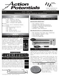
Action Potential Issue #16 - ALA Scientific Instruments
Action Potential Issue #16 - ALA Scientific Instruments
Action Potential Issue #16 - ALA Scientific Instruments

Create successful ePaper yourself
Turn your PDF publications into a flip-book with our unique Google optimized e-Paper software.
“Proudly Celebrating Our 20 th Anniversary”<br />
“Furthering Life Science through Innovative Instrumentation”<br />
PRSRT STD<br />
AUTO<br />
U.S. POSTAGE PAID<br />
Hicksville, NY<br />
Permit No. 73<br />
“Providing electrophysiology<br />
instruments since 1986”<br />
<strong>Issue</strong> <strong>#16</strong> Winter/Spring 2006 <strong>ALA</strong> <strong>Scientific</strong> <strong>Instruments</strong><br />
“Furthering Life Science through Innovative Instrumentation”<br />
<strong>ALA</strong> News Release<br />
In Memoriam<br />
1100 Shames Drive,<br />
Suite 110<br />
Westbury, NY 11590<br />
To:<br />
Special Symposium at Biophysical Society Meeting<br />
Drug Discovery for Ion Channels VI<br />
Date: Friday, February 17, 2006<br />
Time: 8:00 AM - 5:00 PM<br />
Location: ballroom J, Salt Palace Convention Ctr<br />
Salt Lake City, UT<br />
Tel #<br />
516-997-5780<br />
Fax #<br />
516-997-0528<br />
E-Mail:<br />
sales@alascience.com<br />
Internet:<br />
www.alascience.com<br />
In <strong>ALA</strong>’s continuing effort to bring only the finest instruments to our<br />
clients, we have formed an agreement with <strong>Scientific</strong>a Ltd. to supply<br />
the new Patch Star micromanipulator, along with the rest of their<br />
product line. "We're very excited about working with <strong>Scientific</strong>a<br />
because of their vast knowledge and experience in our field,” said<br />
<strong>ALA</strong> President Alan Kriegstein. <strong>ALA</strong> staff is available for installations<br />
and demonstrations in the NY metro area, and other locations with<br />
advanced notice. Let us show you how <strong>Scientific</strong>a products can be<br />
combined with our other fine instruments to build a perfect rig.<br />
Product Update<br />
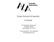
The NEW <strong>Scientific</strong>a PatchStar is a high precision, ultra-stable<br />
micromanipulator, which can move not only in XYZ and but also in<br />
a virtual approach axis.<br />
Key Features...<br />
*Ultra stable - Drifts less than 1µm over 2<br />
hours<br />
*Electrically Silent - even single channel<br />
recordings are routine<br />
*Nanoresolution - 20ηm movement provides<br />
piezo like feel<br />
*20mm working travel - speed of motion for<br />
all applications<br />
*Revolutionary "Smart Sensor" - detects the angle of your headstage<br />
for automatic smooth and straight virtual approach.<br />
*Wide choice of user control device - Control Cube, Joystick or<br />
Patchpad<br />
*Sliding bracket and rotation stages - included at no additional cost<br />
*Designed by and built for electrophysiologists<br />
*Free 2 year warranty for added peace of mind<br />
<strong>Scientific</strong>a’s Movable Top Plate is an ultra stable, smooth moving<br />
mounting platform designed to combine electrophysiology with confocal<br />
and multiphoton microscopy. It offers absolute stability and<br />
precise alignment without disruption to sensitive preparations. The<br />
MTP allows you to search your preparation without moving your<br />
scope while keeping delicate patches in place. Exceptionally<br />
smooth movement, coupled<br />
with a rugged single structure<br />
ensures that there is no movement<br />
once a position has been<br />
reached.<br />
Key Features...<br />
*Extremely stable<br />
*Compatible with ALL major upright fixed stage microscopes.<br />
*Compatible and designed for use with ALL major manipulators<br />
*Can be used to combine electrophysiology with confocal<br />
microscopy.<br />
*Crossed roller bearings for super smooth movement<br />
*Attachable anywhere on a metric or English threaded table<br />
*Height adjustable for different samples.<br />
Jonathan J. Adams: 1927-2006<br />
<strong>ALA</strong> <strong>Scientific</strong> <strong>Instruments</strong> mourns the<br />
passing of our founder Jonathan (John)<br />
Adams. John was a loving husband,<br />
caring father, Holocaust survivor, and<br />
an exceptional human being. John was<br />
a kind gentle soul. Despite living<br />
through the worst that humanity had to<br />
offer he rebuilt his life and lived with dignity<br />
and style and never lost hope that<br />
someday people would live with compassion<br />
and understanding for each<br />
other. He was a mentor, a teacher and<br />
a good friend. He had a positive impact<br />
on all the lives he touched.<br />
He will be missed!<br />
In 1960 John formed Medical Systems Corporation (MSC). MSC<br />
was the very first and only exhibitor at the first Society for<br />
Neuroscience meeting. Each of the following people, now active<br />
in the field of instrumentation, worked at Medical Systems at some<br />
point and has been influenced by John in a positive manner:<br />
Milan Kessler (1940-2001) founder of Instrutech Corp., Andrew<br />
Pomerantz, Vice President of <strong>ALA</strong> <strong>Scientific</strong> <strong>Instruments</strong>, Alan<br />
Kriegstein, President of <strong>ALA</strong> <strong>Scientific</strong> <strong>Instruments</strong>, Harry<br />
Benedict, former President of MSC., and George Kai, President of<br />
Microdata <strong>Instruments</strong>.<br />
After John retired from Medical Systems Corp. his love of science<br />
would lead him to Jurgen List and the famous List patch clamp<br />
amplifiers. In 1986, they combined to form Adams and List<br />
Associates Ltd. to import the new developments in patch clamping<br />
that came out of the Max Planck Institute. That company, now in<br />
its 20th year, is <strong>ALA</strong> <strong>Scientific</strong> <strong>Instruments</strong>. John Adams retired<br />
from <strong>ALA</strong> in 1993 and moved to Florida with is wife Dorothy, who<br />
passed in 2002. He is survived by his daughter Lucy, her husband<br />
Bob, and their two daughters.<br />
<strong>ALA</strong> Around the World<br />
Biophysical Society - Salt Lake City, UT,<br />
February 18 - 22, 2006 - Booth 1014-1016<br />
FENS 2006 - Vienna, Austria<br />
July 8 - 12, 2006 - Booth TBD<br />
Safety Pharmacology - San Diego (Mission Valley), CA<br />
Sept. 26 - 28 - Booth TBA<br />
Society for Neuroscience - 36th Annual Meeting, Atlanta, GA<br />
October 14 - 18 - Booth TBA<br />
American Heart <strong>Scientific</strong> Session - Chicago, IL<br />
Nov. 12 - 15 - Booth TBA<br />
John Adams, founder of<br />
Medical Systems and<br />
<strong>ALA</strong> <strong>Scientific</strong>
Gap Junction Recordings with Two npi SEC Amplifiers<br />
Jose F. Ek Vitorin and Janis M. Burt<br />
Department of Physiology, University of Arizona, Tucson AZ 85724<br />
Special Symposium: Biophysical Society Meeting<br />
Drug Discovery for Ion Channels VI<br />
Date: Friday, February 17, 2006<br />
Time: 8:00 AM - 5:00 PM<br />
Location: Ballroom J, Salt Palace Convention Ctr<br />
We have used dual discontinuous single electrode voltage<br />
clamp amplifiers in combination with fluorescence<br />
microscopy to examine the selectivity of gap junctions<br />
and their comprising channels. For the same junction<br />
we first determine its permeability to fluorescent molecules<br />
and then its macroscopic conductance (g j ); in<br />
most cases the conductance properties of the comprising<br />
channels (γ j ) is then examined. To determine junctional<br />
permeability we introduce dye into one cell of a<br />
pair and monitor its intercellular diffusion as a function<br />
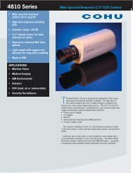
of time (typically 5-10 minutes). Figure 1 shows a pair<br />
of Cx43-expressing rat insulinoma cells before (1A, differential<br />
interference contrast image) and 60 seconds<br />
after (1B, fluorescent image, false colors) accessing<br />
one of them with a dye-containing patch pipette (whole<br />
cell<br />
1A<br />
configuration). The junctional permeability to the dye<br />
was quantified by calculating the rate constant (k 2 ) for<br />
its intercellular diffusion (from donor to recipient). To<br />
determine g j a second patch electrode is then placed in<br />
the recipient cell and transjunctional potential differences<br />
alternately established from either cell such that<br />
g j and γ j could be calculated. The ratio between k 2 and<br />
g j (an estimate of the junctional permeability to small<br />
ions, like K and Cl) defines the selectivity for the fluorescent<br />
probe (see Ek-Vitorin and Burt, 2005).<br />
To better understand how junctional selectivity is determined<br />
by the selectivity of the comprising channels, the<br />
calculated k 2 must be related to the conductances of<br />
the channels constituting the same junction.<br />
Unfortunately, amplifiers suitable for measurement of g j<br />
are typically not ideal for measurement of single channel<br />
activity. Traditional (non-switching) voltage clamp<br />
amplifiers are designed to provide for large current<br />
injection and modest noise handling (suitable for wholecell<br />
voltage clamp of frog oocytes) or for small current<br />
delivery and high signal-to-noise ratio (suitable for voltage<br />
clamp of membrane patches and small mammalian<br />
cells). Neither of these amplifier types is ideal for gap<br />
junction recordings, where large currents can be<br />
1B<br />
Figure 1. Transjunctional diffusion of the fluorescent<br />
dye NBD-M-TMA (see ref. 1) For this<br />
cell pair k 2 =0.675; calibration bar=10µM<br />
required to establish a stable transjunctional voltage<br />
between well-coupled, small mammalian cells.<br />
Discontinuous single electrode voltage clamp (dSEVC)<br />
amplifiers can be used to record in these circumstances.<br />
They are capable of injecting considerable<br />
amounts of current (Strickholm, 1995) when switched to<br />
current injection mode, but still directly evaluate membrane<br />
potential when in voltage recording mode. Thus,<br />
the dSEVC amplifier allows for accurate assessment of<br />
macroscopic g j (Muller et al, 1999) and, importantly,<br />
also allows for channel activity of the same junction to<br />
be determined. Typical examples of such electrical<br />
recordings are shown next.<br />
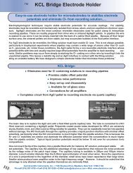
In figure 2, panel A illustrates the voltage protocol used<br />
to determine g j - it consists of a 10mV depolarizing<br />
pulse alternately applied (V 1 , V 2 ) to each side of the<br />
junction, while keeping the opposite side at 0mV. Panel<br />
B shows the membrane currents recorded from donor<br />
(I 1 ) and recipient (I 2 ) cells for the described voltage<br />
pulses; junctional current (I j ) is the downward deflection<br />
of both traces. For this pair (pre-treated with a MAPK<br />
inhibitor) g j was 10.8ηS. In panel C, the same voltage<br />
protocol was implemented (only I 1 and I 2 are shown)<br />
during application of halothane, an agent known to<br />
Figure 2. Determining macroscopic junctional conductance<br />
(g j , as I j /V j ). Voltage (A) and current (B) traces obtained<br />
(from the Rin43 cell pair illustrated in figure 1) shortly after<br />
documenting the rate of intercellular dye diffusion and during<br />
halothane application (C). Traces taken from the SEC<br />
amplifiers with output filtering set at 1kHz (V) and 13kHz (I);<br />
currents were subsequently low-pass filtered at 100Hz. For<br />
all traces, sampling rate was 1kHz. Calibration: x, 2s; y, 10<br />
mV/100pA.<br />
reversibly reduce junctional conductance. Note the fast<br />
reduction of junctional current (I 2 trace during V 1<br />
pulse), and after complete uncoupling the remnant<br />
capacitive artifacts and the non-junctional membrane<br />
current (I 2 trace during V 2 pulse).<br />
After uncoupling, single channel activity appears spontaneously<br />
or can be stimulated by a partial washout of<br />
the halothane. To document at maximum resolution the<br />
detailed behavior of observed channels, we routinely<br />
obtain current signals from the npi SEC amplifiers<br />
(http://www.npielectronic.com/home.cgi?main=products) at high filtering<br />
values (13kHz) for tape storage; these signals<br />
are subsequently filtered as necessary for analysis.<br />
Figure 3. Determining channel conductance (γ j ). A transjunctional<br />
voltage is established (V 1 at ± 40mV, V 2 at 0mV)<br />
while recording transjunctional current (I 2 ). Dotted line<br />
marks zero transjunctional current in I 2 . Calibration: x, 5s; y,<br />
5pA. Output filtering: voltage trace, 1kHz; current 50Hz.<br />
Due to the excessive number of points at the set sampling<br />
rate (2kHz), data were decimated (at 1/5) for illustration purposes.<br />
Alternatively, the filtered (low-pass Bessel filter - LPF)<br />
current signal can be digitized and stored directly. As<br />
shown in Figure 3, it is also possible to record directly<br />
from the amplifiers (without LPF) channels of the<br />
expected amplitude. For this experiment, natural closure<br />
of channels (I 2 ) occurred before the voltage pulses<br />
(V 1 ) were terminated. To compensate for possible tip<br />
potential imbalance, pulses of both polarities were<br />
applied. Notice that single channel events are clearly<br />
defined and their amplitudes can be easily determined.<br />
References:<br />
1. Ek-Vitorin JF, Burt JM. Quantification of Gap Junction Selectivity.<br />
Am.J.Physiol Cell Physiol. 2005; 289:C1535-1546.<br />
2. Strickholm, A. A single electrode voltage, current- and patchclamp<br />
amplifier with complete stable series resistance compensation.<br />
J. Neurosci. Methods 61: 53-66, 1995.<br />
3. Muller, A., M. Lauven, R. Berkels, S. Dhein, H.-R. Polder, and W.<br />
Klaus. Switched single-electrode voltage-clamp amplifiers allow<br />
precise measurement of gap junction conductance. Am.J.Physiol.<br />
276: C980-C987, 1999.<br />
Opening remarks:<br />
Cathy Smith-Maxwell,<br />
Molecular Devices<br />
Keynote address:<br />
Targeting Voltage-Gated Calcium Channels: From Proofof-Concept<br />
to Clinical Candidates.<br />
Terrance Snutch, University of British Columbia and<br />
Neuromed Technologies, Canada<br />
Session I: Automated Drug Discovery with the<br />
Oocyte Expression System<br />
OpusXpress as a Tool for Ion Channel Drug Discovery.<br />
Sean Donovan, Pfizer<br />
The Roboocyte as a Tool for Automated Ion Channel Drug<br />
Screening and Development.<br />
Steven Petrou, University of Melbourne, Australia<br />
Session II: Automated Drug Discovery with<br />
Mammalian Cell Expression Systems Part 1<br />
Development and Validation of Two Novel Automated<br />
Methods of Patch-Clamp Recording.<br />
Mark Bowlby, Wyeth Research<br />
Screening Na + Channel Blockers Using Fluorescence<br />
based and Automated Electrophysiological Assays.<br />
George Ehring, Allergan<br />
Assay Systems and Compound Identification for Potassium<br />
Channels. Min Li, Johns Hopkins University<br />
Session II: Automated Drug Discovery with Mammalian<br />
Cell Expression Systems Part 2<br />
Ion Channel Safety Pharmacology Using Automated<br />
and Manual Electrophysiology.<br />
Clemens Möller, Evotec AG, Germany<br />
Novel Applications of Population Patch Clamp in Ion<br />
Channel Drug Discovery.<br />
Claire Townsend, GlaxoSmithKline<br />
Screening New Drug Candidates for KCNQ1/KCNE1 (IKs)<br />
Activity with PatchXpress 7000A.<br />
Elena Trepakova, Merck Research Labs<br />
Session III: Topics in Solution Delivery<br />
An Automated Electrophysiological Screening System<br />
for Small Sample Volumes.<br />
Daniel Bertrand, University of Geneva, Switzerland<br />
Session IV: Topics in Ion Channel Drug Discovery<br />
Combination screening of ion channel targets to reduce<br />
false positive rates. Chris Fanger, Hydra Biosciences<br />
Small Molecule Libraries as Research Tools for Ion<br />
Channel Drug Discovery: from Design to Drug Candidate.<br />
Victor Panchenko, ChemDiv<br />
CRAC Channel Inhibitors to Treat Acute and Chronic<br />
Inflammation. Michael Xie, Synta Pharmaceuticals<br />
Closing remarks:<br />
Ian M. Herzberg,<br />
<strong>ALA</strong> <strong>Scientific</strong> <strong>Instruments</strong>