PDF(145K) - Wiley Online Library
PDF(145K) - Wiley Online Library
PDF(145K) - Wiley Online Library

Create successful ePaper yourself
Turn your PDF publications into a flip-book with our unique Google optimized e-Paper software.
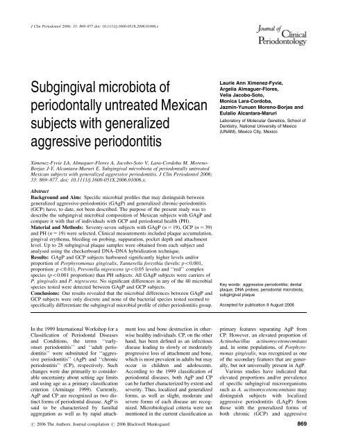
J Clin Periodontol 2006; 33: 869–877 doi: 10.1111/j.1600-051X.2006.01006.x<br />
Subgingival microbiota of<br />
periodontally untreated Mexican<br />
subjects with generalized<br />
aggressive periodontitis<br />
Laurie Ann Ximenez-Fyvie,<br />
Argelia Almaguer-Flores,<br />
Velia Jacobo-Soto,<br />
Monica Lara-Cordoba,<br />
Jazmin-Yunuen Moreno-Borjas and<br />
Eulalio Alcantara-Maruri<br />
Laboratory of Molecular Genetics, School of<br />
Dentistry, National University of Mexico<br />
(UNAM), Mexico City, Mexico<br />
Ximenez-Fyvie LA, Almaguer-Flores A, Jacobo-Soto V, Lara-Cordoba M, Moreno-<br />
Borjas J-Y, Alcantara-Maruri E. Subgingival microbiota of periodontally untreated<br />
Mexican subjects with generalized aggressive periodontitis. J Clin Periodontol 2006;<br />
33: 869–877. doi: 10.1111/j.1600-051X.2006.01006.x.<br />
Abstract<br />
Background and Aim: Specific microbial profiles that may distinguish between<br />
generalized aggressive-periodontitis (GAgP) and generalized chronic-periodontitis<br />
(GCP) have, to date, not been described. The purpose of the present study was to<br />
describe the subgingival microbial composition of Mexican subjects with GAgP and<br />
compare it with that of individuals with GCP and periodontal health (PH).<br />
Material and Methods: Seventy-seven subjects with GAgP (n 5 19), GCP (n 5 39)<br />
and PH (n 5 19) were selected. Clinical measurements included plaque accumulation,<br />
gingival erythema, bleeding on probing, suppuration, pocket depth and attachment<br />
level. Up to 28 subgingival plaque samples were obtained from each subject and<br />
analysed using the checkerboard DNA–DNA hybridization technique.<br />
Results: GAgP and GCP subjects harboured significantly higher levels and/or<br />
proportion of Porphyromonas gingivalis, Tannerella forsythia (levels: po0.001,<br />
proportion: po0.01), Prevotella nigrescens (po0.05 levels) and ‘‘red’’ complex<br />
species (po0.001 proportion) than PH subjects. All GAgP subjects were carriers of<br />
P. gingivalis and P. nigrescens. No significant differences in any of the 40 microbial<br />
species tested were detected between GAgP and GCP subjects.<br />
Conclusions: Our results revealed that the microbial differences between GAgP and<br />
GCP subjects were only discrete and none of the bacterial species tested seemed to<br />
specifically differentiate the subgingival microbial profile of either periodontitis group.<br />
Key words: aggressive periodontitis; dental<br />
plaque; DNA probes; periodontal microbiota;<br />
subgingival plaque<br />
Accepted for publication 9 August 2006<br />
In the 1999 International Workshop for a<br />
Classification of Periodontal Diseases<br />
and Conditions, the terms ‘‘earlyonset<br />
periodontitis’’ and ‘‘adult periodontitis’’<br />
were substituted for ‘‘aggressive<br />
periodontitis’’ (AgP) and ‘‘chronic<br />
periodontitis’’ (CP), respectively. Such<br />
changes were due primarily to considerable<br />
uncertainty about setting age limits<br />
and using age as a primary classification<br />
criterion (Armitage 1999). Currently,<br />
AgP and CP are recognized as two distinct<br />
forms of periodontal disease. AgP is<br />
said to be characterized by familial<br />
aggregation as well as by rapid attachment<br />
loss and bone destruction in otherwise<br />
healthy individuals. CP, on the other<br />
hand, has been defined as an infectious<br />
disease leading to slowly or moderately<br />
progressive loss of attachment and bone,<br />
which is most prevalent in adults but may<br />
occur in children and adolescents.<br />
According to the 1999 classification of<br />
periodontal diseases, both AgP and CP<br />
can be further characterized by extent and<br />
severity. Thus, localized and generalized<br />
forms, as well as slight, moderate and<br />
severe forms of each disease are recognized.<br />
Microbiological criteria were not<br />
mentioned in the current classification as<br />
r 2006 The Authors. Journal compilation r 2006 Blackwell Munksgaard<br />
primary features separating AgP from<br />
CP.However,anelevatedproportionof<br />
Actinobacillus actinomycetemcomitans<br />
and, in some populations, of Porphyromonas<br />
gingivalis, was recognized as one<br />
of the secondary features that are generally,<br />
but not universally present in AgP.<br />
Various studies have indicated that<br />
elevated proportions and/or prevalence<br />
of specific subgingival microorganisms<br />
such as A. actinomycetemcomitans may<br />
distinguish subjects with localized<br />
aggressive periodontitis (LAgP) from<br />
those with the generalized forms of<br />
both chronic (GCP) and aggressive<br />
869
870 Ximenez-Fyvie et al.<br />
periodontitis (GAgP) (Zambon et al.<br />
1983a; Tanner 1992; Muller et al. 1993;<br />
Lopez et al. 1996; Tinoco et al. 1997).<br />
However, whether or not specific subgingival<br />
microbial profiles can distinguish<br />
between individuals with GCP<br />
and GAgP, remains controversial. While<br />
a number of studies have suggested significant<br />
microbial differences between<br />
GCP and GAgP subjects (Dogan et al.<br />
2003; Darby et al. 2005), others have<br />
reported only discrete variations in the<br />
microbial profile of such periodontitis<br />
groups (Mombelli et al. 2002; Lee et al.<br />
2003). For example, Dogan et al. 2003<br />
evaluated by cultural methods the prevalence<br />
and proportion of six periodontal<br />
pathogens in 69 Turkish subjects with<br />
LAgP, GAgP, GCP and periodontal<br />
health (PH). Their findings suggested<br />
that while A. actinomycetemcomitans<br />
was not over-represented in the AgP<br />
groups, a larger percentage of GCP subjects<br />
were colonized by Campylobacter<br />
rectus and Tannerella forsythia than<br />
individuals with either LAgP or GAgP.<br />
In contrast, a different study compared<br />
the prevalence of seven putative periodontal<br />
pathogens in 156 diseased sites<br />
from AgP and 116 sites from CP Korean<br />
subjects, and reported no significant difference<br />
between clinical groups (Lee<br />
et al. 2003). Whether such discrepancies<br />
are due to microbial variations between<br />
subjects from different populations<br />
around the world or to difficulties in<br />
accurately grouping individuals into distinct<br />
clinical categories remains to be<br />
determined.<br />
To our knowledge, no studies have<br />
been published in which the subgingival<br />
microbiota of Mexican subjects with<br />
GAgP has been described. The purpose<br />
of the present study was to determine<br />
the microbial composition of subgingival<br />
plaque samples from periodontally<br />
untreated Mexican subjects with GAgP,<br />
and to compare it with that of individuals<br />
with GCP and PH using the<br />
checkerboard DNA–DNA hybridization<br />
technique.<br />
Material and Methods<br />
Subject population<br />
The present study received approval<br />
from the Ethics Committee for Human<br />
Studies of the Division of Postgraduate<br />
Studies and Research of the School of<br />
Dentistry of the National University of<br />
Mexico (UNAM). All subjects were<br />
asked to sign informed-consent forms,<br />
with which they acknowledged their<br />
willingness to participate.<br />
Nineteen subjects with GAgP, 39<br />
with GCP and 19 with PH were included<br />
in the study (n 5 77 subjects). Subjects<br />
were recruited from the population of<br />
individuals that sought consults and/or<br />
treatment at the Periodontology Department<br />
of the Division of Postgraduate<br />
Studies and Research of the School of<br />
Dentistry of UNAM in Mexico city<br />
from February of 1999 to February of<br />
2004. Every subject that fit the entry<br />
criteria was included in the study. All of<br />
the subjects selected were currently nonsmokers,<br />
who had not received any form<br />
of periodontal therapy in the past other<br />
than professional supragingival plaque<br />
removal and had X20 natural teeth<br />
(excluding third molars). All subjects<br />
were born and lived in Mexico, and<br />
were of Mexican descent, i.e. both of<br />
their parents and X2 of their grandparents<br />
were born and lived in Mexico.<br />
Subjects included in the periodontitis<br />
clinical groups had X18 sites with<br />
attachment level X5 mm. GAgP and<br />
GCP subjects were 12–29 and 435<br />
years of age, respectively. PH subjects<br />
had less than three sites with attachment<br />
level of 4 mm, no sites with attachment<br />
level X5 mm, and were X22 years of<br />
age. Exclusion criteria included pregnancy,<br />
lactation, antibiotic therapy in<br />
the previous 3 months and any systemic<br />
condition which could influence the<br />
course of periodontal disease such as<br />
diabetes, HIV/AIDS or autoimmune diseases.<br />
Clinical monitoring and sample collection<br />
Clinical measurements were taken at six<br />
sites per tooth (mesiobuccal, buccal,<br />
distobuccal, distolingual, lingual and<br />
mesiolingual) at all teeth excluding third<br />
molars (a maximum of 168 sites per<br />
subject) as previously described (Haffajee<br />
et al. 1983). Clinical assessment<br />
included plaque accumulation (0/1,<br />
undetected/detected), gingival erythema<br />
(0/1), bleeding on probing (0/1), suppuration<br />
(0/1), pocket depth and attachment<br />
level. Pocket depth and attachment<br />
level measurements were taken twice by<br />
the same examiner and the average of<br />
the pair of measurements was used for<br />
analysis. Such measurements were<br />
recorded to the nearest millimeter using<br />
a North Carolina periodontal probe<br />
(Hu-Friedy, Chicago, IL, USA). Table 1<br />
summarizes the clinical features of the<br />
77 subjects included in the study.<br />
Samples of subgingival plaque were<br />
obtained from the mesiobuccal site of up<br />
to 28 teeth in each subject. After drying<br />
and isolation with cotton rolls, supragingival<br />
plaque was removed from the<br />
sampled sites and subgingival samples<br />
were taken with individual sterile<br />
Gracey curettes (Hu-Friedy). The samples<br />
were placed in individual tubes<br />
containing 150 ml of TE buffer (10 mM<br />
Tris-HCl, 0.1 mM EDTA, pH 7.6). Samples<br />
were dispersed and 100 ml of 0.5 M<br />
NaOH were added to each tube. All<br />
samples were stored at 201C until<br />
processing.<br />
Microbial assessment<br />
Digoxigenin-labelled whole-genomic<br />
DNA probes were prepared and samples<br />
were processed individually for the<br />
detection and enumeration of 40 microbial<br />
species using the checkerboard<br />
DNA–DNA hybridization technique<br />
(Socransky et al. 1994), following the<br />
procedures previously described (Ximenez-Fyvie<br />
et al. 2006). Table 2 presents<br />
a list of the 40 bacterial strains<br />
employed for the preparation of DNA<br />
probes. Before the microbial detection<br />
in clinical samples, the specificity and<br />
sensitivity of DNA probes were assessed<br />
by hybridizing each DNA probe against<br />
individual pure cultures of all of the test<br />
species adjusted to 10 4 ,10 5 ,10 6 and 10 7<br />
cells. The sensitivity of the assay was set<br />
to allow the detection of approximately<br />
10 4 cells of a given species by adjusting<br />
the concentration of each individual<br />
DNA probe.<br />
Statistical analysis<br />
Microbiological data available for each<br />
subject were the absolute counts of each<br />
of the 40 test species from up to 28<br />
subgingival plaque samples (mean 5<br />
25.6 samples per subject, total 5 1971<br />
samples analysed). The analyses compared<br />
the composition of subgingival<br />
plaque samples between the three clinical<br />
groups. The data are presented as<br />
mean standard error of the mean<br />
(SEM) levels (DNA probe counts 10 5 )<br />
and proportion (percentage of the total<br />
DNA probe count). In order to compare<br />
the levels and proportion of every bacterial<br />
species, each type of data were<br />
recorded at each site, averaged within a<br />
subject and then across subjects in each<br />
clinical group. The percentage of carriers<br />
r 2006 The Authors. Journal compilation r 2006 Blackwell Munksgaard
Table 1. Clinical characteristics of GAgP, GCP and periodontally healthy subjects included in each clinical group<br />
Clinical characteristic GAgP (n 5 19) GCP (n 5 39) Health (n 5 19)<br />
Mean SEM Range Mean SEM Range Mean SEM Range<br />
Age (years) wzk# 21.5 1.2 12–29 48.3 1.7 35–75 27.8 1.4 22–51<br />
Number of missing teeth wzk 1.1 0.4 0–7 3.8 0.3 0–8 0.8 0.3 0–4<br />
Gender (% females) z 84.2 64.1 42.1<br />
Mean pocket depth (mm, full mouth) wknn 3.9 0.2 2.6–6.1 4 0.2 2.8–7.4 2 0.03 1.7–2.3<br />
Mean attachment level (mm, full mouth, AL) wknn 3.9 0.2 2.6–5.9 4.6 0.2 3.1–9 2 0.03 1.7–2.3<br />
Number of sites with ALX5mm wknn 44.9 4.6 18–80 56.8 4.6 8–118 0 0 0–0<br />
Percent sites with:<br />
Plaque accumulation w k 38.3 8.4 0–100 51.7 5.6 0–100 12.2 3.8 0–72<br />
Gingival erythema n§z 26.5 8.2 0–100 26 5 0–100 3.8 2.3 0–38<br />
Bleeding on probing w k nn 44.4 4.8 13.1–95.5 48.8 3.7 4.5–100 2.8 1.2 0–22.7<br />
Suppuration w k nn 5.3 1.4 0–22 6.8 1.5 0–37 0 0 0–0<br />
n po0.01 and<br />
w po0.001 Kruskal–Wallis test between the three clinical groups.<br />
z po0.001 Mann–Whitney test between GAgP and GCP subjects.<br />
§ po0.01 and<br />
k po0.001 Mann–Whitney test between GCP and healthy subjects.<br />
z po0.05,<br />
# po0.01 and<br />
nn po0.001 Mann–Whitney test between GAgP and healthy subjects.<br />
GAgP, generalized aggressive periodontitis; GCP, generalized chronic periodontitis.<br />
Microbiota of Mexican subjects with GAgP 871<br />
Table 2. Reference strains employed for the development of DNA probes<br />
Species Strain n Complex w Species Strain n Complex w<br />
z<br />
Actinobacillus actinomycetemcomitans<br />
Ungrouped Peptostreptococcus micros 33270 Orange<br />
Actinomyces georgiae 49285 Actinomyces Neisseria mucosa 19696 Other<br />
Actinomyces israelii 12102 Actinomyces Porphyromonas endodontalis 35406 Other<br />
Actinomyces naeslundii stp. 1 12104 Actinomyces Porphyromonas gingivalis 33277 Red<br />
Actinomyces odontolyticus 17929 Purple Prevotella intermedia 25611 Orange<br />
Actinomyces viscosus 43146 Actinomyces Prevotella melaninogenica 25845 Other<br />
Campylobacter gracilis 33236 Orange Prevotella nigrescens 33563 Orange<br />
Campylobacter rectus 33238 Orange Propionibacterium acnes 6919 Other<br />
Campylobacter showae 51146 Orange Selenomonas artemidis 43528 Other<br />
Capnocytophaga gingivalis 33624 Green Selenomonas noxia 43541 Ungrouped<br />
Capnocytophaga ochracea 27872 Green Streptococcus anginosus 33397 Yellow<br />
Capnocytophaga sputigena 33612 Green Streptococcus constellatus 27823 Orange<br />
Corynebacterium matruchotii 14266 Other Streptococcus gordonii 10558 Yellow<br />
Eikenella corrodens 23834 Green Streptococcus intermedius 27335 Yellow<br />
Eubacterium saburreum 33271 Other Streptococcus mitis 49456 Yellow<br />
Eubacterium sulci 35585 Other Streptococcus oralis 35037 Yellow<br />
Fusobacterium nucleatum<br />
§<br />
Orange Streptococcus sanguinis 10556 Yellow<br />
Fusobacterium periodonticum 33693 Orange Tannerella forsythia 43037 Red<br />
Gemella morbillorum 27824 Other Treponema denticola 35405 Red<br />
Leptotrichia buccalis 14201 Other Veillonella parvula 10790 Purple<br />
n American Type Culture Collection, Rockville, MD.<br />
w Strains were grouped according to the description of microbial complexes in subgingival plaque (Socransky et al. 1998) with the following exceptions:<br />
A. georgiae, A. israelii, A. naeslundii 1 and A. viscosus were grouped as ‘‘Actinomyces’’; C. matruchotii, E. saburreum, E. sulci, G. morbillorum,<br />
L. buccalis, N. mucosa, P. endodontalis, P. melaninogenica, P. acnes and S. artemidis were grouped as ‘‘Other’’.<br />
z DNA from serotypes a (43717) and b (43718) was combined to generate a single DNA probe.<br />
§ DNA from subspecies nucleatum (25586), polymorphum (10953) and vincentii (49256) was combined to generate a single DNA probe.<br />
was computed by determining the presence<br />
or absence of every species in each<br />
sample. Subjects in which a given species<br />
was detected in at least one sample,<br />
were considered carriers of that particular<br />
microorganism. Percentages for each<br />
microbial species tested were determined<br />
on the basis of the total number of<br />
subjects in each clinical group. The<br />
proportion of groups of microorganisms<br />
was determined for PH and periodontitis<br />
subjects by grouping the 40 test species<br />
as similarly as possible to the description<br />
of subgingival microbial complexes<br />
r 2006 The Authors. Journal compilation r 2006 Blackwell Munksgaard<br />
(Socransky et al. 1998). Significance of<br />
differences between the three clinical<br />
groups and between GAgP and GCP in<br />
the levels, proportion and percentage of<br />
carriers of each species or microbial<br />
complex was determined using the<br />
Kruskal–Wallis and Mann–Whitney
872 Ximenez-Fyvie et al.<br />
GAgP GCP Health<br />
Counts x 10 5 0 5 10 15 0 5 10 15 0 5 10 15<br />
A. naeslundii 1<br />
A. viscosus<br />
C. matruchotii<br />
V. parvula<br />
A. israelii<br />
P. micros<br />
F. nucleatum<br />
L. buccalis<br />
S. intermedius<br />
E. saburreum<br />
† § ⏐ P. gingivalis<br />
S. constellatus<br />
F. periodonticum<br />
S. anginosus<br />
P. intermedia<br />
E. corrodens<br />
S. sanguinis<br />
S. mitis<br />
E. sulci<br />
C. gingivalis<br />
C. sputigena<br />
* ‡ ⏐ P. nigrescens<br />
A. georgiae<br />
‡ T. denticola<br />
P. melaninogenica<br />
A. odontolyticus<br />
† § T. forsythia<br />
C. rectus<br />
P. acnes<br />
G. morbillorum<br />
S. gordonii<br />
S. oralis<br />
N. mucosa<br />
C. ochracea<br />
C. gracilis<br />
S. artemidis<br />
A. a.<br />
C. showae<br />
S. noxia<br />
P. endodontalis<br />
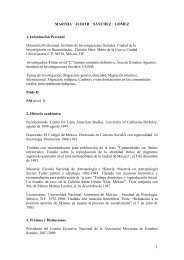
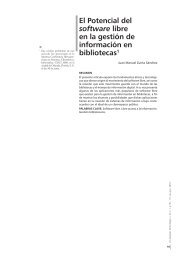
Fig. 1. Bar charts of the mean levels (DNA probe count 10 5 SEM) of each of the 40 test species in 1971 subgingival plaque samples<br />
from 19 generalized aggressive periodontitis (GAgP), 39 generalized chronic periodontitis (GCP) and 19 periodontally healthy subjects. The<br />
levels of each species were computed in each sample, averaged within a subject and then across subjects in each clinical group. The data are<br />
presented in decreasing order based on the levels detected in periodontally healthy subjects. n po0.05 and w po0.001 Kruskal–Wallis test<br />
between the three clinical groups. z po0.05 and § po0.001 Mann–Whitney test between GCP and healthy subjects.<br />
k po0.05 and z po0.01<br />
Mann–Whitney test between GAgP and healthy subjects. Differences between GAgP and GCP subjects were not statistically significant for<br />
any of the species tested after adjusting for multiple comparisons.<br />
tests, respectively, after adjusting for<br />
multiple comparisons as previously<br />
described (Socransky et al. 1991).<br />
Results<br />
Figure 1 summarizes the mean levels<br />
( 10 5 SEM) of the 40 individual test<br />
species in 1971 subgingival plaque samples<br />
from GAgP, GCP and PH subjects.<br />
All of the species tested were detected in<br />
subjects from the three clinical groups.<br />
Actinomyces naeslundii 1, A. viscosus,<br />
Corynebacterium matruchotii, Peptostreptococcus<br />
micros and Veillonella<br />
parvula were the species that presented<br />
the highest mean levels in all three<br />
clinical groups. PH subjects harboured<br />
higher mean levels of only A. naeslundii<br />
1 and Streptococcus intermedius than<br />
subjects in either periodontitis group.<br />
GAgP subjects harboured higher mean<br />
levels of A. israelii, Campylobacter<br />
showae, Neisseria mucosa, P. endodontalis,<br />
Propionibacterium acnes, both<br />
Selenomonas spp. and all Streptococcus<br />
spp. tested except S. sanguinis and S.<br />
oralis, than GCP subjects. However, the<br />
levels of most of the microbial species<br />
tested in both periodontitis groups,<br />
tended to be very similar. A. actinomycetemcomitans<br />
was among the species<br />
detected in the lowest levels in all<br />
clinical groups (GAgP 5 0.5 <br />
0.2 10 5 ; GCP 5 1.2 0.4 10 5 ;<br />
PH 5 0.3 0.1 10 5 ). The differences<br />
between the three clinical groups and<br />
between GAgP and healthy subjects,<br />
were only statistically significant for<br />
P. gingivalis (GAgP 5 5.6 1.4 10 5 ;<br />
GCP 5 9 1.8 10 5 ; PH5 1.6 <br />
0.8 10 5 ; po0.001 and po0.05, respectively),<br />
P. nigrescens (GAgP 5<br />
2.3 0.4 10 5 ; GCP 5 2.5 0.4 <br />
10 5 ; PH5 0.9 0.4 10 5 ; po0.05)<br />
and T. forsythia (GAgP 5 5 2 10 5 ;<br />
GCP 5 5.6 1.5 10 5 ; PH5 0.6 <br />
0.4 10 5 ; po0.001 and po0.01,<br />
r 2006 The Authors. Journal compilation r 2006 Blackwell Munksgaard
Microbiota of Mexican subjects with GAgP 873<br />
GAgP<br />
GCP<br />
Health<br />
%DNA probe count 0 6 12 18 0 6 12 18 0 6 12 18<br />
A. naeslundii 1<br />
A. viscosus<br />
C. matruchotii<br />
V. parvula<br />
A. israelii<br />
E. saburreum<br />
L. buccalis<br />
P. intermedia<br />
F. nucleatum<br />
*† P. gingivalis<br />
P. micros<br />
A. georgiae<br />
S. intermedius<br />
E. corrodens<br />
S. mitis<br />
P. acnes<br />
S. constellatus<br />
N. mucosa<br />
S. sanguinis<br />
C. showae<br />
F. periodonticum<br />
S. anginosus<br />
P. nigrescens<br />
C. gingivalis<br />
*†‡<br />
T. forsythia<br />
E. sulci<br />
T. denticola<br />
C. sputigena<br />
G. morbillorum<br />
P. melaninogenica<br />
A. a.<br />
C. ochracea<br />
P. endodontalis<br />
C. rectus<br />
A. odontolyticus<br />
S. oralis<br />
S. gordonii<br />
C. gracilis<br />
S. noxia<br />
S. artemidis<br />
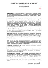
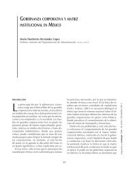
Fig. 2. Bar charts of the mean proportion (% of the total DNA probe count SEM) of each of the 40 test species in 1971 subgingival plaque<br />
samples from 19 generalized aggressive periodontitis (GAgP), 39 generalized chronic periodontitis (GCP) and 19 periodontally healthy<br />
subjects. The proportion of each species was computed in each sample, averaged within a subject and then across subjects in each clinical<br />
group. The data are presented in decreasing order based on the proportions detected in periodontally healthy subjects. n po0.01 Kruskal–Wallis<br />
test between the three clinical groups. w po0.001 Mann–Whitney test between GCP and healthy subjects. z po0.05 Mann–Whitney test<br />
between GAgP and healthy subjects. Differences between GAgP and GCP subjects were not statistically significant for any of the species<br />
tested after adjusting for multiple comparisons.<br />
respectively). Comparing GCP and<br />
healthy subjects, the mean levels of P.<br />
gingivalis (po0.001), P. nigrescens<br />
(po0.05), T. forsythia (po0.001)<br />
and Treponema denticola (GCP 5<br />
3 0.5 10 5 ; PH5 0.8 0.3 10 5 ;<br />
po0.05) were also significantly different.<br />
The differences between GAgP and<br />
GCP subjects were not statistically significant<br />
for any of the species tested.<br />
The mean proportion ( SEM) of<br />
individual species in each clinical group<br />
is summarized in Fig. 2. Samples from<br />
PH subjects harboured larger proportions<br />
of 11 of the 40 test species,<br />
including A. georgiae, A. naeslundii 1,<br />
Capnocytophaga ochracea, N. mucosa,<br />
P. acnes and S. intermedius, than those<br />
from either periodontitis group. The<br />
proportion of a number of putative and<br />
recognized periodontal pathogens, on<br />
the other hand, was higher in both<br />
GAgP and GCP subjects than in healthy<br />
individuals. Such species included<br />
C. rectus, Eikenella corrodens, Fusobacterium<br />
nucleatum, F. periodonticum,<br />
P. micros, P. gingivalis, P. melaninogenica,<br />
P. nigrescens, T. forsythia and<br />
T. denticola. It was notable, that<br />
A. actinomycetemcomitans was among<br />
the species that represented the lowest<br />
proportion in samples from both GAgP<br />
and PH subjects (0.5 0.1% and<br />
0.7 0.3%, respectively). F. periodonticum,<br />
P. gingivalis and P. intermedia,<br />
were among the species that represented<br />
r 2006 The Authors. Journal compilation r 2006 Blackwell Munksgaard<br />
higher mean proportions in samples<br />
from GCP subjects, while P. nigrescens,<br />
T. forsythia and T. denticola, were<br />
among those that represented higher<br />
proportions in samples from subjects in<br />
the GAgP group. The differences<br />
between the three clinical groups, as<br />
well as between GCP and healthy<br />
subjects, were statistically significant<br />
for P. gingivalis (GAgP 5 7.7 1.7%;<br />
GCP 5 9.9 1.4%; PH 5 2.3 1.4%;<br />
po0.01 and po0.001, respectively) and<br />
T. forsythia (GAgP 5 5.9 1.4%;<br />
GCP 5 5 0.9%; PH 5 0.9 0.6%;<br />
po0.01 and po0.01, respectively).<br />
The differences in the mean proportion<br />
of species between GAgP and healthy<br />
subjects, were only statistically significant
874 Ximenez-Fyvie et al.<br />
Table 3. Percentage of carriers of individual species in subjects with generalized aggressive periodontitis (GAgP), generalized chronic periodontitis<br />
(GCP) and periodontal health<br />
Species GAgP GCP Health Species GAgP GCP Health<br />
Actinobacillus actinomycetemcomitans 94.7 89.7 73.7 Peptostreptococcus micros 94.1 85.7 88.2<br />
Actinomyces georgiae 100 87.2 89.5 Neisseria mucosa 94.7 89.7 100<br />
Actinomyces israelii 94.7 100 93.8 Porphyromonas endodontalis 89.5 84.2 68.4<br />
Actinomyces naeslundii stp. 1 100 94.6 100 Porphyromonas gingivalis 100 100 89.5<br />
Actinomyces odontolyticus 94.7 94.4 87.5 Prevotella intermedia 77.8 94.6 72.2<br />
Actinomyces viscosus 100 97.1 94.4 Prevotella melaninogenica 89.5 92.1 72.2<br />
Campylobacter gracilis 73.7 84.2 77.8 Prevotella nigrescens 100 94.9 72.2<br />
Campylobacter rectus 89.5 97.4 84.2 Propionibacterium acnes 84.2 82.1 84.2<br />
Campylobacter showae 89.5 92.3 78.9 Selenomonas. artemidis 89.5 81.1 64.7<br />
Capnocytophaga gingivalis 84.2 88.2 82.4 Selenomonas noxia 89.5 87.2 88.9<br />
Capnocytophaga ochracea 89.5 92.1 89.5 Streptococcus anginosus 100 82.1 89.5<br />
Capnocytophaga sputigena 73.7 92.1 89.5 Streptococcus constellatus 100 87.2 94.1<br />
Corynebacterium matruchotii 94.4 97.3 100 Streptococcus gordonii 94.7 91.9 68.4<br />
Eikenella corrodens 94.7 84.6 88.9 Streptococcus intermedius 84.2 92.3 78.9<br />
Eubacterium saburreum 94.7 81.8 88.9 Streptococcus mitis 83.3 81.1 94.1<br />
Eubacterium sulci 94.7 78.9 100 Streptococcus oralis 94.7 94.6 84.2<br />
Fusobacterium nucleatum 100 92.1 88.2 Streptococcus sanguinis 78.9 89.7 89.5<br />
Fusobacterium periodonticum 94.4 97.1 88.2 Tannerella forsythia 94.4 97.4 89.5<br />
Gemella morbillorum 84.2 82.1 73.7 Treponema denticola 89.5 94.6 78.9<br />
Leptotrichia buccalis 84.2 93.9 88.9 Veillonella parvula 100 92.3 83.3<br />
Subjects in which a given species was detected in at least one sample, were considered carriers of that particular microorganism. Percentages were<br />
determined based on the total number of subjects in each clinical group. No significant differences between the three clinical groups (Kruskal–Wallis<br />
test), GAgP and GCP, GCP and health or GAgP and health (Mann–Whitney test) were found after adjusting for multiple comparisons.<br />
for T. forsythia (po0.05), and no significant<br />
differences were detected<br />
between GAgP and GCP subjects for<br />
any of the microorganisms tested, after<br />
adjusting for multiple comparisons.<br />
Table 3 presents the percentage of<br />
carriers of each individual test species in<br />
the three clinical groups. 73.7–100% of<br />
GAgP, 78.9– 100% of GCP and 64.7–<br />
100% of PH subjects were carriers of<br />
each of the microorganisms tested.<br />
Twenty-two of the 40 test species<br />
(55%) in both the GAgP and GCP<br />
groups, were detected in 90% or more<br />
of subjects. A number of such species<br />
included important periodontal pathogens<br />
like P. gingivalis, T. forsythia and<br />
P. nigrescens. In contrast, only eight of<br />
the 40 test species (20%), in the PH<br />
group, were detected in 90% or more of<br />
subjects. In healthy subjects, none of<br />
such species were putative or recognized<br />
periodontal pathogens. It was<br />
interesting that all GAgP and GCP subjects<br />
were carriers of P. gingivalis, and<br />
that P. nigrescens was also detected in<br />
every subject included in the GAgP<br />
group. GAgP subjects were more frequently<br />
carriers of E. corrodens,<br />
F. nucleatum, P. micros, P. nigrescens<br />
and other species, than subjects included<br />
in the other two clinical groups. On<br />
the other hand, all Campylobacter<br />
spp., F. periodonticum, P. intermedia,<br />
T. forsythia and T. denticola were<br />
among the species that were most frequently<br />
detected in GCP subjects. A<br />
larger percentage of healthy subjects<br />
were carrier of C. matruchotii, Eubacterium<br />
sulci, N. mucosa and S. mitis than<br />
subjects from either periodontitis group.<br />
Although both the levels and proportion<br />
of A. actinomycetemcomitans were low<br />
in GAgP subjects, a larger percentage of<br />
individuals (94.7%) were colonized by<br />
this particular microorganism than<br />
either GCP (89.7%) or PH (73.7%)<br />
subjects. The differences in the percentage<br />
of carriers of all of the species<br />
tested, were not statistically significant<br />
between the three clinical groups, GAgP<br />
and GCP subjects or between either<br />
periodontitis groups and healthy subjects.<br />
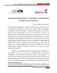
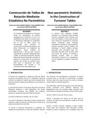
The mean proportion of eight microbial<br />
groups in subjects from each clinical<br />
category is summarized in Fig. 3.<br />
The areas of the pies, were adjusted to<br />
reflect the mean total levels (mean<br />
total DNA probe count) of species in<br />
each clinical category (GAgP 5 93.3<br />
18.4 10 5 ; GCP5 110.7 16.7<br />
10 5 ; PH5 55.3 16.9 10 5 .<br />
po 0.01 between all clinical groups<br />
and GCP versus PH. po0.05 between<br />
GAgP and PH subjects. Not significant<br />
between the GAgP and GCP groups).<br />
The most striking difference in the proportion<br />
of groups of microorganisms<br />
between PH and periodontitis subjects<br />
was a significant increase in the proportion<br />
of ‘‘red’’ complex species observed<br />
in subjects included in either periodontitis<br />
group (po0.001 between the<br />
three groups and GCP versus PH,<br />
po0.01 GAgP versus PH). Additionally,<br />
the proportion of species included in the<br />
Actinomyces group was substantially<br />
lower in periodontitis subjects and in<br />
particular, in GCP individuals. The differences<br />
in the mean proportion of<br />
microbial groups, between all clinical<br />
groups, GCP and healthy subjects, as<br />
well as between GAgP and healthy subjects<br />
were only significant for the ‘‘red’’<br />
complex. No significant differences in<br />
the proportion of either one of the eight<br />
microbial groups were detected between<br />
GAgP and GCP subjects.<br />
Discussion<br />
The present study compared the subgingival<br />
microbial composition of 77 currently<br />
non-smoking Mexican subjects<br />
with no previous history of periodontal<br />
therapy that were either periodontally<br />
healthy or presented two different forms<br />
of periodontal disease (GAgP and GCP).<br />
All of the species detected in both<br />
periodontitis groups, were also present<br />
in PH subjects and the percentage<br />
of healthy and periodontitis carriers<br />
of all of the species tested was not<br />
r 2006 The Authors. Journal compilation r 2006 Blackwell Munksgaard
Microbiota of Mexican subjects with GAgP 875<br />
GAgP<br />
GCP<br />
Health<br />
2.01<br />
17.62<br />
26.23<br />
Other<br />
21.17<br />
Actinomyces<br />
19.96<br />
3.44<br />
6.51<br />
21.28<br />
30.59<br />
16.98<br />
8.28<br />
2.28<br />
Ungrouped<br />
7.46<br />
9.59<br />
15.08<br />
7.02<br />
6.49<br />
17.67<br />
5.73<br />
5.48<br />
18.01<br />
* † ‡<br />
5.90<br />
7.24<br />
Fig. 3. Pie charts of the mean proportion (% of the total DNA probe count) of microbial groups in 1971 subgingival plaque samples from<br />
19 generalized aggressive periodontitis (GAgP), 39 generalized chronic periodontitis (GCP) and 19 periodontally healthy subjects. The<br />
species were organized into 8 microbial groups based on the description of subgingival microbial complexes (Socransky et al. 1998)<br />
(exceptions are noted in Table 2). The areas of the pies were adjusted to reflect the mean total levels of species in each clinical<br />
group. n po0.001 Kruskal–Wallis test between the three clinical groups. w po0.001 Mann–Whitney test between GCP and healthy subjects.<br />
z po0.01 Mann–Whitney test between GAgP and healthy subjects. Differences between GAgP and GCP subjects were not statistically<br />
significant for any of the species tested after adjusting for multiple comparisons.<br />
17.98<br />
significantly different between clinical<br />
groups. Certain microbial species,<br />
including A. naeslundii 1, A. viscosus,<br />
C. matruchotii and V. parvula dominated<br />
in levels and proportion the subgingival<br />
microbiota of both periodontitis and<br />
healthy subjects. The levels and proportion<br />
of P. gingivalis, T. forsythia and<br />
‘‘red’’ complex species as a groups, on<br />
the other hand, were dominant only in<br />
samples from GAgP and GCP subjects.<br />
Low levels and proportion of A. actinomycetemcomitans,<br />
that were not significantly<br />
different between clinical groups,<br />
were detected irrespective of the periodontal<br />
condition of subjects. Taken<br />
together, our results indicated that in the<br />
Mexican population, there were significant<br />
differences in the microbiota of<br />
subgingival plaque samples between<br />
periodontitis and PH subjects. The microbial<br />
differences between GAgP and GCP<br />
subjects, however, were only discrete and<br />
not statistically significant in terms of the<br />
levels, proportion or prevalence of any of<br />
the species or groups of microorganisms<br />
evaluated.<br />
Our findings are in accord with<br />
the results of previous studies that<br />
have suggested that P. gingivalis and<br />
T. forsythia are important pathogenic<br />
species in both GAgP and GCP subjects,<br />
but have failed to determine significant<br />
microbial differences between individuals<br />
with either one of these forms of<br />
periodontal disease (Mombelli et al.<br />
2002; Lee et al. 2003; Takeuchi et al.<br />
2003). Mombelli et al. 2002 systematically<br />
reviewed 33 cross-sectional<br />
and longitudinal studies that provided<br />
microbiological data from both CP and<br />
AgP subjects, to determine if the presence<br />
or absence of five periodontal<br />
pathogens could distinguish between<br />
individuals with either clinical condition.<br />
They concluded that the presence<br />
or absence of A. actinomycetemcomitans,<br />
P. gingivalis, P. intermedia,<br />
T. forsythia or C. rectus could not discriminate<br />
between subjects with CP and<br />
AgP. Takeuchi et al. 2003 employed<br />
polymerase chain reaction to determine<br />
the prevalence and culture to evaluate<br />
the relative proportion of seven subgingival<br />
species in samples from 93 Japanese<br />
subjects with LAgP, GAgP, GCP<br />
and PH. A significantly higher percentage<br />
of GAgP and GCP subjects were<br />
carriers of C. rectus, P. gingivalis,<br />
T. forsythia and T. denticola than PH<br />
subjects. The proportion of A. actinomycetemcomitans,<br />
P. gingivalis and<br />
T. forsythia, however, was similar in<br />
all periodontitis groups.<br />
A. actinomycetemcomitans has been<br />
associated with cases of aggressively<br />
progressing periodontitis in children,<br />
adolescents and adults (Zambon 1985;<br />
Moore 1987; Slots & Listgarten 1988;<br />
Preus et al. 1994). However, its role in<br />
r 2006 The Authors. Journal compilation r 2006 Blackwell Munksgaard<br />
GAgP is still unclear. Our results<br />
revealed that neither the levels, proportion<br />
nor prevalence of A. actinomycetemcomitans,<br />
which were generally low<br />
in all clinical groups, varied significantly<br />
between GAgP, GCP and healthy<br />
subjects. Thus, in Mexican subjects,<br />
A. actinomycetemcomitans did not<br />
appear to play a distinct role in GAgP.<br />
Other studies have also reported low<br />
prevalence and proportion of A. actinomycetemcomitans<br />
in GAgP subjects<br />
from Japan (Ishikawa et al. 2002;<br />
Takeuchi et al. 2003), Brazil (Trevilatto<br />
et al. 2002), Indonesia (Timmerman<br />
et al. 2001) and Greece (Kamma &<br />
Baehni 2003; Kamma et al. 2004). A<br />
number of reports, however, have suggested<br />
that different serotypes of A.<br />
actinomycetemcomitans could be associated<br />
with various forms of periodontal<br />
disease in geographically distinct populations<br />
(Zambon et al. 1983b; Asikainen<br />
et al. 1991; Holtta et al. 1994; Haubek et<br />
al. 1995; Gmur & Baehni 1997;<br />
Socransky et al. 1999). A possible confounder<br />
in our findings with respect to<br />
A. actinomycetemcomitans was the<br />
inability to discriminate between different<br />
serotypes. Separate whole-genomic<br />
DNA probes for serotypes a and b of<br />
A. actinomycetemcomitans were tested<br />
in preliminary studies to determine the<br />
sensitivity and specificity of the DNA<br />
probes used in our ‘‘checkerboard’’
876 Ximenez-Fyvie et al.<br />
assay (data not shown). Significant<br />
cross-reactions between these two particular<br />
DNA probes, however, made it<br />
difficult to distinguish between serotypes<br />
in clinical samples. Therefore a<br />
single DNA probe was generated which<br />
did not exhibit cross-reactions with the<br />
other test species but could not distinguish<br />
between serotypes.<br />
The current classification of periodontal<br />
diseases and conditions describes<br />
GAgP and GCP as two different forms<br />
of disease (Armitage 1999), and while it<br />
is in fact reasonable that GAgP and GCP<br />
represent distinct entities, in cross-sectional<br />
studies, separating GAgP and<br />
GCP subjects into non-overlapping<br />
groups is a difficult challenge. The<br />
classification emphasizes that the diagnosis<br />
of such forms of periodontal disease<br />
should not be based on age or<br />
knowledge of the rate of disease progression.<br />
However, AgP was described<br />
as presenting rapid attachment loss and<br />
bone destruction, usually in persons<br />
under 30 years of age with a pronounced<br />
episodic nature of the destruction. CP<br />
was described as most prevalent in<br />
adults with a slow to moderate rate of<br />
progression. All of such features continue<br />
to be, to a certain extent, agedependant<br />
and require knowledge of<br />
the rate of disease progression. Because<br />
in cross-sectional studies there are no<br />
reliable means of determining the actual<br />
time of disease initiation, rate of progression<br />
or even disease activity, subject<br />
classification is primarily based on the<br />
clinical measurements observed at a<br />
given point in time. Thus, while it is<br />
highly unlikely that GAgP will be misdiagnosed<br />
when only subjects under the<br />
age of 30 years exhibiting severe and<br />
extensive periodontal destructions are<br />
included in such groups, it is impossible<br />
to ascertain what proportion of the individuals<br />
included in GCP groups are<br />
actually GAgP subjects that were evaluated<br />
after the age of 30. While a certain<br />
amount of such overlap cannot entirely<br />
be ruled out in the present study, a<br />
conscious effort was made to minimize<br />
it, e.g., we established an age gap<br />
between GAgP and GCP subjects. Individuals<br />
of up to 29 years of age were<br />
included in the GAgP group and only<br />
subjects that were 35 years of age or<br />
more were selected for the GCP group.<br />
The subgingival microbiota of both<br />
GAgP and GCP Mexican subjects, in<br />
contrast to PH subjects, was characterized<br />
by significant increases in the levels<br />
and/or proportion of certain periodontal<br />
pathogens, including P. gingivalis,<br />
T. forsythia, T. denticola and P. nigrescens.<br />
However, significant microbiological<br />
differences between GAgP and<br />
GCP subjects could not be determined<br />
and none of the 40 bacterial species<br />
tested seemed to specifically characterize<br />
the subgingival microbial profiles of<br />
either periodontitis group. Thus, we<br />
conclude that in Mexican individuals,<br />
changes in the levels, proportion or<br />
prevalence of specific microbial species,<br />
cannot be used to accurately differentiate<br />
between subjects with GAgP<br />
and GCP. Our results warrant further<br />
research of possible non-microbial<br />
determinants in the pathogenesis of<br />
GAgP and GCP in the Mexican population<br />
such as genetic and immunological<br />
factors that may be specifically involved<br />
in these particular forms of periodontal<br />
disease.<br />
Acknowledgements<br />
This study was supported in part by<br />
research Grants J34909-M from the<br />
National Council of Science and Technology<br />
(CONACyT, Mexico city) and<br />
IN205402 from the General Direction of<br />
Faculty Affairs of the National University<br />
of Mexico (DGAPA, PAPIIT, Mexico<br />
city), both to Dr. Ximenez-Fyvie.<br />
The authors wish to acknowledge the<br />
clinical support provided by Dr. Magdalena<br />
Paulin-Perez and Dr. Guadalupe<br />
Marin-Gonzalez of the Periodontology<br />
Department of the Division of Postgraduate<br />
Studies and Research of the School<br />
of Dentistry of UNAM.<br />
References<br />
Armitage, G. (1999) Development of a classification<br />
system for periodontal diseases and<br />
conditions. Annals of Periodontology 4, 1–6.<br />
Asikainen, S., Lai, C. H., Alaluusua, S. & Slots,<br />
J. (1991) Distribution of Actinobacillus actinomycetemcomitans<br />
serotypes in periodontal<br />
health and disease. Oral Microbiology and<br />
Immunology 6, 115–118.<br />
Darby, I., Hodge, P., Riggio, M. & Kinane, D.<br />
(2005) Clinical and microbiological effect of<br />
scaling and root planing in smoker and nonsmoker<br />
chronic and aggressive periodontitis<br />
patients. Journal of Clinical Periodontology<br />
32, 200–206.<br />
Dogan, B., Antinheimo, J., Cetiner, D., Bodur,<br />
A., Emingil, G., Buduneli, E., Uygur, C.,<br />
Firatli, E., Lakio, L. & Asikainen, S. (2003)<br />
Subgingival microflora in Turkish patients<br />
with periodontitis. Journal of Periodontology<br />
74, 803–814.<br />
Gmur, R. & Baehni, P. C. (1997) Serum immunoglobulin<br />
G responses to various Actinobacillus<br />
actinomycetemcomitans serotypes in a<br />
young ethnographically heterogeneous periodontitis<br />
patient group. Oral Microbiology and<br />
Immunology 12, 1–10.<br />
Haffajee, A. D., Socransky, S. S. & Goodson, J.<br />
M. (1983) Comparison of different data analyses<br />
for detecting changes in attachment<br />
level. Journal of Clinical Periodontology<br />
10, 298–310.<br />
Haubek, D., Poulsen, K., Asikainen, S. &<br />
Kilian, M. (1995) Evidence for absence in<br />
northern Europe of especially virulent clonal<br />
types of Actinobacillus actinomycetemcomitans.<br />
Journal of Clinical Microbiology 33,<br />
395–401.<br />
Holtta, P., Alaluusua, S., Saarela, M. &<br />
Asikainen, S. (1994) Isolation frequency<br />
and serotype distribution of mutans streptococci<br />
and Actinobacillus actinomycetemcomitans,<br />
and clinical periodontal status in<br />
Finnish and Vietnamese children. Scandinavian<br />
Journal of Dental Research 102,<br />
113–119.<br />
Ishikawa, I., Kawashima, Y., Oda, S., Iwata, T.<br />
& Arakawa, S. (2002) Three case reports<br />
of aggressive periodontitis associated with<br />
Porphyromonas gingivalis in younger<br />
patients. Journal of Periodontal Research<br />
37, 324–332.<br />
Kamma, J. & Baehni, P. (2003) Five-year<br />
maintenance follow-up of early-onset periodontitis<br />
patients. Journal of Clinical Periodontology<br />
30, 562–572.<br />
Kamma, J., Nakou, M., Gmur, R. & Baehni, P.<br />
(2004) Microbiological profile of early onset/<br />
aggressive periodontitis patients. Oral Microbiology<br />
and Immunology 19, 314–321.<br />
Lee, J., Choi, B., Yoo, Y., Choi, S., Cho, K.,<br />
Chai, J. & Kim, C. (2003) Distribution of<br />
periodontal pathogens in Korean aggressive<br />
periodontitis. Journal of Periodontology 74,<br />
1329–1335.<br />
Lopez, N., Mellado, J. & Leighton, G. (1996)<br />
Occurrence of Actinobacillus actinomycetemcomitans,<br />
Porphyromonas gingivalis and<br />
Prevotella intermedia in juvenile periodontitis.<br />
Journal of Clinical Periodontology<br />
23, 101–105.<br />
Mombelli, A., Casagni, F. & Madianos, P.<br />
(2002) Can presence or absence of periodontal<br />
pathogens distinguish between subjects<br />
with chronic and aggressive<br />
periodontitis? A systematic review. Journal<br />
of Clinical Periodontology 29 (Suppl. 3),<br />
10–21; discussion 37–18.<br />
Moore, W. E. (1987) Microbiology of periodontal<br />
disease. Journal of Periodontal<br />
Research 22, 335–341.<br />
Muller, H., Lange, D. & Muller, R. (1993)<br />
Actinobacillus actinomycetemcomitansrecovery<br />
from extracrevicular locations of the<br />
mouth. Oral Microbiology and Immunology<br />
8, 344–348.<br />
Preus, H. R., Zambon, J. J., Dunford, R. G. &<br />
Genco, R. J. (1994) The distribution and<br />
transmission of Actinobacillus actinomycetemcomitans<br />
in families with established<br />
adult periodontitis. Journal of Periodontology<br />
65, 2–7.<br />
r 2006 The Authors. Journal compilation r 2006 Blackwell Munksgaard
Microbiota of Mexican subjects with GAgP 877<br />
Slots, J. & Listgarten, M. A. (1988) Bacteroides<br />
gingivalis, Bacteroides intermedius and Actinobacillus<br />
actinomycetemcomitans in human<br />
periodontal diseases. Journal of Clinical<br />
Periodontology 15, 85–93.<br />
Socransky, S. S., Haffajee, A. D., Cugini, M. A.,<br />
Smith, C. & Kent, R. L. Jr. (1998) Microbial<br />
complexes in subgingival plaque. Journal of<br />
Clinical Periodontology 25, 134–144.<br />
Socransky, S. S., Haffajee, A. D., Smith, C. &<br />
Dibart, S. (1991) Relation of counts of microbial<br />
species to clinical status at the sampled<br />
site. Journal of Clinical Periodontology 18,<br />
766–775.<br />
Socransky, S. S., Haffajee, A. D., Ximenez-<br />
Fyvie, L. A., Feres, M. & Mager, D. (1999)<br />
Ecological considerations in the treatment of<br />
Actinobacillus actinomycetemcomitans and<br />
Porphyromonas gingivalis periodontal infections.<br />
Periodontology 2000 20, 341–362.<br />
Socransky, S. S., Smith, C., Martin, L., Paster,<br />
B. J., Dewhirst, F. E. & Levin, A. E. (1994)<br />
‘‘Checkerboard’’ DNA–DNA hybridization.<br />
Biotechniques 17, 788–792.<br />
Takeuchi, Y., Umeda, M., Ishizuka, M., Huang,<br />
Y. & Ishikawa, I. (2003) Prevalence of<br />
periodontopathic bacteria in aggressive periodontitis<br />
patients in a Japanese population.<br />
Journal of Periodontology 74, 1460–1469.<br />
Clinical Relevance<br />
Scientific rationale for study: Various<br />
studies have indicated that elevated<br />
proportions and/or prevalence<br />
of specific subgingival microorganisms<br />
may distinguish subjects with<br />
localized AgP from those with the<br />
GCP and GAgP. However, whether<br />
Tanner, A. (1992) Microbial etiology of periodontal<br />
diseases where are we? Where are<br />
we going? Current Opinions in Dentistry 2,<br />
12–24.<br />
Timmerman, M. F., Van der Weijden, G. A.,<br />
Arief, E. M., Armand, S., Abbas, F., Winkel,<br />
E. G., Van Winkelhoff, A. J. & Van der<br />
Velden, U. (2001) Untreated periodontal disease<br />
in Indonesian adolescents. Subgingival<br />
microbiota in relation to experienced progression<br />
of periodontitis. Journal of Clinical<br />
Periodontology 28, 617–627.<br />
Tinoco, E. M., Beldi, M. I., Loureiro, C. A.,<br />
Lana, M., Campedelli, F., Tinoco, N. M.,<br />
Gjermo, P. & Preus, H. R. (1997) Localized<br />
juvenile periodontitis and Actinobacillus actinomycetemcomitans<br />
in a Brazilian population.<br />
European Journal of Oral Science 105,<br />
9–14.<br />
Trevilatto, P., Tramontina, V., Machado, M.,<br />
Goncalves, R., Sallum, A. & Line, S. (2002)<br />
Clinical, genetic and microbiological findings<br />
in a Brazilian family with aggressive periodontitis.<br />
Journal of Clinical Periodontology<br />
29, 233–239.<br />
Ximenez-Fyvie, L. A., Almaguer-Flores, A.,<br />
Jacobo-Soto, V., Lara-Cordoba, M., Sanchez-Vargas,<br />
L. O. & Alcantara-Maruri, E.<br />
(2006) Description of the subgingival microbiota<br />
of periodontally untreated mexican<br />
subjects: chronic periodontitis and periodontal<br />
health. Journal of Periodontology 77,<br />
460–471.<br />
Zambon, J. (1985) Actinobacillus actinomycetemcomitans<br />
in human periodontal disease.<br />
Journal of Clinical Periodontology 12,<br />
1–20.<br />
Zambon, J. J., Christersson, L. A. & Slots, J.<br />
(1983a) Actinobacillus actinomycetemcomitans<br />
in human periodontal disease. Prevalence<br />
in patient groups and distribution of<br />
biotypes and serotypes within families. Journal<br />
of Periodontology 54, 707–711.<br />
Zambon, J. J., Slots, J. & Genco, R. J. (1983b)<br />
Serology of oral Actinobacillus actinomycetemcomitans<br />
and serotype distribution in<br />
human periodontal disease. Infection and<br />
Immunity 41, 19–27.<br />
Address:<br />
Laurie Ann Ximenez-Fyvie<br />
Calz.<br />
Desierto de los Leones<br />
# 5600-L<br />
Col. Tetelpan<br />
Mexico city 01760<br />
Mexico<br />
E-mail: lximenez@post.harvard.edu<br />
or not specific subgingival microbial<br />
profiles can distinguish between individuals<br />
with GCP and GAgP,<br />
remains to be determined.<br />
Principal findings: No significant<br />
differences in the levels, proportion<br />
or prevalence of any of the 40 microbial<br />
species tested were detected<br />
between GAgP and GCP subjects.<br />
Practical implications: Our results<br />
suggested that in Mexican individuals,<br />
specific microbial profiles cannot<br />
accurately differentiate between<br />
subjects with GAgP and GCP.<br />
r 2006 The Authors. Journal compilation r 2006 Blackwell Munksgaard