Medical Solutions - Siemens Healthcare
Medical Solutions - Siemens Healthcare
Medical Solutions - Siemens Healthcare

Create successful ePaper yourself
Turn your PDF publications into a flip-book with our unique Google optimized e-Paper software.
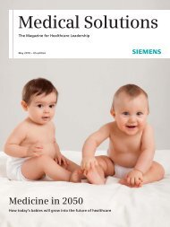
<strong>Medical</strong> <strong>Solutions</strong><br />
The Magazine for <strong>Healthcare</strong> Leadership<br />
December 2008<br />
Breast Cancer<br />
Where are we – and where are we heading?<br />
Cardiology<br />
Diagnosis in the Emergency Room
What’s the difference<br />
between imaging and<br />
imaging greatness??<br />
Ask the Ultimate Power in Imaging.<br />
Every year <strong>Siemens</strong> provides a spectrum of new imaging systems that enhance diagnostic precision. With syngo®<br />
the first unified software interface for all imaging modalities was delivered. Tim® technology revolutionized MRI,<br />
and Dual Source CT continues to drive new clinical possibilities. Talk to us to experience these innovations and new<br />
groundbreaking advancements in imaging excellence.<br />
www.siemens.com/answersforlife +49 69 797 6420<br />
Answers for life.<br />
A91CC-9016-A3-1-7600
Editorial<br />
Advancing Patient Care<br />
The growing and aging population is one<br />
of the most significant challenges facing<br />
healthcare providers. This trend will drive<br />
an increasing demand for healthcare services,<br />
particularly for diseases that occur<br />
later in life and are costly to treat. In<br />
many situations, healthcare delivery has<br />
not evolved to the point where individual<br />
patient’s needs are considered. Our goal<br />
at <strong>Siemens</strong> <strong>Healthcare</strong> is to enable this<br />
high-quality, patient-centered approach<br />
by integrating imaging, laboratory diagnostics,<br />
and healthcare IT, creating seamless<br />
and significantly improved workflow<br />
efficiencies.<br />
Take prostate cancer as an example.<br />
Typically, if a clinician wants to clarify<br />
whether symptoms are signs of prostate<br />
cancer, he or she, in addition to a physical<br />
exam, would order a blood test to<br />
measure Prostate-Specific Antigen (PSA).<br />
A high PSA level is considered to potentially<br />
be caused by prostate cancer. Different<br />
PSA markers have to be combined<br />
and put in relation to each other to identify<br />
men who might show an elevated<br />
PSA, but who, in fact, have a low risk that<br />
this PSA value is a result of prostate<br />
cancer. Also, measuring PSA at just one<br />
point in time is not sufficient.<br />
When the patient’s PSA levels indicate<br />
possible prostate cancer, a biopsy needs<br />
to be performed. The traditional method<br />
involves some measure of patient discomfort,<br />
and possibly anxiety, as a clinician<br />
extracts tissue from different locations<br />
for diagnosis. Ultrasound, however,<br />
allows for an image-guided biopsy, which<br />
enables the physician to target suspicious<br />
areas for extraction, reducing patient<br />
discomfort, long-term complications, and<br />
the amount of time needed for the procedure.<br />
Should the biopsy yield a positive result,<br />
molecular imaging biomarkers enable<br />
the localization of metastatic cancer<br />
cells in the body. And, <strong>Siemens</strong> REMIND<br />
clinical decision support software can<br />
help ’rule out’ other factors that might<br />
lead to higher PSA results, but not automatically<br />
to prostate cancer. When this<br />
solution is applied holistically, up to 50<br />
percent of such misleading diagnoses<br />
can be avoided, saving time and money<br />
and increasing patient comfort and<br />
confidence. An estimated US$5 billion<br />
can be saved annually in the U.S. alone by<br />
applying measures to avoid unnecessary<br />
procedures and interventions. 1<br />
With our recent investment in laboratory<br />
diagnostics, <strong>Siemens</strong> has become the<br />
only integrated healthcare company that<br />
can offer a complete portfolio to better<br />
1<br />
Results may vary. Data on file.<br />
Jim Reid-Anderson,<br />
Member of the Managing Board of <strong>Siemens</strong> AG<br />
and CEO of the <strong>Healthcare</strong> Sector<br />
manage prostate cancer, breast cancer<br />
– as discussed in the cover story of this<br />
edition of <strong>Medical</strong> <strong>Solutions</strong> – and many<br />
other diseases throughout the continuum<br />
of care. With our broad portfolio and<br />
ongoing innovations, we are here to<br />
support your efforts in advancing patient<br />
care: enabling earlier, more efficient,<br />
accurate, and patient-friendly diagnoses<br />
and treatments.<br />
Our goal is simple: to help you save lives<br />
and offer the best possible care to your<br />
patients. It is at the center of everything<br />
we do. <strong>Siemens</strong> <strong>Healthcare</strong> provides<br />
answers for healthcare – answers for life.<br />
Sincerely,<br />
<strong>Medical</strong> <strong>Solutions</strong> · December 2008 · www.siemens.com/healthcare-magazine 3
Content<br />
Content<br />
20<br />
Personalized Medicine:<br />
Novel Imaging and Diagnostic Technologies<br />
38<br />
Cardiac Care:<br />
New Standards<br />
Cover Story<br />
03 Editorial<br />
10 Breast Cancer<br />
Back in the 1980s, a bright young student<br />
completed her PhD dissertation on MRI brain<br />
diffusion imaging. Little did she know that<br />
cutting-edge technology would someday<br />
save her life. As a breast cancer patient, she<br />
now talks about her personal experience<br />
and the needs of affected women. In addition,<br />
<strong>Medical</strong> <strong>Solutions</strong> interviewed three<br />
imaging experts around the world about<br />
how diverse imaging solutions and advanced,<br />
integrated technology are providing a new<br />
level of care for breast cancer patients.<br />
06 News<br />
66 Essay Series: Japan<br />
71 Further Reading<br />
75 Service<br />
76 Imprint<br />
77 Subscription<br />
4 <strong>Medical</strong> <strong>Solutions</strong> · December 2008 · www.siemens.com/healthcare-magazine
Content<br />
44<br />
Med Meets IT:<br />
Project Expert Care<br />
60<br />
Training:<br />
Three Locations, One High Standard<br />
Features<br />
20 With a shared vision of preventive<br />
and personalized medicine,<br />
<strong>Siemens</strong> and National Jewish Health<br />
aim to develop novel imaging<br />
and diagnostic technologies using<br />
genomics, proteomics, integrated<br />
research, and clinical care.<br />
26 To overcome claustrophobia and<br />
positioning difficulties, Clínica<br />
de Diagnóstico por Imagem relies on<br />
Open Bore magnetic resonance<br />
imaging.<br />
30 The ARTISTE integrated radiation<br />
therapy solution helps Baton<br />
Rouge General <strong>Medical</strong> Center’s<br />
Pennington Cancer Center to rapidly<br />
and efficiently deliver radiation<br />
therapy close to home.<br />
35 Offering better return on investment<br />
and workflow efficiency,<br />
Biograph Molecular CT – mCT –<br />
is the imaging crossover that will<br />
change the way hospitals think<br />
about integrated imaging.<br />
38 A new cardiac care and research<br />
institute in Western Canada,<br />
the Mazankowski Alberta Heart<br />
Institute, is setting new standards of<br />
care for both pediatric and adult<br />
patients under one roof.<br />
44 By implementing a comprehensive<br />
IT solution across the entire organization,<br />
MedCentral, a regional<br />
health system in Ohio, became a<br />
world-class institution.<br />
50 Tissue Strain Analytics is an<br />
emerging ultrasound technology<br />
with the potential for quicker,<br />
more accurate diagnoses of tissue<br />
anomalies in the liver.<br />
54 Cardiac biomarkers provide rapid<br />
diagnosis and risk stratification, and<br />
help to improve the quality of care<br />
for chest pain patients at South<br />
Austin Hospital’s Emergency Department.<br />
60 Within a global training concept,<br />
<strong>Siemens</strong> <strong>Healthcare</strong> offers stateof-the-art<br />
training for customers<br />
worldwide in three dedicated<br />
training centers located around the<br />
globe.<br />
<strong>Medical</strong> <strong>Solutions</strong> · December 2008 · www.siemens.com/healthcare-magazine 5
News<br />
CT Scanning in a Flash<br />
The SOMATOM® Definition Flash computed<br />
tomography (CT) scanner sets<br />
new standards in both image acquisition<br />
speed and radiation dose as the world’s<br />
fastest CT with the lowest dose ever.<br />
Like no other scanner, it can image the<br />
entire thorax in less than one second and<br />
complete a cardiac scan in one-fourth<br />
the time of a single heartbeat, with a<br />
radiation dose of less than one millisievert.<br />
“Our goal was to build the most<br />
patient-friendly CT by significantly reducing<br />
dose through faster speed,” says Sami<br />
Atiya, PhD, Chief Executive Officer of the<br />
CT Business Unit of <strong>Siemens</strong> <strong>Healthcare</strong>.<br />
“Lowest radiation dose is important to<br />
physicians and patients. It’s important<br />
to us.”<br />
SOMATOM Definition Flash utilizes Dual<br />
Source technology that consists of two<br />
detectors and two X-ray sources. This configuration,<br />
coupled with a gantry rotation<br />
time of 0.28 seconds, enables a temporal<br />
resolution of just 75 milliseconds, makes<br />
dual energy scanning possible, and allows<br />
the use of 200 kilowatts. Now, <strong>Siemens</strong><br />
scientists and engineers have discovered<br />
how to push acquisition speeds to new<br />
levels. SOMATOM Definition Flash can<br />
scan at a pitch of above three, while still<br />
achieving gapless z-sampling, resulting<br />
in a table speed of more than 40 centimeters<br />
per second. That is because the<br />
two detectors create two complementary<br />
data spirals that, when put together,<br />
include all the information found in a<br />
single spiral acquired at a much lower<br />
table speed.<br />
Together, these features enable lung<br />
scans in 0.6 seconds, taking the burden<br />
of breath-holding off the patients. Fast<br />
scan speeds also eliminate the additional<br />
dose penalty of electrocardiographically<br />
(ECG) gated thoracic studies, so radiologists<br />
can scan the thorax and ’get the<br />
heart for free.’ Dedicated cardiac investigations<br />
can be completed in about<br />
250 milliseconds. But more importantly:<br />
It also reduces dose to unprecedented<br />
levels down to below one millisievert. The<br />
new features also permit pediatric scans<br />
more quickly and safely than ever before.<br />
In addition, a shuttle mode makes it possible<br />
for trauma patients to be scanned<br />
to conduct dynamic time-resolved imaging<br />
over 40 centimeters, the longest<br />
range available today.<br />
Besides the reduced radiation exposure<br />
that directly results from the high table<br />
speed, SOMATOM Definition Flash offers<br />
several other dose-conscious features. In<br />
dual energy scans, a new photon shield<br />
prefilters high kilovoltage X-rays, both<br />
improving material separation and substantially<br />
reducing dose, making it perfect<br />
for routine clinical use. Adaptive Dose<br />
Shielding blocks X-rays that will not be<br />
used in image reconstruction. New organspecific<br />
dose reduction eliminates direct<br />
exposure of radiation-sensitive organs,<br />
such as the breast, thyroid gland, or eye.<br />
And <strong>Siemens</strong> is looking to the future,<br />
developing iterative reconstruction techniques<br />
that promise to further reduce<br />
dose.<br />
Willi Kalender, PhD, Director of the<br />
Institute of <strong>Medical</strong> Physics at the University<br />
of Erlangen-Nuremberg in Erlangen,<br />
Germany, says, “The new scanner is a<br />
true revolution. It picks up on the well<br />
established concept of Dual Source CT<br />
but improves it in several ways. We never<br />
before dared to scan with such a low<br />
dose and such a high speed.”<br />
www.siemens.com/<br />
SOMATOM-Sessions-Flash<br />
6 <strong>Medical</strong> <strong>Solutions</strong> · December 2008 · www.siemens.com/healthcare-magazine
News<br />
Large Display for Artis zee Family<br />
Following the release of the Artis zee®<br />
family for interventional imaging in radiology<br />
and cardiology in 2007, <strong>Siemens</strong><br />
launched a new, full-color large display 1<br />
for integration with the new portfolio at<br />
RSNA 2008. The Artis zee Large Display<br />
is a 56-inch monitor that allows users to<br />
replace up to eight single monitors on<br />
the system. It provides the integration of<br />
multiple modalities on one screen for<br />
greater flexibility and enables the user<br />
to configure the screen during the procedure.<br />
The operator can choose from<br />
different screen layouts directly at the<br />
tableside of the angiography system.<br />
This enables the operator to adapt the<br />
configuration to the individual workflow<br />
steps. Continuing the flexibility and<br />
versatility of the Artis zee systems, the<br />
product can be used for interventional<br />
radiology, interventional cardiology/<br />
electrophysiology, and surgery, and is<br />
particularly valuable for interventional<br />
imaging in hybrid rooms as it tremendously<br />
reduces the number of monitors<br />
in the room. With its high resolution<br />
(4 x HD), the Large Display shows even<br />
the finest details. Up to 200 layout combinations<br />
and the possibility to connect<br />
at least 16 image sources and show up<br />
to ten windows simultaneously enhance<br />
imaging results and workflow in the interventional<br />
suite. Moreover, the reduction<br />
of additional displays and the option<br />
to put other video signals on the Large<br />
Display – for instance, for monitoring<br />
other rooms, telemedicine, or endoscopy<br />
– make it a rewarding investment for the<br />
future.<br />
1<br />
The information about this product is being provided<br />
for planning purposes. The product requires 510(k)<br />
review and is not commercially available in the U.S.<br />
Comprehensive MR Oncology <strong>Solutions</strong><br />
Cycle 1 Cycle 2<br />
Cycle 3 Cycle 4<br />
Cycle 5 Cycle 6<br />
syngo GRACE after every chemotherapy cycle of a patient with<br />
known breast cancer: The efficiency of therapy can be easily seen by the<br />
decreasing choline.<br />
Within the field of oncology, magnetic resonance imaging (MRI)<br />
has proven to be one of the most effective imaging techniques.<br />
For asymptomatic and high-risk patients, MRI enables both<br />
early tumor detection and oncological staging without radiation.<br />
To support precise surgery planning, therapy monitoring,<br />
and follow-up, <strong>Siemens</strong> provides comprehensive MRI oncology<br />
solutions, going far beyond single applications and software<br />
features.<br />
In Women’s Health, the first quantitative MRI breast spectroscopy<br />
application syngo® GRACE is now also available for the<br />
3 Tesla systems MAGNETOM® Verio and Trio. By checking relative<br />
choline concentration during therapy, the efficiency for<br />
monitoring treatment is more reliable. This may also reduce<br />
the number of unnecessary breast biopsies for the women<br />
concerned.<br />
In the field of Men’s Health, syngo Tissue 4D 1 , the new task<br />
card for visualization of 3D dynamic measurements, is particularly<br />
valuable for prostate evaluation. Offering two evaluation<br />
workflows – standard curve evaluation or a pharmacokinetic<br />
model – syngo Tissue 4D supports an efficient oncology workflow<br />
and reliable follow-up studies.<br />
Completed by syngo TimCT Oncology – the hardware and<br />
software solution for seamless whole-body imaging using<br />
Continuous Table move – these new applications and workflow<br />
tools expand the comprehensive <strong>Siemens</strong> solution for<br />
oncology diagnosis and staging.<br />
1<br />
This application is pending 510(k) review and is not yet commercially available in the U.S.<br />
<strong>Medical</strong> <strong>Solutions</strong> · December 2008 · www.siemens.com/healthcare-magazine 7
News<br />
What is your SPECT’S IQ?<br />
<strong>Siemens</strong> demonstrated industry leadership once again with<br />
molecular imaging’s most recent innovation in single photon<br />
emission computed tomography (SPECT) – IQ·SPECT 1 . This<br />
new feature in SPECT enables a comprehensive cardiac evaluation<br />
including perfusion, attenuation correction, and calcium<br />
scoring in as little as five minutes 2 when traditional cardiac<br />
SPECT perfusion studies can average 15 to 20 minutes. Available<br />
for the Symbia® product line, IQ·SPECT allows organspecific<br />
cardiac evaluations with enhanced image quality. For<br />
medical facilities, this innovation means being able to accommodate<br />
more patients in less time and meet a variety of<br />
patient needs.<br />
IQ·SPECT’s ‘intelligence’ is achieved through a combination<br />
of three technologies beginning with SMARTZOOM, a specially<br />
designed smart collimator that magnifies the heart while<br />
imaging the rest of the torso under traditional conditions.<br />
Second, the SMARTZOOM collimator works in a cardio-centric<br />
orbit to maximize the amount of cardiac information collected<br />
from the patient. Finally, unique IQ·SPECT reconstruction<br />
completes the innovation and is seamlessly integrated into<br />
the current Symbia workflows and automation features, giving<br />
physicians access to the most flexible and versatile system<br />
available today.<br />
Calcium scoring with SPECT·CT, extracted from a quick lowdose<br />
spiral CT, has also become a critical element in the cardiac<br />
work-up to evaluate the extent of cardiac disease in patients.<br />
Adding the 30-second CT to the SPECT study cannot only illustrate<br />
any ischemia present in the patient, but also can assess<br />
the buildup of calcium in the coronary arteries. The addition<br />
of IQ·SPECT to these important SPECT·CT studies may lead to<br />
new risk stratification algorithms and workups for patients<br />
with suspected coronary artery disease.<br />
1<br />
Works in Progress. The information about the product is preliminary.<br />
The product is under development and is not commercially available in the U.S.,<br />
and its future availability cannot be assured.<br />
2<br />
New Product Feature based on preliminary internal data. Actual performance<br />
characteristics have not been established.<br />
Klinikum Chemnitz is one of the first hospitals<br />
in Germany to link its teleradiology<br />
service with an electronic health record<br />
(EHR). The EHR makes demographic and<br />
administrative data of a telediagnosis<br />
available. Provided the patient agrees,<br />
images and results from diagnostics can<br />
be shared by different facilities. Thanks<br />
to a special security system, the data are<br />
only accessible to authorized users who<br />
Electronic Health Record for Integrated Care<br />
are involved in the patient’s treatment.<br />
<strong>Siemens</strong> equipped the hospital with an<br />
overall technology system that includes<br />
Soarian® Integrated Care 1 (Soarian IC) for<br />
information exchange, as well as radiological<br />
image communication software.<br />
Soarian IC improves the flow of information<br />
across institutions and sectors without<br />
the need to exchange existing primary<br />
systems. As a result, the system<br />
supports the cooperation between the<br />
individual clinical facilities and simplifies<br />
the patient treatment. Together with<br />
14 regional county hospitals as well as<br />
medical centers and several practicing<br />
physicians, Klinikum Chemnitz sets<br />
standards for integrated healthcare in<br />
Germany. Especially regarding diagnoses<br />
in the fields of neurosurgery, traumatology,<br />
angiology, and radiology, the<br />
hospital supports regional hospitals and<br />
practices with its medical expert knowledge.<br />
For example, a patient who has<br />
been brought into a county hospital after<br />
an accident can be scanned with a computed<br />
tomography system. The image<br />
data is sent electronically to a responsible<br />
physician in Klinikum Chemnitz, who<br />
then diagnoses the patient and sends<br />
back the report.<br />
1<br />
The information about this product is preliminary. The<br />
product is under development and is not commercially<br />
available in the U.S. or in Canada, and its future availability<br />
cannot be assured.<br />
8 <strong>Medical</strong> <strong>Solutions</strong> · December 2008 · www.siemens.com/healthcare-magazine
News<br />
Neonatal Care<br />
<strong>Siemens</strong> RAPIDLab® 1245/1265 blood gas analyzers have been<br />
enhanced to measure total bilirubin on neonatal whole-blood<br />
samples. The RAPIDLab systems determine the neonatal total<br />
bilirubin concentration in 60 seconds using multiple wavelength<br />
spectrophotometry. Bilirubin is the main bile pigment formed<br />
from the degradation of hemoglobin. An increased level of<br />
bilirubin in the blood (hyperbilirubinemia) causes jaundice,<br />
the discoloration of body tissues. Neonatal jaundice is usually<br />
harmless, a consequence of immature liver function and the<br />
breakdown of fetal hemoglobin as it is replaced with adult<br />
hemoglobin. Severe neonatal jaundice may indicate a more<br />
serious condition, including erythroblastosis fetalis, that is<br />
most likely caused by blood incompatibilities between baby<br />
and mother. Extremely high levels of bilirubin in infants may<br />
cause bilirubin encephalopathy or kernicterus, a form of brain<br />
damage.<br />
The Business Partner in MRI<br />
“You can’t run a radiology practice without an MRI [magnetic<br />
resonance imaging] system,” says Franz Walter, MD. “Demand<br />
from referrers and patients for radiation-free imaging is growing<br />
steadily.” This is why the radiologist invested in a MAGNETOM®<br />
ESSENZA immediately after taking over the recently outsourced<br />
radiology practice of the Evangelische Krankenhaus<br />
in Zweibrücken, Germany. With a low total cost of ownership,<br />
MAGNETOM ESSENZA is ideal for setting up and expanding MRI<br />
services. Zhen Jin, MD, agrees. The Director of the MRI Center<br />
at Hospital 306 in Beijing, China, looks back on 13 years of MRI<br />
service at her institute – and installed two additional systems<br />
last year: a 3 Tesla MAGNETOM Trio and a 1.5 Tesla MAGNETOM<br />
ESSENZA. The decision for the MAGNETOM ESSENZA was a<br />
natural one for both physicians: “It is the first system that is<br />
optimized for cost of ownership. Zero helium boil-off and a 50<br />
percent lower electricity consumption compared to conventional<br />
systems are just two examples of its affordability,” says<br />
Walter. “We wanted a robust system for clinical use to allow<br />
more time for research on the MAGNETOM Trio,” Zhen Jin says<br />
as she explains the reason for buying two systems within such<br />
a short timeframe. MAGNETOM ESSENZA offers full diagnostic<br />
capabilities thanks to Tim® (Total imaging matrix). Tim provides<br />
flexibility through versatile coil combinations, accuracy<br />
through high signal strength and spatial resolution, and speed<br />
resulting from parallel imaging. It offers access to the applications<br />
needed for both an outpatient practice and a primary care<br />
hospital. Zhen Jin also appreciates the integrated IsoCenter<br />
Matrix coil of MAGNETOM ESSENZA. Thus, the region of interest<br />
is always at the center of the magnetic field, which makes<br />
coil repositioning and changing obsolete.<br />
With 28 patients a day, she is also happy with the speed of the<br />
exams, as is Walter with 22 exams, which include outpatients<br />
as well as referrals from within the hospital. Walter mentions a<br />
Parkinson’s patient who was referred for an angio exam. First,<br />
he was skeptical about being able to achieve diagnostic image<br />
quality. Thanks to MAGNETOM ESSENZA’s fast sequences,<br />
movement artifacts were reduced and the images were good<br />
for diagnosis. “From any perspective, MAGNETOM ESSENZA is<br />
definitely up to date,” he says.<br />
At Hospital 306 in Beijing, Dr. Zhen Jin is happy with MAGNETOM<br />
ESSENZA’s low cost of ownership.<br />
Dr. Franz Walter of the radiology practice at Evangelische Krankenhaus<br />
Zweibrücken likes the system’s ease of use.<br />
www.siemens.com/ESSENZA<br />
<strong>Medical</strong> <strong>Solutions</strong> · December 2008 · www.siemens.com/healthcare-magazine 9
Breast Cancer<br />
Breast cancer is by far the most common cancer among<br />
women. The numbers have been increasing worldwide,<br />
rising rapidly particularly in younger women.<br />
10 <strong>Medical</strong> <strong>Solutions</strong> · December 2008 · www.siemens.com/healthcare-magazine
Breast Cancer<br />
An Unexpected Encounter<br />
Back in the 1980s, a bright young graduate<br />
student completed her PhD dissertation on MRI<br />
brain diffusion imaging at the Massachusetts<br />
Institute of Technology. Little did she know that<br />
cutting-edge technology would someday save<br />
her life.<br />
By Diana Smith<br />
“I was trained as a scientist and have<br />
always been interested in health and technology,”<br />
says the scientist, who wishes<br />
to remain anonymous. “I became very<br />
interested in MRI [magnetic resonance<br />
imaging] in my first year of graduate<br />
school. My doctoral thesis was on MR<br />
diffusion imaging, and I continued working<br />
on MRI abroad for my post-doctorate<br />
research.”<br />
A passionate scientist, she continued<br />
work in the field and built an impressive<br />
global resume. Since 2006, she has been<br />
doing research collaboration with a leading<br />
hospital in Asia, performing a clinical<br />
research study on breast imaging to<br />
compare breast MRI and breast diffusion<br />
imaging with conventional techniques<br />
such as mammography and ultrasound.<br />
New Imaging Techniques<br />
In the blink of an eye, a speck on a<br />
mammogram or an aberrant lump felt in<br />
the shower can change a woman’s life.<br />
Breast cancer is by far the most common<br />
cancer among women. 1 The numbers<br />
have been increasing worldwide, rising<br />
1<br />
http://www.who.int/cancer/detection/breastcancer/en/.<br />
Last accessed Nov. 4 th , 2008<br />
rapidly, particularly in younger women.<br />
According to the American Cancer Society,<br />
in 2008, 1.3 million cases will be identified,<br />
and almost 500,000 women will<br />
die from the disease. In the U.S., breast<br />
cancer will be diagnosed in one in eight<br />
women. Though rare, men can also get<br />
breast cancer.<br />
New imaging techniques are helping<br />
doctors diagnose tumors with greater<br />
precision and less trauma. Mammograms<br />
can be less effective in women with<br />
dense tissue which makes the images<br />
harder to read. Magnetic resonance<br />
imaging has been shown to find breast<br />
<strong>Medical</strong> <strong>Solutions</strong> · December 2008 · www.siemens.com/healthcare-magazine 11
Breast Cancer<br />
lesions underwent diffusion imaging<br />
and dynamic contrast-enhanced MRI 4<br />
using a 3 Tesla scanner (<strong>Siemens</strong><br />
MAGNETOM® Trio, A Tim® system).<br />
The clinical research study was performed<br />
in Asia, where, increasingly<br />
younger women are being affected by<br />
breast cancer. Younger women tend<br />
to have denser (leaner) breast tissue.<br />
Dense breast tissue can present special<br />
difficulties for disease detection. In<br />
mammography, dense breast tissue and<br />
tumors both appear white. “It’s like finding<br />
a polar bear in a snowstorm,” says<br />
the scientist.<br />
The scientist’s breast cancer was discovered by chance.<br />
“Women need<br />
to know what they<br />
should look for<br />
in getting the<br />
best diagnostic<br />
examination.”<br />
cancers that mammograms miss in a<br />
certain group of patients. 2 A study published<br />
by the Scientific Assembly of the<br />
Radiological Society of North America<br />
(RSNA) reported that the detection rate<br />
for nonpalpable, invasive breast cancers<br />
increased by 42 percent in women with<br />
dense breasts when mammography was<br />
followed by ultrasound. 3<br />
“Taking the technology to a new level,”<br />
the scientist explains, “diffusion is looking<br />
at the water mobility, the movement<br />
of water molecules in tissue. We thought<br />
this technique would be very sensitive<br />
in finding abnormalities in the breast,<br />
and it’s perfect when you have a group<br />
of patients who have proven biopsies,<br />
because then you have histologic comparison.<br />
We have the exact pathological<br />
specimen to compare with what we see<br />
in imaging.”<br />
In a clinical study in Hong Kong, 31<br />
female patients with suspected breast<br />
2<br />
Efficacy of MRI and Mammography for Breast-Cancer<br />
Screening in Women with a Familial or Genetic Predisposition.<br />
N Engl J Med, Vol. 351, No. 5:427-437<br />
3<br />
Mammographic Density and the Risk and Detection of<br />
Breast Cancer. N Engl J Med, Vol. 356, No. 5:227-263<br />
Accidental Discovery<br />
A chance discovery changed everything,<br />
recalls the scientist. “The clinical results<br />
from the patient study were really good.<br />
Out of curiosity, I went in the scanner<br />
for this diffusion technique. No contrast<br />
injection was needed with diffusion<br />
imaging. It was quick and easy, and I was<br />
out of the scanner in five minutes. As<br />
I came out, I saw the stricken face of the<br />
radiologist and knew something was<br />
wrong.”<br />
Since the diffusion technique was new<br />
and is not yet routine for breast imaging,<br />
the radiologist in charge recommended<br />
a follow-up with a complete examination<br />
and conventional diagnostic methods.<br />
These included digital mammography,<br />
ultrasound, contrast-enhanced MRI 4 , and<br />
lymph-node mapping.<br />
Not the Right Destiny<br />
At only age 45, the researcher was in a<br />
low-risk group with no family history of<br />
the disease. Previous mammograms and<br />
ultrasounds were normal. A nonsmoker,<br />
she was slim and followed a healthy diet.<br />
“I was shocked,” she says. “I couldn’t<br />
believe it. I always thought I have been<br />
healthy and active.”<br />
The hours that followed were a roller<br />
coaster of emotions, particularly since<br />
she was thousands of miles from home<br />
and loved ones. “The worst was the<br />
moment when you are told you have<br />
4<br />
Not available in the U.S.<br />
12 <strong>Medical</strong> <strong>Solutions</strong> · December 2008 · www.siemens.com/healthcare-magazine
Breast Cancer<br />
“It’s so important to create and offer well-planned<br />
radiology departments coupled with women’s<br />
health centers to minimize anxiety and suffering<br />
after someone is diagnosed with breast cancer.”<br />
cancer,” she explains. “It feels like you<br />
just got a death sentence.” That evening,<br />
the radiologist in charge, who is also a<br />
caring friend, took the scientist to dinner<br />
and “turned a potentially sad evening<br />
alone after a shocking diagnosis into one<br />
that made me think more about friendship,<br />
people who care, and the life<br />
ahead.”<br />
A Personal Decision<br />
Because of her background in medical<br />
imaging, the scientist was more informed<br />
than most about the diagnostic tools<br />
and treatments used in breast cancer.<br />
Like American actress Christina Applegate,<br />
who elected to have a mastectomy in the<br />
summer of 2008 at age 36 to eliminate<br />
constant fear and onerous exams every<br />
few months, the scientist opted for immediate<br />
surgery.<br />
“I know of women who had lumpectomies<br />
followed by radiation and chemotherapy,<br />
and their cancer reoccurred,” she says.<br />
“Every woman is different. I made my<br />
personal decision to have a mastectomy<br />
to have peace of mind and reduce the<br />
risks of recurrence to almost zero.”<br />
Recovery<br />
“The first couple of weeks were very hard,”<br />
remembers the scientist. “I’ve never<br />
had any kind of surgery before, and of<br />
course, you cannot bathe or even put<br />
on clothing yourself. You are very dependent<br />
on someone to help you.”<br />
She adds, “In a way, I was very lucky that<br />
it [finding the cancer] happened where<br />
I knew the clinicians. Though I was very<br />
far from home when this happened, I was<br />
in a well-known hospital with state-ofthe-art<br />
equipment. I found the hospital<br />
to be well organized with women’s health<br />
and radiology all under the same roof.<br />
Everything was very conveniently located<br />
in the same building and I could get all<br />
the tests done within the first 48 hours.”<br />
“I understand many women are less<br />
fortunate and they are sent to different<br />
places and have to wait weeks before<br />
getting all the tests done. The waiting<br />
can be bewildering. Some hospitals are<br />
still not prepared to offer a streamlined<br />
process for people affected with such a<br />
diagnosis. Sometimes, the hospital may<br />
not have the right imaging equipment or<br />
the latest software. It’s easy for a patient<br />
to be sent to get multiple tests, get lost<br />
in the medical maze, and spend endless<br />
hours and days waiting for the results.<br />
It’s so important to create and offer wellplanned<br />
radiology departments coupled<br />
with women’s health centers to minimize<br />
anxiety and suffering after someone<br />
is diagnosed with breast cancer.”<br />
Speaking Out<br />
Today, the scientist is still consulting<br />
and she is using her experience to be an<br />
advocate and speak to others about the<br />
disease and the imaging modalities used<br />
to diagnose and treat it.<br />
“I’ve learned a lot through this process.<br />
I now teach women about early detection<br />
and how it helps to save lives. It’s<br />
also important to know about the possibilities<br />
in detection and treatment,” she<br />
emphasizes.<br />
“Most people know someone who has<br />
had breast cancer. Most people know of<br />
mammography and breast ultrasound.<br />
Some know about breast MRI, but they<br />
don’t really know enough details, for<br />
example, whether their hospital has the<br />
state-of-the-art scanner with the latest<br />
software or whether it is using a scanner<br />
that is ten years old, which does not offer<br />
the best resolution or the same functionalities<br />
as the newer ones. Women need<br />
to know what they should look for in<br />
getting the best diagnostic examination<br />
and what they should ask their healthcare<br />
providers. When patients are wellinformed,<br />
they have the best chance for<br />
survival and can live a long and healthy<br />
life.”<br />
An Enlightened Path<br />
“I’m back to my previous activities and<br />
actually feel better than before,” says<br />
the scientist. “When you go through an<br />
experience like this, you get to see who<br />
really cares about you and who loves<br />
you for who you are. I have a stronger<br />
appreciation for people who show kindness<br />
despite work pressure and busy<br />
schedules. I particularly remember a lateshift<br />
nurse and her words of kindness<br />
and encouragement when she came<br />
to do an IV [infusion] around midnight.<br />
When you are flat on your back in a<br />
hospital bed, you have time to think, to<br />
ponder, and to feel.”<br />
She concludes, “I have come from this<br />
experience with a greater appreciation<br />
for life. I’m grateful for the early detection<br />
that helped save my life, and I have<br />
certainly grown from the enlightenment<br />
and reflections during this unexpected<br />
journey.”<br />
<strong>Medical</strong> <strong>Solutions</strong> · December 2008 · www.siemens.com/healthcare-magazine 13
Breast Cancer<br />
A Worldwide Challenge<br />
Diverse imaging solutions and advanced, integrated technology<br />
are providing a new level of care for breast cancer patients.<br />
For a global view, <strong>Medical</strong> <strong>Solutions</strong> interviewed three imaging<br />
experts around the world:<br />
Gladys Lo, MD, Chief Radiologist, Department of Diagnostic<br />
and Interventional Radiology, Hong Kong Sanatorium and Hospital,<br />
Hong Kong, China<br />
John F. Nelson, MD, <strong>Medical</strong> Director, Battlefield Imaging,<br />
Battlefield Auxiliary Breast Center, Ringgold, Georgia, U.S.<br />
Karsten Ridder, MD, Radiological Group Practice,<br />
Outpatient Clinic Professor Dr. Uhlenbrock and Partners,<br />
Diagnostic Breast Center, St. Josefs-Hospital,<br />
Dortmund-Hoerde, Germany<br />
14 <strong>Medical</strong> <strong>Solutions</strong> · December 2008 · www.siemens.com/healthcare-magazine
Breast Cancer<br />
Thank you for finding time to talk to<br />
us across many time zones. All of you<br />
provide state-of-the-art breast cancer<br />
care with integrated imaging systems<br />
from <strong>Siemens</strong> that optimize clinical,<br />
operational, and financial workflow.<br />
Let’s discuss how diagnosis and treatment<br />
of breast cancer has changed<br />
since you started in the field.<br />
NELSON: I have been practicing for<br />
about 20 years, so I’ve seen quite a few<br />
changes. Technologically, we’ve obviously<br />
seen huge strides in screening mammography<br />
just in the ability to see and pick<br />
up lesions. In recent years, most of us in<br />
the U.S. and across the world have probably<br />
transitioned to digital mammography.<br />
I think probably everyone on this<br />
panel would agree the improved screenings<br />
have saved lives. So, that has really<br />
changed the way I practice. Secondly,<br />
of course, the different modalities we<br />
use to evaluate patients diagnosed with<br />
suspected breast cancer also have ballooned.<br />
Ultrasound is no longer something<br />
that we do occasionally – it’s something<br />
we do all the time. Additionally,<br />
advanced techniques like breast MRI<br />
[magnetic resonance imaging] have<br />
revolutionized what I do as a diagnostician.<br />
RIDDER: Changing from analog to digital<br />
mammography is like the invention of<br />
rubber for the wheel. It is much faster<br />
and more precise than before, especially<br />
when you are looking at workflow. CAD<br />
[computer-aided diagnosis] is a helpful<br />
support in managing the workload of a<br />
screening center such as ours. But this is<br />
only one advantage. On the other hand,<br />
digital systems help the radiologist and<br />
surgeon communicate with the pathologist.<br />
LO: The incidence of breast cancer in<br />
Hong Kong has increased to one in 23,<br />
and digital mammography is fantastic<br />
because Chinese women have very dense<br />
breasts. So, advanced digital mammography<br />
has really helped to look through<br />
the breast tissue, and also in picking up<br />
the microcalcifications.<br />
How can ultrasound or other modalities<br />
improve the ability to detect cancers?<br />
LO: Ultrasound has always been popular<br />
in Hong Kong because of the very dense<br />
breasts the women have here. We’ve<br />
always found it to be very useful and<br />
complimentary to mammography. MRI, of<br />
course, I think is a breakthrough. Like Dr.<br />
Ridder, we also have a multidisciplinary<br />
approach in our hospital. We communicate<br />
very closely with the breast surgeons,<br />
pathologists, radiation therapists, and<br />
oncologists.<br />
Why is it important to be an early<br />
adopter of technology? What are the<br />
benefits to patients? To the hospital?<br />
RIDDER: Here in Dortmund, where we<br />
are located, we are a city of 1.5 million.<br />
We are part of the hospital’s Radiology<br />
Institute, and we have the pressure<br />
of the free market. Women are free to<br />
decide which institute they want to go<br />
to. Having better technology gives us a<br />
competitive advantage. The second thing<br />
is that with the new techniques, it is better<br />
for the patients. With our advanced<br />
radiology equipment, we get the most<br />
sensitivity and specificity we can.<br />
Mammography is only one small part of<br />
all the basic things that have to be<br />
offered along with the other modalities.<br />
LO: Our hospital is a private hospital and<br />
actually prides itself in getting the best<br />
machines. We have a 3 Tesla MRI breast<br />
unit, and we’ve been doing a special<br />
sequence called diffusion to look at the<br />
breast tissue and had some very good<br />
preliminary results that will be published<br />
in JCAT [Journal of Computer Assisted<br />
Tomography] next year and were presented<br />
in Toronto at the ISMRM [International<br />
Society for Magnetic Resonance<br />
in Medicine] this year [2008] in May.<br />
You had a special case as a result of<br />
the diffusion study. Can you tell us<br />
about that?<br />
LO: One of my patients is a scientist and<br />
is aware of what we are doing. Previously,<br />
she had standard mammography, but it<br />
was not diagnostic because her breasts<br />
were very dense. So, we decided she<br />
should have the diffusion examination<br />
because it doesn’t involve any ionizing<br />
radiation, there’s no injection, and it’s<br />
very quick. What happened was that the<br />
diffusion study unexpectedly turned out<br />
to be abnormal. So, this was followed<br />
with a complete contrast-enhanced MRI<br />
scan, of course, and at the site where<br />
the diffusion abnormality was seen, there<br />
was actually a bilobulated rim-enhancing<br />
mass with type three signal intensity time<br />
graph, quite diagnostic like a BI-RADS<br />
[Breast Imaging Reporting and Data<br />
System] 1 five lesion, and this turned out<br />
to be DCIS [ductal carcinoma in situ].<br />
After the MRI was done, I suggested<br />
doing an ultrasound as well and we saw<br />
the lesion again. I also persuaded her to<br />
do mammography again because I was<br />
afraid she might have an area of DCIS<br />
1<br />
BI-RADS is a quality assurance tool originally designed<br />
for use with mammography. The system is a collborative<br />
effort of many health groups but is published and trademarked<br />
by the American College of Radiology.<br />
<strong>Medical</strong> <strong>Solutions</strong> · December 2008 · www.siemens.com/healthcare-magazine 15
Breast Cancer<br />
“There are a lot of<br />
tools out there<br />
that we can parlay<br />
into what we are<br />
currently doing<br />
to add diagnostic<br />
capabilities.”<br />
John F. Nelson, MD, <strong>Medical</strong> Director,<br />
Battlefield Imaging, Battlefield Auxiliary Breast<br />
Center, Ringgold, GA, USA<br />
that’s only shown with microcalcifications.<br />
Both the MRI and the ultrasound<br />
may not show a certain percentage of<br />
DCIS cases that present with microcalcifications.<br />
Indeed, her tumor was at eight<br />
o’clock, but on the mammography at ten<br />
o’clock, there was a stipulated area that<br />
had some microcalcifications in it.<br />
NELSON: Was the diagnostic MRI also<br />
negative?<br />
LO: No, it wasn’t. It was an irregularly<br />
marginated mass, but it had a type one<br />
graph. So it was indeterminate. It was<br />
like a BI-RADS four at the ten o’clock<br />
lesion, which was seen on mammography,<br />
and a BI-RADS five lesion that was<br />
not seen on mammography for the eight<br />
o’clock.<br />
How are you using other methods<br />
of molecular medicine such as PET·CT<br />
[positron emission tomography/<br />
computed tomography], SPECT·CT<br />
[singe photon emission computed<br />
tomography/computed tomography],<br />
or biomarkers?<br />
NELSON: At our institution, we really<br />
reserve PET·CT for women with suspected<br />
extensive disease. For most of our<br />
women with locally advanced disease, we<br />
evaluate with breast MRI, and I bet that is<br />
true for the other two physicians. We’ve<br />
actually experimented at our institution<br />
with Bruce Porter’s techniques 2 , and what<br />
we’re doing now is a lot more wholebody<br />
MRI for staging and the chest and<br />
abdomen for screening, along with our<br />
breast MRI.<br />
RIDDER: PET·CT is also promising in other<br />
cancers, like ovarian cancer or lymphatic<br />
cancer. Where we use these PET techniques<br />
is also for extended breast cancer<br />
and the staging of treatment.<br />
How does an integrated diagnostic<br />
strategy affect your patients and your<br />
facility’s success?<br />
LO: Patients who all of a sudden find<br />
out they have some abnormality want to<br />
find out the exact extent of the abnormality<br />
and what it is right away. If you<br />
send them to all different types of places<br />
to get it and they have to wait, that’s tremendously<br />
stressful on the patient. We<br />
are lucky that we have everything in one<br />
place, including the hospital.<br />
NELSON: In fact, that’s really why our<br />
facility was built. We are actually in a<br />
breast center, so every modality, including<br />
breast MRI, is available. We even offer<br />
Saturday morning service. We’re also in<br />
a very competitive environment here.<br />
We are motivated at our center to place<br />
the patient at the center of the wheel<br />
and all the spokes go out, but the patient<br />
shouldn’t have to move. It’s our job to<br />
provide all the services that go along<br />
with breast cancer evaluation.<br />
RIDDER: I think my colleagues will agree,<br />
everyone is short of time, and so the<br />
time pressure is extreme. Also, women<br />
need to get their results in a short time.<br />
Why did you choose women’s<br />
health and breast cancer as your field<br />
of expertise?<br />
RIDDER: Honestly, I think it’s one of the<br />
most exciting fields in radiology, with<br />
all the new techniques that have been<br />
2<br />
Refer to, e.g., Beatty, J., Porter, B: Contrast-enhanced<br />
breast magnetic resonance imaging: the surgical perspective.<br />
Am J Surg 193; 5:600-605.<br />
Smith J.P., Hanson J., Dawson J., Porter B., Tickman R.J.:<br />
emerging technologies in surgical planning for breast<br />
cancer. Am J Surg 184; 4:377-9.<br />
16 <strong>Medical</strong> <strong>Solutions</strong> · December 2008 · www.siemens.com/healthcare-magazine
Breast Cancer<br />
developed in the last ten, 20 years, and<br />
are still being developed. Maybe only<br />
comparable to cardiac MRI or multislice<br />
computed tomography.<br />
NELSON: For me, it was a calling. My<br />
sister was diagnosed with breast cancer<br />
back when I was still in medical school.<br />
She had two kids. I saw how breast cancer<br />
affects the patients, their loved ones<br />
and families, and changes the course of<br />
life for everybody. So when I got to the<br />
point of choosing my area of specialization,<br />
it was just natural.<br />
“We are lucky that<br />
we have everything<br />
in one place.”<br />
Gladys Lo, MD, Chief Radiologist,<br />
Department of Diagnostic and Interventional<br />
Radiology, Hong Kong Sanatorium and Hospital,<br />
Hong Kong, China<br />
And why did you choose <strong>Siemens</strong><br />
equipment for your work?<br />
NELSON: Battlefield Imaging was a brandnew<br />
center built from scratch alongside<br />
“Having better<br />
technology<br />
gives us a<br />
competitive<br />
advantage.”<br />
Karsten Ridder, MD, Radiological<br />
Group Practice, Outpatient Clinic<br />
Prof. Dr. Uhlenbrock and Partners,<br />
Diagnostic Breast Center,<br />
St. Josefs-Hospital,<br />
Dortmund-Hoerde, Germany<br />
the rest of our medical facilities about<br />
five years ago. We wanted to go completely<br />
digital at our center – no film. We<br />
weren’t building space for film. I actually<br />
flew to Dortmund to look at the digital<br />
mammography system they had in place<br />
there. We’ve had a long-standing relationship<br />
with <strong>Siemens</strong>, and we have extremely<br />
good <strong>Siemens</strong> service. A lot of what<br />
drove my interest in <strong>Siemens</strong> was that<br />
relationship. When I saw the system and<br />
compared it with the other two systems<br />
available at the time, I just didn’t feel<br />
comfortable that either of the others<br />
could provide me with the image quality<br />
or the back-up service that I knew would<br />
be necessary. The same is true for MRI.<br />
At that time, the Espree [MAGNETOM®<br />
Espree Open Bore MRI system with Tim®<br />
technology] was just coming on the<br />
market. We have a relatively large patient<br />
population; many of our patients are<br />
overweight or obese. The Espree just fit<br />
perfectly with what we were trying to<br />
provide. It was really the first full-field,<br />
high-end machine that offered those sort<br />
of facilities for the patients. <strong>Siemens</strong><br />
really had the technology that worked<br />
well for us.<br />
RIDDER: It’s the whole package you get<br />
from <strong>Siemens</strong>, not limited to just the<br />
image quality. For example, we have a<br />
different machine that is supposed to<br />
have the same detector as the mammography<br />
system from <strong>Siemens</strong>, but there is<br />
no comparison between the two images.<br />
I’m also using our MRI for heart examinations<br />
and work with other <strong>Siemens</strong><br />
systems as well. Thanks to the common<br />
syngo® user interface, it is easy to switch<br />
between the modalities.<br />
LO: Prior to getting our <strong>Siemens</strong> digital<br />
mammography unit, we had one from a<br />
different vendor. We have images from<br />
patients who come for follow-up. The old<br />
images are from the other vendor and<br />
the new images are <strong>Siemens</strong>, and it’s<br />
like night and day. The new <strong>Siemens</strong> unit<br />
is seeing so much more, and I’m very<br />
pleased with that.<br />
NELSON: I would add that the <strong>Siemens</strong><br />
digital unit had several filter combinations,<br />
some of which use a considerably<br />
lower dose. Compared to our screen<br />
film, we were seeing 30 to 40 percent<br />
lower doses. We have marketed that<br />
very strongly in our community, and it<br />
has been very well received.<br />
Are you excited about any new<br />
trends or innovative leading-edge<br />
imaging solutions for the future?<br />
RIDDER: We have just started with<br />
ultrasound automated breast volume<br />
<strong>Medical</strong> <strong>Solutions</strong> · December 2008 · www.siemens.com/healthcare-magazine 17
Breast Cancer<br />
scanning [ABVS] 3 . We haven’t used the<br />
technology for a very long time, but<br />
what I can say now is that we are looking<br />
at a very promising technique that<br />
holds a huge potential for breast imaging<br />
in the future.<br />
NELSON: I would echo that there are<br />
some other things on the horizon. I think<br />
all of us are interested to see if breast<br />
tomosynthesis 4 is really going to take off.<br />
Certainly, we’ve found elasticity imaging<br />
in ultrasound very useful. And I’m really<br />
excited about diffusion imaging on MRI.<br />
There are a lot of tools out there that<br />
we can parlay into what we are currently<br />
doing to add diagnostic capabilities.<br />
Diana Smith is a freelance writer based in<br />
Liberty Hill, TX, USA.<br />
3<br />
The information about this product is being provided<br />
for planning purposes. The product is pending 510(k)<br />
review, and is not yet commercially available in the U.S.<br />
4<br />
Caution: Investigational Device. Limited by U.S. Federal<br />
Law to investigational use. The information about Digital<br />
Breast Tomosynthesis is preliminary. This product is<br />
under development and not commercially available in<br />
the U.S., and its future availability cannot be assured.<br />
Further Information<br />
www.siemens.com/breastcare<br />
www.siemens.com/<br />
news-breastcare<br />
Breast Cancer:<br />
Where are we – and where are we heading?<br />
Challenge:<br />
“It was easy, and I was out of the scanner in five minutes,” says the scientist. “As I<br />
came out, I saw the stricken face of the radiologist and knew something was wrong.”<br />
Working on MRI diffusion, a promising breakthrough imaging technique for the<br />
breast, a scientist unexpectedly discovers her own disease. One chance test completely<br />
changed her life, but that was just the beginning of an arduous emotional<br />
and physical journey.<br />
Solution:<br />
Today, physicians and clinicians are using an arsenal of integrated diagnostics that<br />
have revolutionized the management of breast cancer. “I think probably everyone<br />
would agree that improved screenings have saved lives,” says John F. Nelson, MD,<br />
<strong>Medical</strong> Director of Battlefield Auxiliary Breast Center in Ringgold, Georgia, U.S.<br />
“That has really changed the way I practice.”<br />
Integrated diagnostics have other benefits, including improved workflow and patient<br />
convenience. Gladys Lo, MD, Chief Radiologist at Hong Kong Sanatorium, emphasizes<br />
how new approaches to diagnosis and treatment have positive emotional ramifications.<br />
“For patients who all of the sudden find out they have some abnormality, they<br />
would like to find out the exact extent of what it is right away. If you send them to<br />
all different types of places and they have to wait, that’s tremendously stressful on<br />
them.”<br />
Result:<br />
Technologically, huge strides have been made in the imaging field in the last two<br />
decades. Integrating laboratory diagnostics, advanced imaging, and information<br />
technologies can improve a patient’s outcome at every stage of care. In addition,<br />
integrated technology affects workflow. “It is much faster and more precise than<br />
before,” says Dr. Karsten Ridder of St. Josefs-Hospital, Dortmund, Germany.<br />
The journey of detecting, coping with, and beating breast cancer resulted in an<br />
enlightened new perspective for the scientist. Now, this survivor gives real advice,<br />
not only on early detection and treatment, but also because of her background,<br />
specifically on what to look for in hospital imaging equipment and how the level of<br />
technology may make a difference in a person’s life. All scanners are not created<br />
equally.<br />
The ACUSON S2000 ABVS<br />
Automated Breast Volume Scanner<br />
reduces operator dependence and<br />
variability.<br />
Patient with a 2.8-centimeter,<br />
grade 3, invasive ductal carcinoma<br />
in the right breast imaged with<br />
digital mammography (left) and<br />
breast tomosynthesis. The mediolateral<br />
oblique (MLO) digital<br />
mammography view shows dense<br />
breast tissue with subtle distortion<br />
in the lower breast. The MLO<br />
tomosynthesis slice shows a spiculated<br />
mass in the lower breast.<br />
18 <strong>Medical</strong> <strong>Solutions</strong> · December 2008 · www.siemens.com/healthcare-magazine
MAGNETOM Espree – Pink is a dedicated MR Breast Scanner with a 70-centimeter Open Bore at 1.5T and an ultra-short 125-centimeter<br />
system length.<br />
Diverse Imaging <strong>Solutions</strong><br />
In a multipronged, comprehensive approach, <strong>Siemens</strong> combines<br />
laboratory diagnostics, advanced imaging, and information<br />
technologies to help physicians detect, diagnose, and treat<br />
breast cancer earlier, faster, and with greater precision. New<br />
technology offers a range of breast care solutions – all designed<br />
to contribute to successful disease management.<br />
MAGNETOM Espree – Pink<br />
<strong>Siemens</strong> announced the latest innovation in breast MRI,<br />
MAGNETOM® Espree – Pink, the new dedicated MRI Breast Scanner<br />
with a 70-centimeter Open Bore at 1.5 Tesla and an ultrashort<br />
125-centimeter system length. Both the 70-centimeter<br />
Open Bore scanner and the new breast coil (Sentinelle Vanguard<br />
for <strong>Siemens</strong>) offer an enhanced level of patient comfort, especially<br />
for obese and claustrophobic patients. The system has<br />
the capability to position the patient feet-first or head-first<br />
and provides excellent access to perform biopsies. Sentinelle<br />
Vanguard for <strong>Siemens</strong> offers excellent image quality and optimized<br />
biopsy access for higher accuracy in intervention and<br />
faster examination time. The dedicated workplace includes<br />
syngo® BreVis 1 for flexible reading and reporting and syngo<br />
BreVis Biopsy 1 for fast and accurate MR breast biopsy workflow<br />
with automatic calculation of target coordinates.<br />
ACUSON S2000 ABVS Automated Breast Volume<br />
Scanner<br />
The ACUSON S2000 ABVS Automated Breast Volume Scanner 2<br />
streamlines workflow and reduces operator dependence and<br />
variability by quickly and comfortably surveying and acquiring<br />
full-field sonographic volumes for comprehensive review and<br />
diagnosis of the breast. ACUSON S2000 ABVS features an<br />
integrated room suite design that combines the advanced<br />
ACUSON S2000 ultrasound system and a column stand with<br />
an arm assembly, which holds a transducer pod specially<br />
designed for automated ultrasound breast imaging. It supports<br />
a high patient load with 250 to 400 single images acquired<br />
in one scan to calculate the volumes, which are sent to a<br />
dedicated ABVS Workplace for analysis and manipulation. The<br />
system features the anatomical coronal plane, which is not<br />
available using conventional ultrasound and includes semiautomated<br />
reporting features and comprehensive BI-RADS<br />
report capabilities.<br />
Breast Tomosynthesis<br />
The latest technology now under development in full-field<br />
mammography, breast tomosynthesis 3 , is a 3D imaging technology<br />
that acquires 2D projection images of a compressed<br />
breast at multiple angles during a sweep of the X-ray tube.<br />
Poised to enhance mammography, the new technology will<br />
take the two-dimensional images and reconstruct them to<br />
reveal depth – the third dimension of anatomy. Tomosynthesis<br />
slices have the potential to show tumors that remain invisible<br />
in individual images.<br />
1<br />
This information about this product is preliminary. The product is under development<br />
and not commercially available in the U.S., and its future availability cannot be ensured.<br />
2<br />
The information about this product is being provided for planning purposes. The product<br />
is pending 510(k) review and is not yet commercially available in the U.S.<br />
3<br />
Caution: Investigational Device. Limited by U.S. Federal Law to investigational use.<br />
The information about Digital Breast Tomosynthesis is preliminary. This product is<br />
under development and not commercially available in the U.S., and its future availability<br />
cannot be assured.<br />
<strong>Medical</strong> <strong>Solutions</strong> · December 2008 · www.siemens.com/healthcare-magazine 19
“National Jewish Health and<br />
<strong>Siemens</strong> share a common vision<br />
of bringing laboratory diagnostics<br />
together with imaging in<br />
order to give our patients the<br />
best care possible.”<br />
Michael Salem, MD, President and CEO,<br />
National Jewish Health, Denver, CO, USA<br />
Bringing Personalized Medicine<br />
into Focus<br />
For 109 years, National Jewish Health has sought to offer<br />
patients the best possible care. U.S. News & World Report has<br />
ranked National Jewish Health the number one respiratory<br />
hospital in the nation for 11 years straight. It is continuing its<br />
legacy by launching a personalized medicine initiative, seeking<br />
to become a nationally recognized clinical thought leader<br />
among American healthcare providers.<br />
By Amy K. Erickson<br />
20 <strong>Medical</strong> <strong>Solutions</strong> · December 2008 · www.siemens.com/healthcare-magazine
“<strong>Siemens</strong> Financial representatives<br />
were very responsive to our<br />
questions. They took the time to<br />
understand National Jewish Health<br />
and our financing needs.”<br />
Christine Forkner, CFO,<br />
National Jewish Health,<br />
Denver, CO, USA<br />
In collaboration with <strong>Siemens</strong>, National<br />
Jewish Health’s individualized medicine<br />
strategy is aimed at merging research and<br />
clinical efforts to improve and develop<br />
novel imaging and laboratory diagnostic<br />
technologies.<br />
<strong>Medical</strong> <strong>Solutions</strong> sat down with Michael<br />
Salem, MD, President and Chief Executive<br />
Officer of National Jewish Health, and<br />
Christine Forkner, Chief Financial Officer<br />
at National Jewish Health, to discuss their<br />
strategic alliance with <strong>Siemens</strong>, their<br />
shared vision for collaborative research,<br />
improved diagnostic imaging, and financing.<br />
It is an ambitious endeavor: National<br />
Jewish Health will integrate <strong>Siemens</strong> technology<br />
throughout the facility’s 19-acre<br />
campus in Denver, Colorado, U.S. – in<br />
order to offer patients advanced diagnoses<br />
and treatments.<br />
Personalized medicine is a new therapeutic<br />
approach that uses genetic<br />
and other information about a person<br />
to tailor the prevention, detection,<br />
treatment, and monitoring of disease.<br />
How does partnering with <strong>Siemens</strong><br />
<strong>Healthcare</strong> bolster your strategic plan<br />
to advance the field of personalized<br />
medicine and use those advances to<br />
better care for your patients?<br />
SALEM: Personalized and preventive<br />
medicine is about the right diagnosis and<br />
treatment for the individual patient. By<br />
combining our strengths in technology,<br />
patient care, and research, <strong>Siemens</strong> and<br />
National Jewish will advance the idea of<br />
early detection and prevention. We will<br />
be able to provide more accurate diagnoses<br />
that lead to more targeted and<br />
effective therapies for our patients and<br />
patients around the world. To do this,<br />
we set up three pillars of infrastructure:<br />
the Integrated Bioinformation and Specimen<br />
Center, the Center for Genetics<br />
and Therapeutics, and the Institute for<br />
Advanced Biomedical Imaging 1 .<br />
Why did National Jewish Health<br />
select <strong>Siemens</strong> <strong>Healthcare</strong> as a clinical<br />
and <strong>Siemens</strong> Financial Services, Inc.<br />
[<strong>Siemens</strong> Financial] as a financial<br />
partner?<br />
SALEM: <strong>Siemens</strong> is a world-class company<br />
and our collaboration brings together<br />
the best of the best in terms of faculty,<br />
staff, and technology. We share a common<br />
vision of bringing laboratory diagnostics<br />
together with imaging in order to<br />
1<br />
The Institute for Advanced Biomedical Imaging is a<br />
registered trademark of National Jewish Health.<br />
<strong>Medical</strong> <strong>Solutions</strong> · December 2008 · www.siemens.com/healthcare-magazine 21
Patient-centered Medicine<br />
give our patients the best care possible.<br />
FORKNER: From a financial standpoint,<br />
when we looked at <strong>Siemens</strong> Financial,<br />
we were looking for a broad agreement<br />
with a broad scope and a wide range of<br />
criteria. We considered how easy they<br />
are to work with, their clear and straightforward<br />
documents, their experience in<br />
dealing with local authorities, and their<br />
expertise in tax-exempt lending. We compared<br />
their offers with several national<br />
and large regional banks, equipmentlending<br />
companies, and their competition.<br />
<strong>Siemens</strong> Financial came out on top.<br />
How will National Jewish Health and<br />
<strong>Siemens</strong> <strong>Healthcare</strong> benefit from this<br />
joint effort?<br />
Summary<br />
Challenge:<br />
• Moving away from a reactive trialand-error<br />
method of practicing<br />
medicine to a predictive, personalized<br />
model<br />
• Practically implementing better<br />
diagnostics that lead to more effective<br />
treatments<br />
• Obtaining financial assistance to<br />
fund state-of-the-art technologies<br />
Solution:<br />
• Collaborating with healthcare<br />
institutions to improve and develop<br />
novel imaging and diagnostic<br />
technologies<br />
• Integrating <strong>Siemens</strong> technologies<br />
throughout National Jewish Health<br />
to help diagnose respiratory, cardiac,<br />
and rheumatologic diseases<br />
• Merging the institution’s research<br />
and clinical efforts at the point of<br />
care for the benefit of the patient<br />
• Providing easy, affordable financing<br />
options to healthcare entities<br />
Result:<br />
• Improved patient care<br />
• Development of a practical model of<br />
proactive personalized healthcare<br />
• Advancement of molecular medicine<br />
• Better diagnostic and imaging<br />
technologies<br />
SALEM: We think National Jewish is<br />
very well positioned to help <strong>Siemens</strong><br />
advance its technologies. In our laboratories,<br />
we have invented and are committed<br />
to a number of novel diagnostic<br />
tests – whether they are genetic tests,<br />
biomarkers, or new predictive tests that<br />
can track patient progress – and we think<br />
a partnership with a leader like <strong>Siemens</strong><br />
will allow us to bring these things to<br />
patients a lot sooner than we otherwise<br />
would have. Additionally, we have tremendous<br />
expertise and access to samples<br />
and patients. With technology from<br />
<strong>Siemens</strong> and great minds on both sides,<br />
we have the opportunity to be very successful.<br />
Why did you choose <strong>Siemens</strong> Financial<br />
to finance the equipment and technology?<br />
In other words, what differentiates<br />
<strong>Siemens</strong> from other lending<br />
sources?<br />
SALEM: This is a competitive business and<br />
we were looking for a business partner<br />
and a research partner.<br />
FORKNER: We went with <strong>Siemens</strong> Financial<br />
because our market analysis indicated<br />
that they had the best rate and some of<br />
the easiest processes. <strong>Siemens</strong> Financial<br />
representatives were very responsive<br />
to our questions. They took the time to<br />
understand National Jewish Health and<br />
our financing needs. They had excellent<br />
forms, which minimized lawyer time and<br />
expense. Overall, among the companies<br />
we considered, <strong>Siemens</strong> Financial came<br />
in as the number one frontrunner from<br />
a financial standpoint and in the broader<br />
relationship. On top of that, they were<br />
nice people to work with, which is important,<br />
especially in the most complicated<br />
financial transactions. I think we came<br />
together to make an excellent deal.<br />
How important is it that <strong>Siemens</strong><br />
Financial provide equipment financing<br />
options to healthcare providers?<br />
FORKNER: It’s imperative that they do so<br />
for several reasons. <strong>Healthcare</strong> financing<br />
is always complicated. A lot of hospitals<br />
are experiencing a credit crunch at a time<br />
when radiology equipment continues to<br />
advance. You have to be very competitive<br />
in today’s healthcare world. It is beneficial<br />
to everyone that <strong>Siemens</strong> Financial<br />
not only has the financing, but also the<br />
expertise to make a lot of different financ-<br />
<strong>Siemens</strong> Provides Personalized<br />
Financing Options<br />
<strong>Siemens</strong> Financial Services, Inc. provides innovative financial solutions<br />
to healthcare providers such as National Jewish Health. With expertise in<br />
asset-based lending, capital markets, equipment financing, commercial<br />
trade finance, and vendor financing, each transaction is tailored to fit the<br />
specific borrowing needs of the client.<br />
The financing arm for healthcare at <strong>Siemens</strong> Financial Services has been<br />
in existence for over 20 years. “We offer a turnkey approach,” says Lynn<br />
Beckham, Vice President of Tax-Exempt <strong>Healthcare</strong> Corporate Finance for<br />
<strong>Siemens</strong> Financial Services. “The customer not only looks to us for equipment,<br />
but also for financial assistance, as was the case with National Jewish<br />
Health.”<br />
After <strong>Siemens</strong> identified the best funding avenues for National Jewish<br />
Health, the US$13 million transaction was completed in about five weeks<br />
from start to finish.<br />
“We offer excellent customer service,” says Beckham. “Our rates are very<br />
attractive and we are always accessible. With this equipment, National<br />
Jewish Health will be able to expand and do more research, which in the<br />
end, means helping more people.”<br />
22 <strong>Medical</strong> <strong>Solutions</strong> · December 2008 · www.siemens.com/healthcare-magazine
ing options available – from tax-exempt<br />
to not tax-exempt, from an operating<br />
lease to a capital lease, there are a lot of<br />
options out there. <strong>Siemens</strong> Financial not<br />
only makes those available, they also<br />
make them easy.<br />
How are current U.S. market conditions<br />
affecting your ability to fund projects?<br />
FORKNER: The market has impacted<br />
budgets across the country and ours is<br />
no exception. <strong>Siemens</strong> Financial worked<br />
wonderfully with us to help get our program<br />
financed. National Jewish has a<br />
dedicated donor base and, with relationships<br />
with industry leaders like <strong>Siemens</strong>,<br />
I think National Jewish will weather this<br />
crunch just fine.<br />
National Jewish Health’s mission of<br />
personalized medicine is aligned with<br />
<strong>Siemens</strong> strategy of providing cuttingedge<br />
technology to improve patient<br />
care. Can you highlight a few examples<br />
of how this collaborative effort benefits<br />
patients?<br />
SALEM: I think the benefit to patients<br />
will be this transition to more preventive<br />
care – this notion or idea of early detec-<br />
Collaboration Strengthens Patient Care<br />
A cornerstone of National Jewish Health’s mission to<br />
advance personalized medicine is the integration of<br />
<strong>Siemens</strong> technology within the existing medical infrastructure.<br />
Currently, physicians and clinicians at National Jewish<br />
Health are supported by a wide range of <strong>Siemens</strong> in vivo<br />
diagnostics and information technology, including<br />
SOMATOM® Definition, SOMATOM Sensation 64,<br />
MAGNETOM® Avanto, syngo® Imaging and syngo Workflow,<br />
Biograph TM 40, ARCADIS TM Avantic, and c.cam. Additionally,<br />
National Jewish Health plans to install several <strong>Siemens</strong><br />
solutions for in vitro diagnostics in the near future.<br />
The Institute for Advanced Biomedical Imaging, which<br />
opened its doors in the spring of 2008, houses two <strong>Siemens</strong><br />
computed tomography (CT) systems and one <strong>Siemens</strong><br />
PET·CT (positron emission tomography-CT) system.<br />
The institute also has an integrated radiology information<br />
system (RIS) and picture archiving and communication<br />
system (PACS) that uses the same syngo architecture as<br />
the imaging modalities, leading to streamlined data<br />
reconciliation and consistency. Additionally, National<br />
Jewish Health and <strong>Siemens</strong> are launching several collaborative<br />
research projects that seek to improve the diagnostic<br />
capabilities of several imaging technologies. The overarching<br />
goal of these projects is to detect disease earlier<br />
and make more precise diagnoses, leading to improved<br />
patient care.<br />
These projects include:<br />
• Using technology from <strong>Siemens</strong> to develop techniques<br />
for detecting very small lung nodules (four to eight<br />
millimeters in diameter) in individuals at a high risk for<br />
lung cancer<br />
• Developing a model of the lung that can be used by<br />
others to calibrate CT systems in order to produce comparable<br />
images<br />
• Developing a Quantitative Imaging Laboratory at<br />
National Jewish to improve quantitative imaging techniques<br />
for better integration of radiologic and molecular<br />
imaging<br />
<strong>Medical</strong> <strong>Solutions</strong> · December 2008 · www.siemens.com/healthcare-magazine 23
Patient-centered Medicine<br />
tion. How do you practically implement<br />
the idea of prescribing medications<br />
more precisely? Patients suffering from<br />
COPD [Chronic Obstructive Pulmonary<br />
Disease] offer one example. COPD is<br />
really a syndrome, a broad group of conditions<br />
grouped together through a fairly<br />
crude diagnostic tool – the amount of<br />
air a person can exhale in one second.<br />
Advanced imaging is increasingly able to<br />
distinguish the two main characteristics<br />
of COPD – lung destruction and airway<br />
inflammation. As we learn to distinguish<br />
the varieties of COPD and couple that<br />
with information gleaned from biomarkers<br />
and genetics, physicians will have<br />
improved knowledge about a patient’s<br />
disease. Physicians will be able to target<br />
treatments to address specific situations<br />
within the COPD spectrum, and patients<br />
will have better outcomes.<br />
We also expect to improve imaging of<br />
the right side of the heart, which is less<br />
advanced than imaging of the left side<br />
of the heart. It can often be difficult to<br />
determine whether a patient’s shortness<br />
of breath – a common complaint among<br />
our patients – is caused by problems in<br />
the lung or in the right side of the heart,<br />
which pumps blood to the lungs. By<br />
developing improved tools to measure<br />
right-heart function, we will better understand<br />
our patients’ needs and address<br />
them more effectively.<br />
One of our collaborative research projects<br />
also seeks to detect potentially cancerous<br />
lung nodules when they are smaller than<br />
we can currently detect. Earlier detection<br />
and removal of cancerous nodules<br />
could significantly improve survival in<br />
lung cancer.<br />
What are the measurable outcomes<br />
of the partnership between National<br />
Jewish Health and <strong>Siemens</strong>?<br />
24 <strong>Medical</strong> <strong>Solutions</strong> · December 2008 · www.siemens.com/healthcare-magazine
Patient-centered Medicine<br />
SALEM: Research results and patient<br />
outcomes. As our collaboration produces<br />
new methods and tools for diagnostic<br />
imaging and healthcare solutions by<br />
bringing together imaging and the clinical<br />
reference labs, we will share that knowledge,<br />
which may change the way medicine<br />
is practiced. And, as the broader<br />
diagnostic imaging and laboratory communities<br />
adopt those new methods and<br />
tools, patient outcomes will improve.<br />
Providing testing using cutting-edge<br />
diagnostics and targeted therapies is a<br />
real challenge. We think if we put<br />
together the best of what the industry<br />
has to offer and the best of what academia<br />
and medicine have to offer in this<br />
environment here at National Jewish<br />
Health, then we have the potential to<br />
really help patients.<br />
Amy K. Erickson is a Chicago-based writer<br />
specializing in medicine, science, and biotechnology.<br />
Further Information<br />
www.siemens.com/<br />
personalized-medicine<br />
<strong>Medical</strong> <strong>Solutions</strong> · December 2008 · www.siemens.com/healthcare-magazine 25
Magnetic Resonance Imaging<br />
Expects to improve diagnosis for nervous and circulatory systems: Dr. Romeu Côrtes Domingues<br />
Room to Breathe<br />
<strong>Siemens</strong> 70-centimeter Open Bore technology makes magnetic<br />
resonance imaging less claustrophobic and more comfortable, enabling<br />
faster and better imaging for delicate cases.<br />
By Reinaldo José Lopes<br />
26 <strong>Medical</strong> <strong>Solutions</strong> · December 2008 · www.siemens.com/healthcare-magazine
Magnetic Resonance Imaging<br />
It’s been a long wait. In October 2007,<br />
Romeu Côrtes Domingues, MD, and his<br />
colleagues at CDPI (Clínica de Diagnóstico<br />
por Imagem, a major imaging diagnosis<br />
facility in Rio de Janeiro, Brazil) visited<br />
a factory in Erlangen, Germany, and<br />
were able to have a ’sneak peek‘ view of<br />
MAGNETOM® Verio, <strong>Siemens</strong> new 3 Tesla<br />
(3T) Open Bore magnetic resonance<br />
imaging (MRI) device. “It was still a top<br />
secret project back then,” recalls Domingues.<br />
“We had almost closed a deal with<br />
another manufacturer, but when we saw<br />
MAGNETOM Verio, we realized that this<br />
was a completely different game – it was<br />
surely going to become the new benchmark<br />
in the market. We simply had to buy<br />
it – and that’s what we did last year.”<br />
Since it was a novel technology, though,<br />
MAGNETOM Verio still had to wait in<br />
order to be registered and approved by<br />
Anvisa (Agência Nacional de Vigilância<br />
Sanitária), the Brazilian counterpart to<br />
the FDA. “They took almost six months<br />
to do it, but thank goodness the device<br />
is finally about to be installed,” says<br />
Domingues with palpable relief.<br />
The team at CDPI now expects to employ<br />
MAGNETOM Verio to reduce rejection by<br />
claustrophobic patients and significantly<br />
improve the diagnosis of a variety of<br />
conditions, especially those involving the<br />
nervous and circulatory systems. “We are<br />
happy and very proud to be the first in<br />
Brazil, and among the first in the world,<br />
to have this kind of technology at our<br />
disposal. Because we focus so much on<br />
MRI – we have 50 doctors working on<br />
it, with thousands of exams per month<br />
– we need the best. And there’s no<br />
question MAGNETOM Verio is the best<br />
option.”<br />
The Brazilian radiologist and his colleagues<br />
have been familiar with <strong>Siemens</strong><br />
MAGNETOM systems for quite a while and<br />
have been working with a MAGNETOM<br />
Symphony and a MAGNETOM Avanto.<br />
“<strong>Siemens</strong> became the world leader in the<br />
market share of MRI devices, thanks also<br />
to their MAGNETOM Espree’s Open Bore<br />
concept,” states Domingues. “Still, mag-<br />
“There’s no question MAGNETOM<br />
Verio is the best option.”<br />
Romeu Côrtes Domingues, MD,<br />
Clínica de Diagnóstico por Imagem, Rio de Janeiro, Brazil<br />
netic resonance can be a difficult method<br />
when you take into account the patients’<br />
rejection rate due to claustrophobia or<br />
lack of comfort. Conventional small bores<br />
may frighten off about three percent of<br />
patients – sometimes we even need to<br />
hold a patient’s hand to calm him or her<br />
down while the session proceeds,” he<br />
says.<br />
Ten Centimeters that Matter<br />
Open Bore MRI changes that by providing<br />
ten centimeters of additional breathing<br />
room for the patients. “It may not seem<br />
so at first, but ten centimeters matter a<br />
lot. You’ll never want to be examined in<br />
a standard-bore device after that,” says<br />
Domingues. Combined with the Open<br />
Bore approach, MAGNETOM Verio will<br />
allow the team at CDPI to handle difficult<br />
procedures in a much more flexible way<br />
and, thanks to 3T, to provide them with<br />
a welcome enhancement in resolution<br />
and precision, and also with a bigger<br />
chance of an early diagnosis in a number<br />
of conditions.<br />
“Of course, we’re talking about doubling<br />
the magnetic field. And that means<br />
doubling the signal-to-noise ratio, too.<br />
Some lesions that are nearly invisible for<br />
a 1.5T machine – in a patient with a case<br />
of epilepsy that’s difficult to control, for<br />
instance, or breast cancer at the earliest<br />
stages – are sure to show up with 3T.”<br />
But at least in most systems, the precision<br />
of 3T comes at the price of “unfriendly<br />
60-centimeter bore systems,” as Domingues<br />
puts it – precisely the ones that<br />
look the most oppressive to patients.<br />
“The unique combination of 3T with<br />
Open Bore in MAGNETOM Verio will help<br />
us enjoy the best of both worlds,” he says.<br />
Domingues explains that both claustrophobic<br />
patients and children, who normally<br />
have a hard time undergoing MRI,<br />
stand to benefit from Open Bore technology.<br />
“Depending on the kind of procedure,<br />
you can actually let the mother<br />
caress the child during the exam. Obese<br />
patients, up to 250 kilograms, can also<br />
be redirected to it. And there are positioning<br />
advantages for those with chronic<br />
pain or limited mobility.”<br />
Even for patients who do not suffer from<br />
any disability and feel just fine in a tight<br />
place, Open Bore technology can make<br />
a difference, according to the Brazilian<br />
radiologist. “Let’s say you need to image<br />
a patient’s wrist. In any conventional<br />
machine he would have to go in with<br />
his wrist first, and we all know how unbearable<br />
it is to keep your arm stretched<br />
for 15 minutes. If you need to examine<br />
someone’s knees, lumbar vertebrae or<br />
abdomen, the patient’s head can stay<br />
outside the tunnel, as we say. And that’s<br />
a lot less stressful.”<br />
Total Imaging<br />
According to Domingues, another important<br />
factor for the success of <strong>Siemens</strong> MRI<br />
systems is Tim® (Total imaging matrix)<br />
technology. “This is crucial, because MRI<br />
procedures have now become so common<br />
that between 20 and 30 percent<br />
of our patients arrive here with requests<br />
to image two or three different areas,”<br />
he says. With Tim, up to ten coils can be<br />
used at the same time. That means<br />
there is no need for the patient to get in<br />
and out of the system, or to change<br />
position inside it. At the end of a typical<br />
working day of 15 hours, the result is<br />
that about two or three extra patients<br />
<strong>Medical</strong> <strong>Solutions</strong> · December 2008 · www.siemens.com/healthcare-magazine 27
Open Bore systems open up new possibilities: MAGNETOM Espree and MAGNETOM Verio facilitate exams of claustrophobic, obese,<br />
and immobile patients, or patients in pain.<br />
Summary<br />
Challenge:<br />
• Patients’ rejection to MRI due to<br />
claustrophobia<br />
• Difficult or impossible imaging of<br />
obese patients, patients that are<br />
immobilized, or people with chronic<br />
pain<br />
• Lack of Open Bore MRI systems with<br />
a 3T magnetic field<br />
Solution:<br />
• Investment in MAGNETOM Espree, a<br />
1.5T, 70-centimeter Open Bore system,<br />
and its newly released 3T counterpart,<br />
MAGNETOM Verio<br />
Result:<br />
• Improvement in workflow – up to<br />
60 MRI examinations in a single day<br />
• More comfort for patients<br />
• Increased precision in difficult exams<br />
• Better research capabilities<br />
have been examined. “We broke our own<br />
record a couple of months ago, doing<br />
60 sessions in a single day. One would<br />
never be able to reach the same amount<br />
with a different machine,” reports<br />
Domingues.<br />
With MAGNETOM Verio, the team at CDPI<br />
hopes to strike a rewarding balance<br />
between faster imaging and higher resolution.<br />
“By doubling the magnetic field,<br />
going from 1.5T to 3T, you could in theory,<br />
image a brain tumor in ten minutes,<br />
instead of spending 20 minutes on it. But<br />
with 3T, in those cases, we can produce<br />
a complete study of the relevant brain<br />
area, including perfusion, spectroscopy,<br />
and functional data, that enables us to<br />
classify the tumor as malignant or benign<br />
with a very high degree of certainty. That<br />
would take about 15 minutes, but the<br />
gain in diagnostic quality would more<br />
than compensate for the additional time<br />
we spend,” he says.<br />
Besides, the combination of Tim, 3T,<br />
and Open Bore technology is also suited<br />
to dramatically improve the diagnosis<br />
of numerous conditions, explains Domingues.<br />
In the case of breast tumors, MRI<br />
can both bring to light nodules that are<br />
hard to detect through mammography,<br />
and guide doctors to obtain biopsies of<br />
the affected tissue with a high degree<br />
of precision. He says: “You are able to<br />
obtain ‘slices’ of breast tissue that are<br />
0.5 millimeters thin, so absolutely nothing<br />
eludes us.”<br />
Thanks to Tim, an interesting trend in<br />
recent times is the use of MRI to image<br />
a patient’s body from head to toe. “It<br />
can be very useful to detect metastasis.<br />
In patients with diabetes, where there’s<br />
systemic damage to blood vessels, you<br />
can inject the contrast in the whole body<br />
in order to have a global picture. The<br />
same goes for myositis, a condition<br />
that affects the whole musculoskeletal<br />
system,” Domingues says.<br />
“In angiographies with 3T, you’re able to<br />
do dynamic studies where the contrast<br />
is literally seen arriving at the artery and<br />
coming back through a vein real fast.”<br />
The same praise goes to neurological<br />
exams. According to Domingues, smaller<br />
and more precocious lesions tend to<br />
appear in better detail and, with the<br />
help of spectroscopy, it is easier to say<br />
whether a given abnormality is a tumor,<br />
an inflammatory lesion, or a stroke. And<br />
28 <strong>Medical</strong> <strong>Solutions</strong> · December 2008 · www.siemens.com/healthcare-magazine
Magnetic Resonance Imaging<br />
“Once a patient is examined with<br />
MAGNETOM Verio, he won’t think of being<br />
imaged in another machine.”<br />
Romeu Côrtes Domingues, MD,<br />
Clínica de Diagnóstico por Imagem, Rio de Janeiro, Brazil<br />
there is also the possibility of dynamic<br />
imaging – a knee in movement, for<br />
example, a kind of exam that is often<br />
sought by Brazil’s top soccer players.<br />
Research<br />
Apart from the benefits for patients,<br />
Domingues sees MAGNETOM Verio as a<br />
boost for CDPI’s research capabilities.<br />
“We’ve been able to forge a strong partnership<br />
with universities in Brazil and<br />
abroad. It’s also a strong motivating factor<br />
for our doctors. It’s always good for<br />
them to get away from cases of headaches<br />
and meniscus lesions every now and<br />
then.” The research output has been so<br />
great, says Domingues, that his group<br />
has had 21 papers accepted for presentation<br />
at the 2008 annual meeting of the<br />
Radiological Society of North America<br />
(RSNA) – more than all the Brazilian<br />
research groups put together in previous<br />
meetings. A paper by Domingues and his<br />
colleagues that has just been accepted<br />
for publication in the American Journal<br />
of Roentgenology shows how positron<br />
emission tomography (PET) and MRI can<br />
be combined to get a clearer picture of<br />
lesions in the nervous system, abdomen,<br />
and bones when the data from PET·CT<br />
(computed tomography) is somewhat<br />
doubtful.<br />
Domingues hopes that MAGNETOM Verio<br />
will keep his team at the top of their<br />
game and, more importantly, the patients<br />
will feel like it is the best solution for<br />
them. “I believe that, once a patient is<br />
examined with MAGNETOM Verio, he<br />
won’t think of being imaged in another<br />
machine. The difference between it<br />
and any other device is just staggering.<br />
That’s why we’re sure that this is an<br />
investment that will pay off. If you do<br />
the math, you’ll find out that, at the<br />
end of five years of work, you can buy<br />
another machine thanks to the time you<br />
saved with the first one,” he concludes.<br />
Reinaldo José Lopes is a science and health<br />
writer at G1, Brazil’s largest news website.<br />
Further Information<br />
www.siemens.com/Verio<br />
www.siemens.com/Espree<br />
Dynamic Duo<br />
In the U.S., MAGNETOM Espree and MAGNETOM Verio also<br />
help to image children, claustrophobic and obese patients,<br />
and body areas that are difficult to image. The medical team<br />
at South Jersey Radiology Associates, New Jersey, like its<br />
colleagues in Rio de Janeiro, are witness to the flexibility<br />
and precision of both systems. “They enable us to capture<br />
patients we would not have been able to image with conventional<br />
MRI,” says William F. Muhr, MD, Director of Body<br />
Imaging at the private practice. In two of their locations<br />
east of Philadelphia, they decided to replace conventional<br />
1.5 Tesla systems with the <strong>Siemens</strong> Open Bore technology.<br />
Muhr says MAGNETOM Espree and MAGNETOM Verio help<br />
to improve workflow at their facilities by easing the positioning<br />
of patients and diminishing the number of image<br />
retakes caused by anxiety-related movements. Thanks to<br />
MAGNETOM Verio’s 3 Tesla field, the New Jersey team also<br />
obtains high-quality images of difficult body areas. “We get<br />
really good image quality in abdominal exams for obese<br />
patients, and also in challenging exams of small structures<br />
like the wrist,” Muhr remarks.<br />
For orthopedic exams, MAGNETOM Verio is fast, around<br />
15 minutes on average, compared to 30 minutes in conventional<br />
systems, and that is probably a factor in the high<br />
acceptance rate among patients: For their next exam, around<br />
90 percent of patients ask to be imaged on MAGNETOM<br />
Verio again.<br />
<strong>Medical</strong> <strong>Solutions</strong> · December 2008 · www.siemens.com/healthcare-magazine 29
Designed for Adaptive Radiation Therapy, the<br />
ARTISTE solution enables radiation oncologists<br />
to create treatment plans that include IGRT,<br />
conformal radiation therapy, IMRT, high-precision<br />
radiation therapy, and gated treatments.
Radiation Therapy<br />
The Future of Oncology:<br />
The ARTISTE Solution<br />
At Baton Rouge General <strong>Medical</strong> Center’s Pennington Cancer Center<br />
in Louisiana, U.S., the staff shares the belief that cutting-edge cancer<br />
care should not be limited to patients at larger hospitals and academic<br />
medical centers.<br />
By Sameh Fahmy, MS<br />
Pennington Cancer Center partnered<br />
with <strong>Siemens</strong> to rapidly and efficiently<br />
deliver advanced and routine radiation<br />
therapy close to home, providing a wider<br />
range of options for a larger scope of<br />
patients than ever before.<br />
“We need to provide state-of-the-art<br />
cancer care for the people of this state so<br />
that they don’t need to go elsewhere,”<br />
says Director of Radiation Oncology Zack<br />
Smith, RT, MBA. “They should stay here<br />
– where their families are, where their<br />
community is, where their jobs are, and<br />
where they have all of the support mechanisms<br />
that will make their treatment<br />
easier.”<br />
To that end, the 544-bed, communityowned<br />
hospital became the first in the<br />
United States to install the <strong>Siemens</strong><br />
ARTISTE integrated radiation therapy<br />
solution. By combining a range of<br />
advanced imaging options, rapid image<br />
acquisition and processing, and precise<br />
treatment delivery, the system has given<br />
the Pennington Cancer Center the flexibility<br />
to treat routine cases as well as<br />
those that require complicated treatment<br />
plans, such as Intensity-Modulated Radiation<br />
Therapy (IMRT), while maintaining<br />
a quick and efficient workflow.<br />
Enhanced Flexibility<br />
Pennington Cancer Center installed<br />
ARTISTE in February of 2008. <strong>Medical</strong><br />
Director William Russell, MD, explains<br />
that its flexibility was a key factor in their<br />
decision. The solution gives Russell and<br />
his colleagues the ability to create treatment<br />
plans that include Image-Guided<br />
Radiation Therapy (IGRT), conformal radiation<br />
therapy, IMRT, high-precision radiation<br />
therapy, and also gated treatments.<br />
“We chose ARTISTE because it gives us<br />
the full spectrum of treatment options,”<br />
Russell says. “It allows us to efficiently<br />
and rapidly deliver routine radiation therapy<br />
for patients who don’t require overly<br />
sophisticated plans, while also enabling<br />
us to deliver more complex treatments<br />
using the same platform.”<br />
ARTISTE is engineered specifically for<br />
Adaptive Radiation Therapy (ART), which<br />
aims to precisely deliver dose to the target<br />
while sparing surrounding healthy tissue.<br />
Russell points out this is particularly<br />
important because the size and shape<br />
of tumors change during treatment and<br />
because tumors can shift in response to<br />
factors such as weight loss, inflammation<br />
in nearby tissues, and normal physiological<br />
functions – for example, lung tumors<br />
move as the patient breathes, and the<br />
prostate shifts in response to fullness in<br />
the bladder and rectum.<br />
To help ensure that the treatment dose<br />
is delivered to the target and not healthy<br />
tissue, this radiation therapy solution<br />
allows physicians to image the patient<br />
just prior to treatment, verify that the<br />
patient position is correct, and adapt to<br />
any anatomical changes immediately<br />
before – or in some cases during – treatment.<br />
ARTISTE also offers the ability to<br />
incorporate the dose used for pretreatment<br />
imaging into the treatment plan<br />
so that clinicians can accurately monitor<br />
the dose delivered to the patient.<br />
“<strong>Siemens</strong> has always been a leader in<br />
healthcare solutions, so for us, it made the<br />
most sense to stay with a company that<br />
had a proven track record in both therapy<br />
<strong>Medical</strong> <strong>Solutions</strong> · December 2008 · www.siemens.com/healthcare-magazine 31
and imaging for a combined modality<br />
machine,” Russell says. “This system is the<br />
future of radiation oncology.”<br />
The ability to choose between multiple<br />
imaging options is one of the features<br />
that makes ARTISTE so unique. Russell<br />
explains that for patients with simple<br />
treatment plans, ARTISTE offers twodimensional<br />
OPTIVUE portal imaging<br />
for low-dose, high-resolution image<br />
quality. In situations where additional<br />
imaging information is required, it offers<br />
its powerful and unique 3D MVision<br />
Megavoltage Cone Beam Imaging.<br />
MVision uses the treatment beam to<br />
provide 3D target imaging with excellent<br />
soft-tissue resolution. MVision also<br />
allows clinicians to incorporate dose distributions<br />
from cone beam imaging into<br />
patient treatment plans.<br />
Rapid Workflow, Improved<br />
Outcomes<br />
The images ARTISTE produces are of<br />
exceptionally high quality, while maintaining<br />
acquisition speed. “The speed with<br />
which the megavoltage cone beam<br />
image is acquired and the speed at which<br />
the software arrives at a solution for adaptive<br />
targeting is three minutes,” Russell<br />
explains, “and that’s very fast.”<br />
The system’s rapid speed increases<br />
patient comfort by decreasing their time<br />
on the treatment table and, Russell says,<br />
can improve outcomes by minimizing<br />
the likelihood that the patient or the<br />
target will shift while images are being<br />
acquired. Smith notes that another<br />
benefit of MVision is that it is fully integrated<br />
and therefore requires no addon<br />
hardware. “When you bolt on accessories,<br />
you introduce the possibility<br />
of set-up errors and need extra quality<br />
assurance steps,” Smith says. “Because<br />
everything is in line with MVision, what<br />
you’re seeing is a Beam’s-Eye-View<br />
[BEV].”<br />
ARTISTE’s In-Line Technology also<br />
streamlines workflow and increases<br />
patient comfort. Therapists are afforded<br />
clear access to the patient during setup,<br />
and the risk of collision between the<br />
linear accelerator and objects in the room,<br />
such as a patient’s wheelchair, is minimized.<br />
“But the biggest plus for me is<br />
that when patients walk into the room,<br />
they see a sleek system that is not going<br />
to enclose them with a bunch of imaging<br />
apparatus coming out of the sides,” Smith<br />
says. “It’s a very unthreatening environment,<br />
and that makes the patients very<br />
relaxed and allows us to take care of<br />
them quickly.”<br />
ARTISTE includes the 160 MLC Multileaf<br />
Collimator to provide highly accurate<br />
and precise field shaping. Its leaves<br />
move at four centimeters per second<br />
to quickly deliver treatment, and its low<br />
transmission and leakage minimizes dose<br />
to healthy tissue. It has a small, fivemillimeter<br />
leaf thickness over the full field<br />
to improve conformity to the tumor shape.<br />
Smith says most patients will only need<br />
a fraction of ARTISTE’s capabilities, but<br />
for some patients, even a small increase<br />
in precision can result in significantly improved<br />
outcomes. He recalls one patient<br />
who was treated for a spinal metastasis<br />
in her upper thorax but suffered a recurrence<br />
several months later. The cancer<br />
came back in the same region and had<br />
started to deteriorate a vertebral body,<br />
causing pain. The spinal column had<br />
already received a near-maximum radiation<br />
dose, Smith says, and without the<br />
new technology, the sole treatment<br />
option would have been analgesics and<br />
a treatment that would only have slowed<br />
the progression temporarily.<br />
Using MVision cone beam guidance,<br />
ARTISTE allowed the patient’s physician<br />
to pursue a more aggressive treatment<br />
plan with a degree of precision that Smith<br />
describes as “almost like a surgeon’s<br />
knife.” He adds, “By the third treatment<br />
she was pain free, and today she’s still<br />
pain free. So her outcome was better.<br />
The physician having that ability to make<br />
a difference in this patient’s outcome<br />
just because of ARTISTE tells me that it’s<br />
the right technology.”<br />
Another feature that increases treatment<br />
options for patients is its 550 TxT<br />
Treatment Table, which accommodates<br />
patients of up to 550 pounds (250 kilograms).<br />
Russell says the combination<br />
of ARTISTE and <strong>Siemens</strong> SOMATOM®<br />
Sensation Open large-bore computed<br />
32 <strong>Medical</strong> <strong>Solutions</strong> · December 2008 · www.siemens.com/healthcare-magazine
Radiation Therapy<br />
“ARTISTE is cutting edge now and<br />
it’ll still be cutting edge next year,<br />
the year after that, and for years<br />
to come.”<br />
Zack Smith, RT, MBA, Director of Radiation Oncology,<br />
Pennington Cancer Center,<br />
Baton Rouge General <strong>Medical</strong> Center,<br />
Baton Rouge, LA, USA<br />
tomography (CT) system, which Pennington<br />
Cancer Center uses for planning,<br />
gives them the ability to effectively treat<br />
obese patients, using the same table for<br />
both imaging and treatment – helping<br />
avoid shifts in patient positioning.<br />
Treating Challenging<br />
Cases in Europe<br />
“It used to be that when patients weighed<br />
over 300 pounds [136 kilograms], not<br />
only could we not treat them well, but<br />
we couldn’t plan the treatment well,”<br />
he explains. “Now, with the combination<br />
of <strong>Siemens</strong> wide-bore CT scanner and<br />
In Europe, MAASTRO Clinic in the Netherlands and the German Cancer<br />
Research Center (DKFZ) in Heidelberg were the first to install ARTISTE.<br />
Clinicians at both centers say its flexibility has allowed them to confidently<br />
treat a number of challenging cases.<br />
ARTISTE was used at DKFZ, for example, to treat an inoperable esophagus<br />
tumor. “Treatment for this type of tumor demands a very complicated<br />
radiotherapy approach,” says Professor Peter Huber, MD, Head of the<br />
Radiation Oncology Clinical Cooperation Unit at DKFZ. “Using the<br />
ARTISTE 160 MLC Multileaf Collimator, we were able to significantly<br />
improve the precision of the dose delivery while protecting immediate<br />
surrounding healthy tissue.”<br />
MAASTRO Clinic has treated challenging clinical cases such as a metastasized<br />
tumor in the abdominal region and a patient with two separate<br />
metastases: one in the head and neck region, and one in the knee cap.<br />
“ARTISTE’s imaging flexibility and simplified workflow help us to confidently<br />
treat proliferated tumors in a wide range of areas of the body,” says<br />
Bas Nijsten, MSc, <strong>Medical</strong> Physicist in the Maastricht Radiation Oncology<br />
Department. “The advanced, high-end imaging capabilities of ARTISTE<br />
allow us to fully integrate all our Image-Guided Radiation Therapy and<br />
MAASTRO-developed Dose-Guided Radiation Therapy methods in one<br />
clinical workflow.”<br />
ARTISTE’s treatment table, we can offer<br />
high-quality, precise treatments to our<br />
larger patients.”<br />
Large patients, as well as patients with<br />
tumors located off-isocenter, also benefit<br />
from MVision’s extended field-of-view<br />
(FOV) option.<br />
Ease of Operation, Financial<br />
Rewards<br />
Patient setup, imaging, verification, and<br />
treatment delivery are controlled via<br />
<strong>Siemens</strong> intuitive syngo® RT Therapist<br />
workspace. Adaptive Targeting on the<br />
software quickly and reliably registers<br />
pretreatment images with the planning<br />
CT.<br />
syngo RT Therapist is a component of<br />
syngo Suite for Oncology 1 , a streamlined,<br />
scalable workspace solution that provides<br />
members of the clinical team with<br />
the tools and data they need to efficiently<br />
accomplish their tasks. syngo Suite for<br />
Oncology also includes syngo RT Oncologist,<br />
syngo RT Physicist, and syngo RT<br />
Dosimetrist. Smith says that using syngo<br />
software across the Cancer Center creates<br />
efficiency by giving clinicians a common<br />
operating platform from which to work.<br />
“So even if I don’t operate the CT very<br />
often, I can go to it and the browsers are<br />
the same and the buttons are familiar<br />
and intuitive,” he says.<br />
Smith says the syngo platform makes it<br />
easier for new users to operate ARTISTE<br />
and has simplified its integration into the<br />
Cancer Center’s entirely paperless environment.<br />
As a <strong>Siemens</strong> partner and the<br />
first ARTISTE site in the United States,<br />
Pennington Cancer Center hosts clinicians<br />
from across the nation and shares its<br />
expertise with centers that are adopting<br />
the solution.<br />
Russell and Smith say that as a community-owned<br />
hospital, Baton Rouge General<br />
has a duty to be a good financial steward.<br />
The addition of ARTISTE accomplishes this<br />
goal, they say, by providing measurable<br />
financial benefits. Smith says its rapid<br />
1<br />
The COHERENCE Suite of Oncology workspaces is<br />
currently being rebranded to syngo Suite for Oncology.<br />
The mentioned workspaces are available for purchase<br />
under the COHERENCE brand name.<br />
<strong>Medical</strong> <strong>Solutions</strong> · December 2008 · www.siemens.com/healthcare-magazine 33
Radiation Therapy<br />
Summary<br />
Challenge:<br />
• Maintaining rapid workflow while<br />
delivering increasingly complex<br />
radiation therapy treatments<br />
• Achieving high image quality in<br />
challenging situations<br />
• Treating irregular tumors and<br />
tumors near critical structures<br />
• Effectively imaging and treating<br />
obese patients<br />
Solution:<br />
• Expanded treatment options with<br />
the ARTISTE integrated imaging and<br />
radiation therapy solution<br />
• Rapid image acquisition and<br />
Adaptive Targeting help ensure that<br />
treatment begins within three<br />
minutes after positioning<br />
• MVision Megavoltage Cone Beam<br />
Imaging delivers exceptional 3D<br />
soft-tissue resolution with extended<br />
field of view<br />
• In-room CTVision allows direct<br />
comparison of daily patient anatomy<br />
with planning data<br />
• Fine-leaf resolution of 160 MLC<br />
allows exceptional large-field<br />
conformity and minimal dose to<br />
organs at risk<br />
• 550TxT Treatment Table accommodates<br />
patients up to 550 pounds<br />
(250 kilograms)<br />
Result:<br />
• Flexibility helps ensure that all<br />
patients receive the treatment best<br />
suited to their needs<br />
• Precise treatment delivery<br />
maximizes dose to target while<br />
minimizing dose to healthy<br />
tissue, improving patient outcomes<br />
• Rapid image acquisition and treatment<br />
delivery enhance workflow<br />
and allow clinicians to treat more<br />
patients, increasing financial rewards<br />
“This system<br />
is the future<br />
of radiation<br />
oncology.”<br />
William Russell, MD,<br />
<strong>Medical</strong> Director,<br />
Pennington Cancer Center,<br />
Baton Rouge General <strong>Medical</strong><br />
Center, Baton Rouge, LA, USA<br />
throughput – even complex treatments<br />
such as IMRT can be accomplished in<br />
ten minutes – allows them to treat more<br />
patients in a day.<br />
“If you have 35 patients undergoing treatment<br />
and you shave off 120 seconds<br />
from each patient, that’s more than an<br />
hour saved every day,” Smith says. “From<br />
an administrator’s perspective, that<br />
means we can treat four or five more<br />
patients in the same amount of time.”<br />
He adds that having a single system that<br />
can accomplish multiple tasks reduces<br />
staff training costs as well as engineering,<br />
maintenance, and vault costs.<br />
Russell points out that the installation<br />
of a technologically advanced linear<br />
accelerator is tangible evidence for the<br />
general public and physicians that<br />
Pennington Cancer Center is committed<br />
to excellence. “Physicians know that we<br />
have the ability to deliver highly sophisticated<br />
treatment plans with a state-ofthe-art<br />
<strong>Siemens</strong> solution,” Russell says,<br />
“and that has certainly resulted in more<br />
patient referrals to this facility.” Smith<br />
says he is confident that ARTISTE will<br />
retain its value over time because it is a<br />
platform for which <strong>Siemens</strong> is continuing<br />
to develop technology.<br />
Upgrades currently available include<br />
diagnostic CT imaging in the treatment<br />
room with the CTVision solution.<br />
Smith also anticipates future advances<br />
such as kVision Kilovoltage Cone Beam<br />
Imaging 2 , which delivers excellent 3D<br />
soft-tissue contrast, particularly for<br />
pelvic and thoracic targeting, and Dose-<br />
Guided Radiation Therapy (DGRT)<br />
Solution 2 . “ARTISTE is cutting edge now<br />
and it’ll still be cutting edge next year,<br />
the year after that, and for years to<br />
come,” Smith says.<br />
2<br />
kVision Kilovoltage Cone Beam Imaging and<br />
DGRT Solution are works in progress and are not<br />
commercially available in the U.S.<br />
Sameh Fahmy, MS, is an award-winning<br />
freelance medical and technology journalist<br />
based in Athens, GA, USA.<br />
Further Information<br />
www.siemens.com/ARTISTE<br />
34 <strong>Medical</strong> <strong>Solutions</strong> · December 2008 · www.siemens.com/healthcare-magazine
Molecular Imaging<br />
Thinking Outside the Box<br />
Offering better return on investment and<br />
workflow efficiency, <strong>Siemens</strong> <strong>Healthcare</strong> unveils<br />
Molecular CT – the next evolution in multimodality<br />
imaging. More than just a hybrid, Biograph mCT<br />
is the imaging crossover that will drive change in<br />
the way hospitals think about integrated imaging.<br />
By Claudette Yasell, MBA<br />
<strong>Siemens</strong> <strong>Healthcare</strong> recently invited<br />
imaging opinion leaders from around the<br />
globe to discuss the future of integrated<br />
imaging and showcase an innovative new<br />
<strong>Siemens</strong> solution: Biograph Molecular<br />
CT – mCT. As the first scanner developed<br />
specifically for an integrated imaging<br />
environment, Biograph mCT sheds light<br />
on how to maximize workflow efficiencies<br />
while improving diagnostic capabilities<br />
for better patient care.<br />
“As the forerunner in integrated imaging<br />
solutions, <strong>Siemens</strong> wants to lead the<br />
evolution of PET·CT [positron emission<br />
tomography – computed tomography] to<br />
maximize these innovations in imaging<br />
and make them available where they will<br />
have the most impact. What we’ve done<br />
with Biograph mCT is taken the best<br />
<strong>Siemens</strong> has to offer in CT and enabled<br />
the introduction of ’smart‘ contrast –<br />
molecular contrast – using the most<br />
advanced PET technology available,”<br />
says Bernd Montag, CEO of <strong>Siemens</strong><br />
<strong>Healthcare</strong>’s Imaging and IT Division.<br />
While hybrid imaging is not new to<br />
molecular imaging experts, the availability<br />
of molecular contrast in radiology<br />
opens doors to increased cooperation<br />
and, potentially, new standard protocols<br />
that provide diagnostic information<br />
which is unachievable using independent<br />
imaging modalities. Financially, using<br />
one all-encompassing scanner can make<br />
the most of an institution’s imaging equipment<br />
and strengthen the existing workflows<br />
between radiology and molecular<br />
imaging; all of which target the patient<br />
as the ultimate beneficiary.<br />
Smaller Footprint, Larger<br />
Impact<br />
Biograph mCT was designed to obtain<br />
functional, anatomical, and molecular<br />
information from one noninvasive diag-<br />
<strong>Medical</strong> <strong>Solutions</strong> · December 2008 · www.siemens.com/healthcare-magazine 35
Molecular Imaging<br />
Summary<br />
Challenge:<br />
• Obtaining functional, anatomical,<br />
and molecular information in a fast,<br />
efficient, and economical way<br />
Solution:<br />
• Adding advanced PET functionality<br />
to a premium CT system<br />
• Placing PET·CT in the radiology<br />
suite<br />
• Enabling molecular contrast in<br />
radiology<br />
Result:<br />
• Increased return on investment<br />
on imaging equipment<br />
• Maximized workflow efficiencies<br />
and patient comfort<br />
• Improved diagnostic capabilities<br />
for better patient care<br />
nostic exam. Using <strong>Siemens</strong> premium<br />
CT technology, it adapts to virtually any<br />
patient and any clinical need with higher<br />
resolution, contrast, and speed.<br />
Biograph mCT comes together in one<br />
powerfully small package. It boasts a large<br />
bore, short tunnel, and small footprint<br />
for unparalleled patient care and comfort.<br />
Biograph mCT is offered with up to<br />
128 slices. With a table that can accommodate<br />
patients up to 500 pounds<br />
(227 kilograms), it makes the technology<br />
available so that many more patients can<br />
benefit from the valuable information it<br />
provides.<br />
In addition to cutting-edge CT technology,<br />
Biograph mCT maximizes the most<br />
advanced PET technology available,<br />
including features such as a 33-percent<br />
increase in the PET field of view, highdefinition<br />
imaging technology with increased<br />
spatial resolution, and time-offlight<br />
functionality. It offers the ultimate<br />
in PET image quality and count rates for<br />
faster, more comprehensive scanning.<br />
It can complete routine five-minute PET<br />
scans, which provide maximum patient<br />
comfort and workflow efficiency. Applications<br />
in oncology include the ability to<br />
delineate lesions for diagnosis, staging,<br />
and restaging of cancer, providing exquisite<br />
anatomical detail plus a measurement<br />
of cell metabolism.<br />
Patient-centric<br />
The ability to provide a high-quality<br />
imaging environment for patients that is<br />
accommodating, comfortable, and reliable<br />
plays a key role in the success of a<br />
hospital’s imaging center. The investment<br />
made in imaging equipment such as this<br />
will provide physicians the information<br />
necessary to better diagnose and treat<br />
patients.<br />
With this innovation, patient care can<br />
also be optimized. With the fastest PET<br />
acquisition times available and ultrafast<br />
CT scanning, patient movement is<br />
decreased, leading to better image quality.<br />
In addition, higher patient comfort<br />
is achieved due to shorter scan times.<br />
Biograph mCT also offers low-dose scanning<br />
1 in both PET and CT; a very important<br />
feature in imaging, as more concerns<br />
are raised with respect to radiation<br />
dose and increased frequency of tests.<br />
1<br />
Data on file<br />
Working Better Together<br />
This type of imaging innovation and<br />
integration is taking a front-row seat in<br />
the eyes of hospital administrators.<br />
Facilities can boast major cost savings,<br />
return on investment, and excellence in<br />
patient care as well as patient and staff<br />
satisfaction when innovation and integration<br />
are optimized. The establishment<br />
of new paradigms such as molecular CT<br />
for integrated imaging diagnostics using<br />
state-of-the-art CT and PET technologies<br />
allows patient data to flow seamlessly<br />
and swiftly among departments, harmonizing<br />
departmental cooperation.<br />
Biograph mCT is the quintessential<br />
definition of efficiency: one team, one<br />
room, one machine, and one comfortable<br />
patient. It offers the potential of diagnosing<br />
disease earlier and of more effectively<br />
managing disease at reduced costs.<br />
“And speaking directly to the bottom<br />
line,” says Montag, “with Biograph mCT,<br />
an institution may only need to purchase<br />
one imager instead of two, representing<br />
a huge cost savings potential at a time<br />
when healthcare budgets are tight.”<br />
Replacing a two scanner purchase with<br />
just one can lead to savings in space,<br />
construction costs, operating costs, and<br />
life-cycle costs. This scanner is also upgradeable<br />
to higher slice configurations<br />
and increased molecular capabilities such<br />
as high-definition PET and time-of-flight<br />
capabilities, so the investment made<br />
today stands firm well into the future.<br />
A smart new solution, Biograph mCT<br />
offers increased benefits for patients<br />
and represents an intelligent solution<br />
for physicians and administrators who<br />
want to provide the best patient care<br />
available and get the most out of their<br />
investment.<br />
Optimizing the Gold Standard<br />
The first choice in imaging diagnostics,<br />
CT provides ultimate imaging capabilities<br />
in anatomical and functional evaluations.<br />
Historically, radiologists were first able<br />
to visualize anatomical structures using<br />
axial CT, then spiral, multislice, and now<br />
Dual Source and adaptive CT. They have<br />
come to rely on the wealth of information<br />
provided by the growing speed and<br />
evolving capabilities of dynamic CT. In<br />
fact, 28 million CT scans were completed<br />
in the U.S. in 2006 for oncological evaluations,<br />
making CT the most widely used<br />
technology to offer insight into diagnosis<br />
and treatment for cancer. 2 But as the prevalence<br />
of diseases and conditions such<br />
as cancer and heart disease increases,<br />
the question becomes, “How can we<br />
offer even better diagnostic information<br />
with CT?”<br />
Currently, information from CT scans<br />
can visualize abnormalities such as<br />
blood clots, cysts, fractures, infections,<br />
and tumors in internal structures (for<br />
example, bones, muscles, organs, and<br />
soft tissue). CT is also used to guide the<br />
placement of instruments within the<br />
2<br />
IMV 2006 CT Market Summary Report<br />
36 <strong>Medical</strong> <strong>Solutions</strong> · December 2008 · www.siemens.com/healthcare-magazine
Molecular Imaging<br />
body, for example, to perform a biopsy.<br />
The addition of an iodine contrast agent<br />
also allows organs and structures to be<br />
seen more visibly. And with the latest CT<br />
technology, tumor perfusion is also possible.<br />
Crossing Over to Molecular<br />
Resolution<br />
Obtaining even better diagnostic information<br />
comes not from the ability to<br />
visualize a tumor or abnormality through<br />
the use of a contrast agent, but from the<br />
ability to delineate the metabolic activity<br />
within the tumor and to determine<br />
whether or not it is responding to treatment.<br />
To move oncology forward, CT<br />
will break out of being a black-and-white<br />
modality and capture this type of information<br />
through the use of a molecular<br />
contrast agent. This concept, using<br />
molecular contrast with PET and CT, has<br />
been applied in the molecular imaging<br />
arena with unprecedented success. “For<br />
years now, we’ve seen how molecular<br />
imaging has influenced the diagnosis<br />
and treatment of cancer. Hybrid molecular<br />
imaging with PET·CT has made<br />
significant inroads in everything from<br />
diagnosis and staging in oncology to<br />
determining the effectiveness of cancer<br />
treatments. It has even been used in<br />
the development of new drugs,” says<br />
Montag.<br />
The information offered by cellular<br />
molecular activity using PET·CT has been<br />
proven to change the management of<br />
oncology cases. Using colorectal cancer<br />
as a prime example, PET·CT changed the<br />
management of the disease in 66 percent<br />
of patients, and additionally, new<br />
disease was found in 43 percent of the<br />
cases. 3<br />
Using a radiolabeled tracer, or molecular<br />
imaging agent, physicians can visualize<br />
metabolic information in tumors from<br />
initial diagnosis through the patient’s<br />
treatment and follow-up care. The most<br />
common molecular imaging agent,<br />
fluorodeoxyglucose, or 18F-FDG, is used<br />
to illustrate metabolic activity within<br />
cancerous tumors. Unavailable using CT<br />
alone, information from the molecular<br />
imaging agent in conjunction with the<br />
PET scan can also determine if metastases<br />
are developing as a result of the primary<br />
cancer. This information can be pivotal<br />
in the management of disease, as this<br />
type of metastatic activity may be too<br />
small to be seen on conventional CT.<br />
Other imaging agents are currently in<br />
the process of being developed. These<br />
agents are being created to capture<br />
disease-specific information. There are<br />
also a number of imaging agents being<br />
developed that can differentiate between<br />
3<br />
Scott et al. PET Changes Management and Improves<br />
Prognostic Stratification in Patients with Recurrent<br />
Colorectal Cancer: Results of a Multicenter Prospective<br />
Study. J Nucl Med, 2008; DOI:<br />
10.2967/jnumed.108.051615<br />
active and inactive tumor cells within a<br />
single tumor, to help the radiation planning<br />
physician determine where to best<br />
target therapy.<br />
The correlated PET·CT images provided<br />
in a multifaceted imaging environment<br />
offer a level of information not previously<br />
available. Taking this information and<br />
applying it in a new arena, asserting that<br />
every CT can have molecular imaging<br />
capabilities, clearly addresses the need<br />
for more effective imaging in oncology<br />
and makes it more widely available;<br />
offering personalized and very specific<br />
information about patients’ disease. So<br />
many advances are being made in the<br />
development of imaging agents that<br />
the most commonly used imager for<br />
oncology studies – CT – should be better<br />
equipped to handle them.<br />
Claudette Yasell holds an MBA from Dominican<br />
University, River Forest, IL, USA.<br />
Further Information<br />
www.siemens.com/mCT<br />
“What we’ve done with Biograph mCT<br />
is taken the best <strong>Siemens</strong> has to offer<br />
in CT and enabled the introduction of<br />
‘smart’ contrast – molecular contrast.”<br />
Bernd Montag, CEO,<br />
<strong>Siemens</strong> <strong>Healthcare</strong>, Imaging & IT Division,<br />
Erlangen, Germany<br />
<strong>Medical</strong> <strong>Solutions</strong> · December 2008 · www.siemens.com/healthcare-magazine 37
Facility Planning
Facility Planning<br />
The Mazankowski Alberta Heart Institute’s development centered on patient experience.<br />
One result: a healing garden at the center of the institute for patients to enjoy a relaxing atmosphere.<br />
Canadian Innovation with Heart<br />
With the support of <strong>Healthcare</strong> Consulting, <strong>Siemens</strong> is helping a<br />
new state-of-the-art cardiac care and research institute in Western<br />
Canada to set new standards of care. Their approach is simple:<br />
clinical and operational transformation. Their efforts will not only<br />
transform the medical options available to patients, but also the<br />
way in which healthcare providers approach patient care.<br />
By Richard Cairney
Facility Planning<br />
“<strong>Healthcare</strong> Consulting<br />
took it all down to what’s<br />
best for the patient.”<br />
Carol Manson McLeod, Senior Operating Officer,<br />
Mazankowski Alberta Heart Institute, Edmonton, Alberta, Canada<br />
Summary<br />
Challenge:<br />
• Identify performance measures and<br />
introduce technological and architectural<br />
improvements to support a<br />
patient-centered culture in a new<br />
cardiovascular institute<br />
Solution:<br />
• Employ <strong>Healthcare</strong> Consulting with<br />
its portfolio of room and functional<br />
planning, IT & technology<br />
consulting, process and performance<br />
optimization, concepts to increase<br />
patient satisfaction, and patientcentric<br />
care concepts<br />
• Examine existing and planned<br />
cardiology practices as they relate<br />
to patients and introduce technological,<br />
structural, and cultural<br />
changes to the way care is delivered<br />
Result:<br />
• Intensive Care Unit constructed<br />
in a way that provides patient and<br />
family privacy while reducing the<br />
risk of spreading infection<br />
• Integrated bedside terminals that<br />
allow for easier admitting, physician’s<br />
access to electronic patient<br />
records, and patient education<br />
• Decentralized nursing stations and<br />
wireless communication devices<br />
that eliminate stress-inducing overhead<br />
pages, bringing patients and<br />
the care team closer together<br />
• Coaster-sized pagers that allow<br />
clinic patients a new degree of freedom<br />
when waiting for treatment<br />
• A sea change in culture necessary to<br />
deliver patient-centric care<br />
The 600,000-square-foot Mazankowski<br />
Alberta Heart Institute in Edmonton,<br />
Alberta, Canada, is one of few North<br />
American heart institutes to handle both<br />
pediatric and adult patients under one<br />
roof. It will take on the most challenging<br />
cases, including transplants and artificial<br />
hearts, and houses a world-class cardiac<br />
research center to help speed the benchto-bedside<br />
process. Other innovations<br />
include hybrid operating rooms where<br />
cardiologists and surgeons, armed with<br />
the latest medical technology, will work<br />
together to conduct minimally invasive<br />
procedures, while being prepared to<br />
change course and conduct a more invasive<br />
procedure if necessary.<br />
The publicly funded Heart Institute,<br />
adjacent to the University of Alberta Hospital,<br />
the Stollery Children’s Hospital and<br />
the University of Alberta itself, will reduce<br />
patient wait times for clinical visits and<br />
surgical cases visits, meeting new guidelines<br />
established by the Canadian Cardiovascular<br />
Society. The former waiting time<br />
for nonurgent cardiac surgeries was<br />
about 14.7 weeks, and the new benchmark<br />
is six to eight weeks; the urgent outpatient<br />
electrophysiology waiting list of<br />
120 days is expected to be cut dramatically,<br />
to 14 days. The new facility offers<br />
greater capacity – it is estimated that it<br />
could handle 600 to 700 more surgeries<br />
annually, up significantly from the 1,140<br />
it performed during the past year.<br />
Beyond increasing capacity to its community,<br />
the Heart Institute represents a<br />
radically different type of treatment center.<br />
By bringing diverse disciplines and<br />
opinions together, the new institute will<br />
be changing the very culture of cardiac<br />
care. And while the institute is equipped<br />
with the very best in medical technology,<br />
it is also designed to achieve a gold<br />
standard of care, where patients’ needs<br />
come first.<br />
“The most difficult thing is to get people<br />
to think outside the box,” says Carol<br />
Manson McLeod, the institute’s Senior<br />
Operating Officer. <strong>Healthcare</strong> Consulting,<br />
she says, has helped manage change and<br />
inspire a cultural shift. “The <strong>Healthcare</strong><br />
Consultants helped people start thinking<br />
of ways they can do things differently –<br />
so the solutions are homegrown.”<br />
Prior to the institute’s opening in 2009,<br />
some of the challenges practitioners<br />
faced were the result of their efforts to<br />
provide innovative treatment in the<br />
previous location, which created high<br />
demands on the existing space. “We had<br />
been scaling up our capacity to provide<br />
new services and increase volumes<br />
in the same space,” says Patient Care<br />
Manager Terry Hogan. “We added a lot<br />
of new services and new concepts that<br />
required new staff. Everyone was vying<br />
for the same space to do their work.”<br />
Patient-centered Care<br />
The leadership team at the Heart Institute<br />
recognizes that moving into the new<br />
institute in 2009 involves much more<br />
than simply new equipment or treating<br />
patients the same way with more room.<br />
It means a sea change in the way the<br />
institute cares for patients. <strong>Siemens</strong><br />
has installed one single-plane and two<br />
40 <strong>Medical</strong> <strong>Solutions</strong> · December 2008 · www.siemens.com/healthcare-magazine
Facility Planning<br />
biplane cath labs, as well as a magnetic<br />
resonance imaging (MRI) system. While<br />
this technology provides patients with<br />
the highest diagnostic capability on the<br />
market today, the Heart Institute engaged<br />
<strong>Healthcare</strong> Consulting to work with the<br />
team. <strong>Siemens</strong> experienced healthcare<br />
professionals helped to develop optimized<br />
workflows, where processes are refined<br />
and technology levers are planned. In<br />
addition, the facility was designed to<br />
create an environment focused on the<br />
highest quality of safe patient care, efficient<br />
utilization of valuable resources,<br />
and a positive experience for cardiac<br />
patients and the care team. The mutual<br />
goal was to create a center of excellence<br />
and an environment where optimized,<br />
patient-centered processes are in place.<br />
Nurses, for example, will carry wireless<br />
phones to eliminate overhead pages.<br />
This enables patients to contact nurses<br />
directly. In clinics, coaster-sized pagers<br />
will be given to patients, allowing them<br />
to leave the waiting area. “They’ll be able<br />
to go anywhere they want to without<br />
worrying about missing their appointment,”<br />
says Patient Care Manager Donna<br />
Daniec. Nursing stations, traditionally<br />
the hub of a treatment unit, have been<br />
removed. Instead, nurses are situated<br />
at alcoves adjacent to patient rooms,<br />
according to Daniec.<br />
“We’re taking everything we need to care<br />
for the patient directly to the patient,”<br />
she says. “When you look at the spectrum<br />
of inpatient and outpatient care, we were<br />
making patients travel around to get the<br />
services they needed.”<br />
Even information will be brought directly<br />
to patients via bedside terminals provided<br />
by <strong>Siemens</strong>, says Manson McLeod.<br />
“We’ve talked about being more focused<br />
on the patient. We want to do the same<br />
thing if you are a planned or unplanned<br />
admission. If you’re coming from a clinic<br />
and need to be admitted, you would normally<br />
have to go hither and yon to find<br />
admitting. But with the terminals, we’re<br />
able to provide bedside registration –<br />
the patients will actually be admitted at<br />
the bedside in their room,” she says. “In<br />
the future, we’ll even be able to have<br />
access to electronic patient records at<br />
the bedside terminals.”<br />
With the greater space and improved workflows, the Heart Institute hopes to handle<br />
50 percent more cardiac surgeries and thus, reduce wait times for patients.<br />
<strong>Medical</strong> <strong>Solutions</strong> · December 2008 · www.siemens.com/healthcare-magazine 41
Facility Planning<br />
“We’re taking<br />
everything we need<br />
to care for the<br />
patient directly to<br />
the patient.”<br />
Donna Daniec, Patient Care Manager,<br />
Mazankowski Alberta Heart Institute,<br />
Edmonton, Alberta, Canada<br />
Turned to <strong>Siemens</strong> to help optimize the University of Alberta’s new cardiac hospital<br />
(from left): David Johnstone, MD, Clinical Director; Donna Daniec, Patient Care Manager;<br />
Terry Hogan, Patient Care Manager; Carol Manson McLeod, Senior Operating Officer<br />
The Heart Institute is working with<br />
<strong>Siemens</strong> to develop a system in which<br />
doctors can call up a patient’s medical<br />
images on the bedside screen to explain<br />
diseases and treatment plans. “The bedside<br />
terminal is a device for us to teach<br />
our patients – we are uploading teaching<br />
materials onto the system and at the<br />
same time, we’re uploading the same<br />
content on our website so that what they<br />
see on the screen at bedside is what they<br />
will be able to see from home, after<br />
they’ve been discharged,” Manson McLeod<br />
explains. The terminals will also serve as<br />
the patient’s television, telephone, and<br />
allows them to surf the net or communicate<br />
via email. She acknowledges that<br />
some patients will be tentative about<br />
using the technology, but adds that the<br />
patient-centric outlook at the institute<br />
will help them become comfortable with<br />
it. “We are going to make sure that all<br />
the members of our staff and volunteers<br />
– from housekeeping to nutrition personnel<br />
– are able to help patients use<br />
this technology.”<br />
Designed for Safety, Comfort,<br />
and Synergies<br />
Manson McLeod credits <strong>Healthcare</strong> Consulting<br />
with helping ensure that the institute<br />
adopts a commitment to patient<br />
care that is embraced by all staff. Advice<br />
from the Consulting team even resulted<br />
in a significant architectural change in<br />
design of the Intensive Care Unit (ICU).<br />
Initially, part of the ICU was to be built<br />
in a traditional way, with eight of the<br />
beds sharing a common space. But with<br />
new concerns about infectious disease,<br />
42 <strong>Medical</strong> <strong>Solutions</strong> · December 2008 · www.siemens.com/healthcare-magazine
Facility Planning<br />
“Having kids under the same roof<br />
really changes the focus of the institute,<br />
in that you need to pay attention to<br />
their unique needs.”<br />
David Johnstone, MD, Clinical Director,<br />
Mazankowski Alberta Heart Institute,<br />
Edmonton, Alberta, Canada<br />
<strong>Siemens</strong> suggested a design change.<br />
“The <strong>Healthcare</strong> consultants looked at<br />
our ICU, and even though we were in<br />
the midst of construction, the organization<br />
agreed that it was important enough<br />
to make the changes,” Manson McLeod<br />
says. “<strong>Healthcare</strong> Consulting took it all<br />
down to what’s best for the patient.”<br />
That’s not surprising, considering that<br />
the priority on the patient experience was<br />
at the forefront of many other elements<br />
in the design of the building, which<br />
does not resemble a traditional hospital.<br />
Every patient room, for example, has<br />
natural light coming either from exterior<br />
windows or from the institute’s indoor<br />
Healing Garden – a placid environment<br />
that has an immediate soothing effect.<br />
Steps are being taken to improve the<br />
experience of outpatients, too. “<strong>Siemens</strong><br />
was very influential – they helped us deal<br />
with some fundamental issues,” adds<br />
David Johnstone, MD, Clinical Director<br />
for the institute. Armed with a worldwide<br />
perspective and experience, <strong>Healthcare</strong><br />
Consulting helped the institute develop<br />
performance indicators. “They got us<br />
thinking on that level, and as a result,<br />
we’ve adopted targets for our services,<br />
allowing information to drive policy.”<br />
Johnstone notes that the institute’s location,<br />
at the heart of Edmonton’s nationally<br />
respected health and medical research<br />
Bedside terminals are used for everything from<br />
patient admission to patient education and<br />
entertainment, to file replacement during ward<br />
rounds.<br />
center, is another of its strengths that<br />
lends to its distinct nature. Many cardiac<br />
patients, for example, are also diabetics.<br />
The Heart Institute has the advantage<br />
of being affiliated with the Alberta Diabetes<br />
Institute, a world-leading diabetes<br />
research center. Similarly, the institute<br />
and University of Alberta Hospital are<br />
attached to the Stollery Children’s Hospital.<br />
“Having kids under the same roof<br />
really changes the focus of the institute,<br />
in that you need to pay attention to<br />
their unique needs and not just replicate<br />
what is done in the Stollery,” he says.<br />
Johnstone adds that the focus on patient<br />
needs is also being applied through new<br />
services designed to keep patients close<br />
to their home communities. The Heart<br />
Institute serves a population scattered<br />
across a vast geographic area, with some<br />
patients coming from remote settings.<br />
“Heart clinics work, but does it make<br />
sense that patients have to wait months<br />
for an appointment and need to drive<br />
several hours to get here?” he asks. “Is it<br />
fair that they have to park so far from<br />
the building that they’re having chest<br />
pain by the time they make it through<br />
the door?”<br />
Telehealth sessions solve some problems,<br />
and Manson McLeod says the institute’s<br />
staff is brushing up on the principles of<br />
adult learning in order to eliminate eighthour<br />
days for patients in presurgery<br />
assessments and education. “We were<br />
doing that because it suited us – not<br />
the patient,” she says. “The <strong>Healthcare</strong><br />
Consultants helped us to change our<br />
way of thinking. We’re now creating a<br />
culture where we think of the patient<br />
first. We are on the cusp of some fundamental<br />
changes.”<br />
<strong>Medical</strong> and technology writer Richard Cairney<br />
also serves as the Communications Officer<br />
for the Engineering Faculty at the University of<br />
Alberta in Edmonton, Alberta, Canada.<br />
Further Information<br />
www.siemens.com/<br />
healthcare-consulting<br />
<strong>Medical</strong> <strong>Solutions</strong> · December 2008 · www.siemens.com/healthcare-magazine 43
Nurses have immediate electronic access to each<br />
patient’s examination and test results. Barcoding helps<br />
prevent risks for patient safety.<br />
Med Meets IT at MedCentral<br />
MedCentral deployed a complete <strong>Siemens</strong> solution suite across its<br />
entire enterprise in an effort to increase patient safety and decrease costs<br />
through operational efficiencies. It ended up with even more, including<br />
dramatic improvements in workflows, processes, and interdisciplinary care.<br />
By Kevin Self<br />
‘Med Meets IT’ is the concept of seamlessly<br />
blending two independent fields –<br />
medicine and information technology –<br />
into a single, fully-integrated, harmonious<br />
solution that is easily accessible and conveniently<br />
deployed in a clinical setting,<br />
resulting in cutting-edge efficiencies and<br />
patient safeguards that were not even<br />
possible in healthcare until recently.<br />
Competition in today’s healthcare markets<br />
is fierce. As consumers become more<br />
informed and begin shopping for hospitals,<br />
health systems are forced to reexamine<br />
their operations to find new ways<br />
to introduce efficiencies and position<br />
themselves as world-class organizations.<br />
As anyone involved with healthcare<br />
management knows, this is easier said<br />
than done.<br />
Add regional and statewide competitive<br />
pressures into the mix and it is enough<br />
to make even the strongest provider turn<br />
and run quickly in the other direction.<br />
For this reason, it is all the more impressive<br />
when – despite mounting obstacles<br />
– a mid-sized, regional health system<br />
demonstrates forward-thinking innovation<br />
to gain competitive advantage.<br />
Meet MedCentral Health System, a nonprofit<br />
health system located in Mansfield,<br />
Ohio, U.S., that services a city population<br />
of approximately 50,000 and a countywide<br />
potential patient pool of nearly<br />
120,000. With 351 beds, 2,600 employees,<br />
two hospitals, and four other local<br />
facilities, MedCentral is situated between<br />
Cleveland and Columbus – larger cities<br />
home to two of the country’s best cardiac<br />
treatment centers. So how is MedCentral<br />
44 <strong>Medical</strong> <strong>Solutions</strong> · December 2008 · www.siemens.com/healthcare-magazine
Integrated <strong>Healthcare</strong><br />
able to compete to the point of earning<br />
a number one state ranking by<br />
HealthGrades 1 for its cardiac surgery<br />
program?<br />
The answer to this question resides in<br />
a number of system-wide solutions, but<br />
there is no debate that the organization’s<br />
IT initiatives are at the heart of its recent<br />
rise to the top. With its tagline of ‘Expert<br />
Care, Close to Home,’ MedCentral epitomizes<br />
the concept of Med Meets IT across<br />
its entire enterprise.<br />
Expert Care, Close to Home<br />
Physicians practicing at MedCentral are<br />
more confident by the ability to deliver<br />
accurate information to the right person<br />
at the right time. Repeat patients are<br />
delighted they do not need to re-do<br />
paperwork before admission. Clinician<br />
recruitment and retention have never<br />
been higher. All are recent benefits of a<br />
strategic effort to improve patient safety.<br />
But this was not always the case. Four<br />
years ago, MedCentral was essentially in<br />
the dark ages as far as fully utilizing IT<br />
to deliver better patient care. The entire<br />
health system was operating on a manual,<br />
paper-based system, and the technology<br />
it had in place offered limited deployment<br />
and integration capabilities. When its<br />
financial system was nearing sunset<br />
at the end of 2004, MedCentral started<br />
looking for other solutions. The need to<br />
replace a single system quickly grew from<br />
a simple upgrade into a much larger<br />
initiative.<br />
“We wanted to find a solution that could<br />
replace our financial system, but also<br />
expand to encompass all of MedCentral<br />
Health System’s needs,” says Chief Executive<br />
Officer James E. Meyer. “We wanted<br />
to enhance our ability to use IT to improve<br />
patient care and safety, and provide<br />
clinicians with better data at the point<br />
of care.”<br />
Enter ’Project Expert Care’ – MedCentral’s<br />
system-wide IT initiative.<br />
Redefining Expert Care<br />
If there was a single mantra for those<br />
involved with Project Expert Care, it was<br />
’provide clinicians with better data, more<br />
efficiently, and at the point of care.’ None<br />
of these goals were achievable with the<br />
old paper-based systems.<br />
In 2004, MedCentral was inefficient and<br />
relied too heavily on manual processes<br />
that exposed the health system to human<br />
error and excess costs. And it was not an<br />
isolated problem. Nearly every department<br />
was plagued by delayed laboratory<br />
results, inaccurate data entry, or lost<br />
images. This environment was not conducive<br />
to maximizing quality patient care.<br />
In radiology, MedCentral was still handsigning<br />
reports, and it was not uncommon<br />
for physicians to wait as long as 48 hours<br />
to receive them. In the laboratory, labels<br />
were printed and manually sorted – waiting<br />
17 hours for hard-copy test results<br />
was considered acceptable. The same was<br />
true for nursing, where the staff was<br />
spending inordinate amounts of time<br />
manually extracting data from records to<br />
accommodate MedCentral’s reporting<br />
obligations.<br />
The objectives of Project Expert Care<br />
were simple: increase patient safety<br />
through improved workflow and decrease<br />
costs through greater operational efficiencies.<br />
The answer was to build an<br />
enterprise-wide solution suite that would<br />
embrace the entire spectrum of care.<br />
A New Nervous System<br />
After an extensive evaluation of multiple<br />
healthcare IT vendors, MedCentral selected<br />
<strong>Siemens</strong> for two primary reasons: its<br />
vision for the future and its robust technology<br />
offering.<br />
“We bought into <strong>Siemens</strong> philosophy of<br />
Med Meets IT because it was extremely<br />
compatible with where we wanted to<br />
go in the future,” says Michael Mistretta,<br />
Vice President of Information Services<br />
(IS) and Chief Information Officer at<br />
MedCentral. “MedCentral’s decision to go<br />
with <strong>Siemens</strong> has been validated many<br />
times over. The incorporation of clinical<br />
and imaging data at the point of care<br />
has been almost transformational in our<br />
delivery for patients.”<br />
In addition to similar overarching philosophies,<br />
the tactical approach to developing<br />
a quality, state-of-the-art IT solution<br />
was the same – taking an enterprise<br />
approach over best-of-breed.<br />
The two new partners decided on a comprehensive<br />
<strong>Siemens</strong> solution that included<br />
Soarian® Clinicals and Financials,<br />
syngo® Suite, a picture archiving and<br />
1<br />
Health Grades, Inc. is a U.S. healthcare ratings<br />
organization, providing ratings and profiles of hospitals,<br />
nursing homes, and physicians.<br />
Thanks to Project Expert Care, MedCentral’s cardiac surgery program earned a number one state<br />
ranking by HealthGrades.<br />
<strong>Medical</strong> <strong>Solutions</strong> · December 2008 · www.siemens.com/healthcare-magazine 45
Integrated <strong>Healthcare</strong><br />
communication system (PACS) and radiology<br />
information system (RIS), Patient<br />
Identification Check, NOVIUS® Lab, Med<br />
Administration Check (MAK), and <strong>Siemens</strong><br />
Pharmacy.<br />
“Workflow was the backbone of the<br />
<strong>Siemens</strong> philosophy and one of the greatest<br />
successes of Project Expert Care.<br />
<strong>Siemens</strong> helped us to look at our workflows,<br />
how we were operating, and how<br />
Soarian would impact those workflows,”<br />
says Claudette Brown, RN, BSN, and<br />
Clinical Applications Manager in the IS<br />
department.<br />
Brad Peffley, Vice President of Clinical<br />
Services, agrees. “We used <strong>Siemens</strong> consulting<br />
services along with our own staff<br />
to tear apart our existing processes and<br />
look at ways we could gain the biggest<br />
advantage from the new system.”<br />
Although the initiative elevated the role<br />
of the IS department, senior management<br />
made it clear this was more than a technology<br />
upgrade. Project Expert Care was<br />
a system-wide initiative that was integral<br />
to the future of MedCentral.<br />
“IT itself doesn’t touch a patient or deliver<br />
care, but it’s the nervous system for<br />
all the different activities involved with<br />
patient care,” says Fred Crowgey, Projects<br />
Director for IT. MedCentral’s nervous system<br />
– its core for patient care delivery –<br />
was Soarian Clinicals.<br />
“Soarian made us nimble and made<br />
results available in a timely manner,”<br />
says Michael David Patterson, MD, Vice<br />
President of Quality and Performance<br />
Excellence and Chief <strong>Medical</strong> Officer.<br />
“Before that, we were reporting lab work<br />
on a paper. Nurses then used that paper<br />
chart for documentation. And the same<br />
was true for medications – we used a<br />
paper MAR [medication administration<br />
record]. It is hard to believe we were<br />
operating like that – and it was only a<br />
few years ago.”<br />
Maximizing Efficiencies in<br />
Patient Care<br />
As part of Project Expert Care, one of<br />
the first metrics baselined and monitored<br />
– as an indication of the efficiencies<br />
MedCentral hoped to gain from the<br />
Soarian solution suite – was reducing<br />
length of stay (LOS).<br />
In 2003, the average LOS was 5.4 days.<br />
Today, that number has been reduced to<br />
4.6 days – the equivalent of having an<br />
extra 30 to 35 beds annually. Plus,<br />
MedCentral’s patient volume is 60 percent<br />
Medicare/Medicaid, which means dealing<br />
with diagnosis-related group (DRG)<br />
reimbursement. Under DRGs, the facility<br />
receives a set dollar amount regardless<br />
of the amount of time a patient stays in<br />
the hospital.<br />
“If we can safely discharge patients –<br />
safety being our primary objective – each<br />
day that we can reduce that stay is a<br />
dollar savings for the organization,” says<br />
Janene Yeater, Assistant Vice President<br />
for Accreditation and Utilization Management.<br />
“Soarian, in part, enables us to<br />
speed that delivery of care, helping to<br />
directly improve our bottom line.”<br />
One of the biggest benefits of Soarian<br />
Clinicals is rapid access to test results,<br />
particularly radiology reports and laboratory<br />
results.<br />
The turnaround time in radiology improved<br />
dramatically when MedCentral<br />
moved from film to <strong>Siemens</strong> syngo Suite<br />
for RIS and PACS. Prior to the implementations,<br />
its average time for a final report<br />
was 24 hours – although 48 hours was<br />
not uncommon. Immediately after implementing<br />
<strong>Siemens</strong> digital imaging systems<br />
A Microcosm of Project Expert Care:<br />
The Emergency Department<br />
There is little doubt of the impact that Soarian and other<br />
<strong>Siemens</strong> solutions have had across the MedCentral enterprise.<br />
This, in fact, can be realized simply by examining its<br />
Emergency Department (ED) – an ideal microcosm of the<br />
benefits and efficiencies realized through MedCentral’s<br />
greater Project Expert Care initiative.<br />
The ED is a primary patient entry point where data capture<br />
is essential, as well as one of the most stressful and fastpaced<br />
areas within any hospital. Small inefficiencies are<br />
amplified within the ED and can lead to problems further<br />
down the continuum of care. As such, it was a focal point<br />
at MedCentral when implementing its Soarian solutions.<br />
Here are some of the efficiencies realized:<br />
• Reduced ED capacity from over 100 percent to around<br />
85 to 90 percent<br />
• Reduced triage-to-admission time from almost eight<br />
hours to 4.5 hours<br />
• Reduced triage-to-discharge time from more than five<br />
hours to 3.2 hours<br />
• Eliminated the pre-implementation practice of boarding<br />
patients – some intensive care unit patients – in the ED<br />
MedCentral’s ED also continues to invest in IT solutions,<br />
the most recent being the Emergency Department Tracking<br />
Board, which provides ED clinicians the ability to automate<br />
portions of the admissions process. For example, if<br />
a patient is admitted in the ED and needs an X-ray, clinicians<br />
simply enter the appropriate data and Soarian will automatically<br />
send a notification to radiology. The radiologist,<br />
in turn, can monitor the ED Tracking Board to assess the<br />
incoming caseload from the ED – introducing time efficiencies<br />
and workflow improvements.<br />
The ED’s end result – representative of the greater Med-<br />
Central enterprise – is improved patient safety, decreased<br />
costs, and better overall quality of care for the patient.<br />
46 <strong>Medical</strong> <strong>Solutions</strong> · December 2008 · www.siemens.com/healthcare-magazine
and PACS throughout the Radiology<br />
Suite, turnaround time was reduced to<br />
six hours. In recent months – despite the<br />
retirement of a radiologist and taking<br />
on volume from MedCentral’s outpatient<br />
center – turnaround time was further<br />
reduced to an average of two hours while<br />
maintaining quality levels.<br />
“With PACS and our voice recognition software,<br />
the final validated radiology report<br />
– not the preliminary report – many times<br />
beats the patient back to the room from<br />
the X-ray department,” says Patterson.<br />
The story is the same for transcriptions.<br />
Despite moving all transcriptions inhouse<br />
and adding the volume from another<br />
facility, <strong>Siemens</strong> solutions enabled a<br />
reduction in radiology staff from six fulltime<br />
equivalents (FTE) to 4.5 FTEs.<br />
“Without a doubt, the PACS solution of<br />
syngo Suite has made radiology more<br />
efficient and drastically improved our<br />
throughput,” says Peffley.<br />
According to Patterson, the same trend<br />
can be found examining the NOVIUS Lab<br />
implementation, which is a testament<br />
to the integration planning of the deployment.<br />
MedCentral’s laboratory is comprised<br />
of many independent systems and<br />
instruments operating in two different<br />
facilities, including an ADVIA WorkCell®<br />
CDX Automation Solution with ADVIA<br />
Centaur® Immunoassay Systems, ADVIA®<br />
1800 Chemistry Systems, and ADVIA<br />
CentraLink® Networking Solution. With<br />
these <strong>Siemens</strong> solutions in place, “Laboratory<br />
results are completed within an<br />
hour or two. Before the implementation,<br />
it was 24 hours before a physician could<br />
even look at that result.”<br />
The reduction in turnaround time enabled<br />
the lab to increase its labor efficiency<br />
as well. The number of lab procedures<br />
completed prior to the implementation<br />
was 9,975 per FTE. After the implementation,<br />
this metric jumped to 10,791 per<br />
FTE – almost a ten percent increase in<br />
productivity. In addition, all data is automatically<br />
verified before it is forwarded<br />
to Soarian Clinicals, helping to assure<br />
consistency and increased quality in the<br />
results review process, further enhancing<br />
patient safety.<br />
“One person can now do the work of<br />
two under the old system,” says Debra<br />
Real-time information on their handhelds helps ensure phlebotomists know about test changes –<br />
and provide the correct sample at the correct time.<br />
Ruckman, Phlebotomy Supervisor. In<br />
the end, these efficiencies in the lab add<br />
up to better and more timely care for<br />
the patient.<br />
“Our physicians now have greater expectations<br />
for lab results and availability.<br />
The effect is a completely different interaction<br />
with the lab,” says Terry Weston,<br />
MD, Vice President of Physician Services.<br />
“The conversation is not ‘where’s the<br />
report and when will I get it,’ but rather<br />
physicians are free to ask deeper questions<br />
and explore other possibilities.”<br />
Enterprise Cost Reductions<br />
The solutions now in place at MedCentral<br />
enable direct (i.e. reduced material costs)<br />
and indirect (i.e. time efficiencies) cost<br />
savings across the enterprise. The radiology<br />
department offers textbook examples<br />
of both.<br />
By transitioning from a film-based system<br />
to PACS, MedCentral has eliminated the<br />
cost of film almost completely. Within<br />
the first month of implementation it was<br />
95 percent filmless. This translates to<br />
an annual savings of about US$450,000.<br />
What is more, that number does not<br />
include the additional savings of chemicals,<br />
canceled maintenance contracts,<br />
and other ancillary costs associated with<br />
film.<br />
MedCentral has also realized indirect<br />
cost savings in radiology, including<br />
the elimination of transcription services,<br />
as well as the intangible benefits of<br />
<strong>Siemens</strong> RIS/PACS integration.<br />
“The integration of RIS and PACS saves<br />
us from doing many mundane tasks,”<br />
says Philip Calendine, MD, Chair of the<br />
Department of Radiology. “When we<br />
select a patient name from our worklist,<br />
all the demographics and patient information<br />
is automatically transferred.<br />
There is no manual data entry. When<br />
you’re reading 200 to 250 studies every<br />
day, saving an extra 20 seconds per<br />
study translates to hours over the course<br />
of a week.”<br />
Automating Patient Safety<br />
Patient safety was the underlying reason<br />
for Project Expert Care, and no two solutions<br />
implemented at MedCentral are<br />
more directly associated with patient safety<br />
than the <strong>Siemens</strong> barcoding solutions:<br />
Patient Identification Check and MAK.<br />
“Prior to go-live with Patient Identification<br />
Check, I would do three or four disciplines<br />
a month with people misdrawing<br />
a patient,” says Project Expert Care<br />
Director Crowgey, also formerly the Lab<br />
Director. “For the first year after implementation,<br />
in every place that Patient<br />
Identification Check was used, I didn’t<br />
do a single discipline.”<br />
In fact, since the implementation of<br />
Patient Identification Check in March<br />
2006, there has not been a single<br />
patient identification error. This is due,<br />
<strong>Medical</strong> <strong>Solutions</strong> · December 2008 · www.siemens.com/healthcare-magazine 47
Integrated <strong>Healthcare</strong><br />
Summary<br />
Challenge:<br />
• Inefficient and time-consuming<br />
paper reporting system<br />
• Inadequate paper-based documentation<br />
• Increased risk for patient<br />
safety due to human error in<br />
manual processes<br />
• Medication errors that<br />
threatened patient safety<br />
• Excess turnaround times for<br />
lab and radiology reports<br />
• High costs associated with filmbased<br />
imaging technology<br />
• Extensive back-logs in the<br />
Emergency Department<br />
Solution:<br />
• <strong>Siemens</strong> Soarian Clinicals<br />
workflow management technology<br />
enables best-practice<br />
implementations and continuing<br />
process improvements<br />
• NOVIUS Lab streamlines and<br />
standardizes laboratory processes,<br />
reducing turnaround<br />
times<br />
• An ADVIA automation system,<br />
along with ADVIA chemistry<br />
and immunoassay systems,<br />
and the ADVIA CentraLink data<br />
management system automates<br />
the laboratory<br />
• Patient Identification Check<br />
automates patient safety<br />
through point-of-care barcoding<br />
technology – helping<br />
ensure the five rights of<br />
patient safety<br />
• Med Administration Check<br />
(MAK) automates medication<br />
administration through pointof-care<br />
barcoding technology<br />
– helping ensure the five<br />
rights of medication administration<br />
• syngo Suite improves communication<br />
through the use of<br />
complete imaging management<br />
workflow, increasing efficiency<br />
and access to digital information<br />
in large part, to the quick adoption of<br />
the technology by phlebotomists.<br />
“We had a strict policy for labeling tubes<br />
among the phlebotomists – basically<br />
three strikes in a year and you’re out,”<br />
says Crowgey. “They quickly recognized<br />
that using technology would preserve<br />
their jobs, as well as help reduce the<br />
potential to make an error.”<br />
Patient safety is also reinforced by using<br />
mobile devices, according to Karen<br />
Phalor, ASCP, an IT analyst and medical<br />
technologist at MedCentral. “By simply<br />
using their handhelds, phlebotomists<br />
can be fed real-time data when there are<br />
changes or tests added – this enables<br />
the correct sample at the correct time.”<br />
If Patient Identification Check is the safety<br />
net for patient identification – satisfying<br />
the five rights of patient safety – then<br />
Med Administration Check (MAK) is the<br />
equivalent for medication – satisfying<br />
the five rights of medication administration.<br />
Patient Identification Check coordinates<br />
the efforts of the lab with that of<br />
the phlebotomists. MAK coordinates the<br />
efforts of the pharmacy with that of the<br />
nurses.<br />
The data generated shortly after go-live<br />
with the integrated <strong>Siemens</strong> Pharmacy<br />
and MAK solution speaks volumes to the<br />
layer of safety the technology provides.<br />
“The first week that we launched the<br />
solution, we caught 383 medication errors<br />
– the wrong patient about to receive the<br />
wrong medication, or about to receive it<br />
at the wrong time, or at the wrong dose,”<br />
says Patterson. “That was a humbling<br />
experience. I can’t imagine a healthcare<br />
system even considering a future without<br />
something like Pharmacy MAK in place.”<br />
Core and Quality Measures<br />
MedCentral has seen a dramatic turnaround<br />
in its ability to share information<br />
because of Soarian. “Our core measures<br />
have improved so much that we’ve not<br />
only hit our goals, but we’re also talking<br />
about raising the bar,” says Patterson.<br />
“You can pick any core measure and I can<br />
directly relate how Soarian assisted with<br />
that quality-of-care improvement.”<br />
When MedCentral first started capturing<br />
core measurement data:<br />
• Acute myocardial infarction (AMI) was<br />
in the 50 th percentile. Today, MedCentral<br />
is at 93 percent<br />
• Pneumonia was in the mid-30 th percentile.<br />
Currently, is it at 90 percent<br />
• Congestive heart failure was one<br />
of MedCentral’s lowest baseline scores.<br />
Currently, it is at 94 percent<br />
“We’re on the upper level of our stretch<br />
goal for congestive heart failure and<br />
looking to extend that beyond the national<br />
average,” says Michael Schwartz,<br />
Executive Director of Cardiovascular<br />
Medicine, and adds that the functionality<br />
and flexibility of Soarian Cardiology –<br />
<strong>Siemens</strong> cardiovascular information solution<br />
– and syngo Dynamics – <strong>Siemens</strong><br />
cardiology PACS – have also helped<br />
recruit and retain the best cardiologists.<br />
The department is a source of pride for<br />
MedCentral. In 2007, 2008, and now<br />
also for 2009, HealthGrades named its<br />
cardiac surgery program the best in Ohio<br />
– awarding it a Number One ranking.<br />
In an effort to enhance reporting functionality<br />
to the already robust Soarian<br />
offering, MedCentral recently launched<br />
Soarian Quality Measures, which will<br />
streamline the quality improvement process<br />
by automating chart abstraction and<br />
help expedite the submission of quality<br />
measures – as defined by the Centers for<br />
Medicare and Medicaid Services and The<br />
Joint Commission.<br />
Interdisciplinary Care<br />
The ability for various departments and<br />
disciplines to work together and easily<br />
share information was a priority from<br />
the beginning – the primary reason for<br />
taking an enterprise approach to Project<br />
Expert Care. “When we examined the<br />
Soarian solution from an integration perspective<br />
– bringing together pharmacy,<br />
lab, radiology, et cetera – it was our top<br />
pick,” says Phalor.<br />
Clinicians at MedCentral credit two features<br />
of the Soarian implementation for<br />
enabling this interaction: the functional<br />
screen and online access.<br />
The functional screen is part of the nursing<br />
admission assessment that identifies<br />
criteria points from each discipline.<br />
Nurses mark each criteria point exhibited<br />
48 <strong>Medical</strong> <strong>Solutions</strong> · December 2008 · www.siemens.com/healthcare-magazine
y a new admission. Soarian processes<br />
the completed functional screen and<br />
automatically prompts a consult referral<br />
to the relevant discipline.<br />
“The functional screen has been significant<br />
in improving our patient care, but<br />
it also helps decrease our length of stay,”<br />
says Joann Plaster, Vice President of<br />
Nursing and Social Work Services. “The<br />
sooner you can get all the necessary disciplines<br />
involved in the care, the faster<br />
that patient gets better.”<br />
In the first month of operation with<br />
Soarian’s rules and workflow technology<br />
– the system on which the functional<br />
screen is based – MedCentral saw a<br />
double-digit percentage increase in the<br />
number of consults delivered to the<br />
various disciplines.<br />
“This functionality also goes a long way<br />
with Joint Commission and other compliance<br />
reporting,” says Yeater. “This process<br />
is now automated and patient care<br />
is done consistently and in a standardized<br />
fashion, which directly impacts accreditation.”<br />
Access to information has been, perhaps,<br />
the single greatest benefit of the Soarian<br />
solution. “There’s a lot of data available<br />
that we didn’t have before,” says Peffley.<br />
“Whether it’s patient statistical information,<br />
departmental statistical information,<br />
or financial information and budget<br />
reports, I can get it myself and it’s available<br />
almost immediately.”<br />
The result is better communication<br />
between departments, particularly with<br />
the nursing and the medical staff. “Nurses<br />
have access to results immediately and<br />
electronically. The physicians – even from<br />
home – also have online access,” says<br />
Weston. “This is a vast improvement and<br />
makes a nurse’s reporting capability exponentially<br />
stronger and faster.” Expressing<br />
similar sentiments, Crowgey adds:<br />
“Online access to the right information<br />
at the right time improves patient care<br />
and supports faster clinical decisionmaking.<br />
It is just good quality patient<br />
care.”<br />
A Foundation for the Future<br />
MedCentral, with its suite of <strong>Siemens</strong><br />
solutions in place, is only beginning its<br />
Turnaround time for radiology reports has decreased from 24 hours to two hours with the<br />
implementation of PACS and voice recognition.<br />
Project Expert Care initiative. The health<br />
system already has plans to expand the<br />
use of existing technology to remote<br />
facilities and has a list of new technologies<br />
that are in some stage of planning.<br />
These include Computerized Physician<br />
Order Entry (CPOE), further utilization<br />
of its Emergency Department Tracking<br />
Board, <strong>Siemens</strong> critical care application,<br />
Soarian Plans of Care, Decision Support<br />
with embedded analytics, Soarian<br />
Cardiology and AXIOM® Sensis, Soarian<br />
Quality Measures, radio frequency identification<br />
(RFID) capabilities, and Soarian<br />
Health Information Management (HIM).<br />
“The goal is to have our entire enterprise<br />
on one Soarian system,” says Patterson.<br />
“<strong>Siemens</strong> will beat every other vendor<br />
and win the race to a complete enterprise<br />
solution. This is the secret to our great<br />
partnership. MedCentral will help <strong>Siemens</strong><br />
to achieve that goal, because we want<br />
to be the first health system to implement<br />
the solution.”<br />
Kevin Self is a writer for Launch International,<br />
a company specializing in strategic marketing<br />
and sales enablement for technology companies.<br />
Further Information<br />
www.siemens.com/syngo<br />
www.siemens.com/Soarian<br />
• The PACS, part of syngo Suite,<br />
helps provide security as well<br />
as remote and easy access to<br />
archived digital images across<br />
various disciplines<br />
• The RIS, part of syngo Suite,<br />
enhances imaging communications<br />
to and from the radiology<br />
department and integrates<br />
seamlessly with the PACS<br />
• Soarian Cardiology and syngo<br />
Dynamics aid in moving from<br />
modality-focused to patientcentered<br />
care<br />
Result:<br />
• Workflow efficiencies resulting<br />
in reduced length of stays<br />
• Increased patient safety by<br />
automating processes and<br />
minimizing human error<br />
• Increased laboratory efficiency,<br />
decreased laboratory turnaround<br />
time, improved laboratory<br />
efficiency, and increased<br />
laboratory capacity<br />
• Significant improvements in<br />
core measures<br />
• Reduced costs through greater<br />
productivity and elimination<br />
of materials and outsourced<br />
services<br />
• Increased interdisciplinary<br />
communication<br />
• Enhanced accessibility to<br />
information<br />
<strong>Medical</strong> <strong>Solutions</strong> · December 2008 · www.siemens.com/healthcare-magazine 49
Ultrasound<br />
50 <strong>Medical</strong> <strong>Solutions</strong> · December 2008 · www.siemens.com/healthcare-magazine
Ultrasound<br />
Tissue Strain Analytics –<br />
A Breakthrough for Ultrasound<br />
Liver Examinations<br />
A new era is dawning for quicker, more accurate<br />
diagnoses of pathologies in the liver, thanks to<br />
a research partnership between The University<br />
College London Hospital and <strong>Siemens</strong> <strong>Healthcare</strong>.<br />
By Nils Lindstrand<br />
A member of the <strong>Siemens</strong> <strong>Healthcare</strong><br />
Ultrasound Research Advisory Board,<br />
Professor William Lees, MD, of the University<br />
College London Hospital (UCLH),<br />
UK, started researching the clinical potential<br />
of ultrasound Acoustic Radiation Force<br />
Impulse (ARFI) imaging about one year<br />
ago. He soon realized that this technology<br />
might be a way to increase the clinical<br />
diagnostic information that results from<br />
conventional sonographic examinations.<br />
“Tissue Strain Analytics 1 adds an independent<br />
parameter to our existing morphological<br />
diagnostic process. No single<br />
parameter is going to enable us to characterize<br />
tissue with any degree of accuracy,<br />
but the more parameters we have,<br />
the more confident our diagnosis can be,”<br />
says Lees of the emerging application.<br />
Virtual Touch Tissue Imaging 1 , the first<br />
commercially available implementation<br />
of ARFI, uses an acoustic ’push pulse’ to<br />
interrogate the mechanical strain prop-<br />
1<br />
This product is not commercially available in the U.S.<br />
<strong>Medical</strong> <strong>Solutions</strong> · December 2008 · www.siemens.com/healthcare-magazine 51
A member of the <strong>Siemens</strong> <strong>Healthcare</strong> Ultrasound Research Advisory Board, Professor Dr. Lees examines the capabilities<br />
of Virtual Touch Tissue Imaging and Quantification in liver fibrosis.<br />
Summary<br />
Challenge:<br />
• Obtain qualitative visual or quantitative<br />
value measurements of the<br />
mechanical stiffness properties of<br />
tissue<br />
Solution:<br />
• Tissue Strain Imaging allows<br />
visualization of differences in the<br />
stiffness of tissues and pathologies<br />
that may otherwise appear very<br />
similar using conventional ultrasound<br />
imaging<br />
• Tissue Strain Imaging has the<br />
potential for immediate results<br />
• Tissue Strain Imaging is userindependent<br />
Result:<br />
• Virtual Touch applications, together<br />
with conventional sonographic<br />
scans, may enable physicians to<br />
avoid unnecessary biopsies<br />
• Grey scale image presents a map of<br />
regions and localized areas which<br />
shows relative stiffness in the tissue<br />
• Numeric value provides a good<br />
understanding of the general condition<br />
of the tissue<br />
erties or stiffness of tissue, a method<br />
similar to a physical palpation exam. Virtual<br />
Touch images provide complementary<br />
information to the standard B-mode<br />
image by supplying insights into changes<br />
in tissue stiffness, which are often associated<br />
with pathology.<br />
A Virtual Touch image is formed by applying<br />
a push pulse, which results in the<br />
relative displacement of tissue elements.<br />
The degree of displacement will vary<br />
with the specific stiffness properties. For<br />
example, soft tissue will experience<br />
greater displacement than very stiff tissue,<br />
which may displace a very small amount<br />
or not at all. Conventional ultrasound<br />
beams track the displacement of tissue.<br />
This information is compared to the<br />
baseline image, resulting in a qualitative<br />
elastogram, which visually represents<br />
the variation in stiffness within a region<br />
of interest.<br />
Today, this technology is only available<br />
by <strong>Siemens</strong> with the ACUSON S2000<br />
ultrasound system. “Virtual Touch Tissue<br />
Imaging is an important evolution of the<br />
ultrasound scanning technologies,” says<br />
Lees. “The technology works, it is reliable<br />
and robust. Virtual Touch Tissue Imaging<br />
has a minimal impact on the tissue, yet<br />
it has the ability to extract an impressive<br />
amount of vital information out of a<br />
single procedure.”<br />
Breakthrough for Liver<br />
Examinations<br />
Another application of ARFI technology is<br />
the measurement of shear-wave velocity.<br />
This is implemented by Virtual Touch<br />
Tissue Quantification 1 . Shear waves,<br />
which travel at greater speeds in stiff<br />
tissue compared with soft tissue, are<br />
generated, and travel perpendicular to<br />
the push pulse. While they do not interact<br />
directly with the transducer, their<br />
movement may be tracked by detecting<br />
tissue displacement perpendicular to<br />
the transmitted, conventional ultrasound<br />
beam.<br />
“It is like hitting a board on the upside<br />
and feeling the effect at the ends,” says<br />
Lees, “only with an extremely high accuracy<br />
in the measurement.” Virtual Touch<br />
Tissue Quantification may prove to be a<br />
major breakthrough in identifying early<br />
stages of liver diseases causing cirrhosis.<br />
In early studies, the application proved<br />
extremely sensitive in diagnosing fibrosis<br />
and distinguishing it from normal liver<br />
and cirrhosis. Conventional ultrasound,<br />
on the other hand, cannot detect fibrotic<br />
changes prior to cirrhosis. “We will need<br />
more data to determine whether this new<br />
technology is also capable of tracking<br />
progression of fibrosis or responses to<br />
treatment, but I am very optimistic that<br />
it will have this capability,” says Lees.<br />
The robustness of Virtual Touch applications<br />
is an important advantage of this<br />
method. “We have been examining<br />
obese patients during the clinical studies<br />
already completed, and Virtual Touch<br />
Imaging showed very good accuracy,”<br />
says Lees.<br />
1<br />
This product is not commercially available in the U.S.<br />
52 <strong>Medical</strong> <strong>Solutions</strong> · December 2008 · www.siemens.com/healthcare-magazine
Ultrasound<br />
Lees stresses the advantage of Virtual<br />
Touch applications as a user-independent<br />
method. “Both with traditional physical<br />
palpations and preceding ultrasound<br />
technologies, it is easier than you would<br />
imagine for a physician to press harder<br />
to get the result he or she was expecting<br />
before the examination. With Virtual<br />
Touch applications, however, it is just<br />
pushing a button. You get the same<br />
accuracy every time, regardless of the<br />
operator, time, or expectations.”<br />
A Way to Avoid Unnecessary<br />
Biopsies<br />
Virtual Touch applications may offer a<br />
way to reduce unnecessary biopsies<br />
and other invasive procedures otherwise<br />
needed to give an accurate diagnosis<br />
through easy evaluation of pathology.<br />
“These kinds of anomalies may be difficult<br />
to separate from malign tissue with<br />
other kinds of scanning technologies,”<br />
says Lees. “This is typical of how Virtual<br />
Touch Imaging is helping us: We can avoid<br />
many biopsies and other uncomfortable<br />
and unnecessary examinations.”<br />
Using this technology together with<br />
conventional sonographic scans and<br />
traditional biochemical examination,<br />
physicians may also be able to give a<br />
more reliable answer whether a pathology<br />
is malignant or benign.<br />
“I believe Virtual Touch applications will<br />
be an integral part of scanning procedures<br />
in the near future,” says Lees. “Our clinical<br />
tests up until now have shown a very<br />
high accuracy in separating malignant,<br />
benign, and healthy tissue. The method<br />
is quick and user-independent, and is<br />
totally unnoticeable for the patient.”<br />
More Validation Needed<br />
“My present experience today is built on<br />
more than 200 cases,” says Lees, “and it<br />
indicates that Virtual Touch applications<br />
can detect fibrosis in an otherwise<br />
normal-appearing liver.” Now this needs<br />
to be validated against liver biopsy and<br />
biochemical testing for chronic liver<br />
disease. Experience shows that up to a<br />
thousand validated cases may be needed<br />
to be able to define its role. Lees is convinced<br />
that Virtual Touch applications<br />
offer improved patient care on an individual<br />
basis. Physicians will be able to tell<br />
patients that they have nothing to worry<br />
about, or to proceed faster to additional<br />
examinations and treatment if this<br />
proves necessary. “The benefit of Virtual<br />
Touch applications is that we can be<br />
more convinced of doing the right things<br />
and giving correct information to the<br />
patient.”<br />
Lees and the UCLH are now organizing<br />
clinical tests for Virtual Touch applications,<br />
collaborating with a number of<br />
other clinics and hospitals in Great Britain.<br />
They will start as soon as the last details<br />
in the methodology are established and<br />
agreed upon. “We should be able to conduct<br />
these clinical tests within a couple<br />
of months,” says Lees. “We have already<br />
submitted an abstract to the European<br />
Congress of Radiology 2009 on Virtual<br />
Touch applications, and I think it will be<br />
sufficiently validated for wider clinical<br />
application towards the end of 2009.”<br />
The interest for Virtual Touch applications<br />
has so far largely been confined to<br />
the liver. It may, however, prove to be<br />
a valuable tool also in examinations of<br />
other organs, for example, the kidneys<br />
and thyroid and maybe the lung. It will<br />
probably not work as well in rapidly<br />
moving structures such as the heart and<br />
vascular system. “Apart from this, we<br />
will need to establish a much larger database<br />
of Virtual Touch Tissue Quantification<br />
measurements in other diseases<br />
of organs,” says Lees. “I think it will be<br />
another year or two before we fully<br />
understand the potential of this technology.”<br />
Nils Lindstrand is a freelance business and<br />
technology writer based in Stockholm, Sweden.<br />
Further Information<br />
www.siemens.com/strain<br />
Tissue Strain Analytics<br />
at a Glance<br />
Tissue Strain Analytics is a new ultrasound application that enables visual<br />
or numerical measurements of the mechanical stiffness of tissue. This<br />
new dimension of information, which is not available using conventional<br />
sonographic imaging, represents the most significant advancement in<br />
ultrasound technology since the advent of Doppler imaging. Tissue Strain<br />
Analytics features three applications:<br />
• Virtual Touch Tissue Imaging allows clinicians to create a relative<br />
stiffness map (elastogram) for any region of interest.<br />
• Virtual Touch Tissue Quantification is the first and only application<br />
to provide a numerical value of shear-wave speed related to tissue<br />
stiffness at a precise anatomical location.<br />
• eSie Touch elasticity imaging, available on both the ACUSON<br />
Antares and ACUSON S2000 systems, enables high-resolution<br />
elastography using both superficial and endocavity transducers.<br />
<strong>Medical</strong> <strong>Solutions</strong> · December 2008 · www.siemens.com/healthcare-magazine 53
In a busy hospital emergency department such as<br />
that of St. David’s South Austin Hospital, a part of<br />
St. David’s HealthCare in Austin, Texas, U.S., it is a race<br />
against time to diagnose acute coronary syndrome.<br />
Shearing unprecedented time off the diagnostic process<br />
contributes to faster, more appropriate therapy,<br />
better asset utilization and lower costs. Most importantly<br />
– it saves lives.<br />
By Diana Smith<br />
Rush Hour<br />
’Time is muscle‘ is a common adage<br />
among cardiologists, referring to the critical<br />
moments after a myocardial infarction<br />
(MI), or heart attack, when even minutes<br />
without intervention can translate into<br />
muscle damage. However, not all patients<br />
present to emergency departments with<br />
obvious MI symptoms. For these patients,<br />
the need to rapidly determine if a heart<br />
attack has occurred, obviously, is critical.<br />
Yet evaluation can be a clinical challenge<br />
when blood must be sent to a busy central<br />
hospital laboratory where even<br />
immediate tests can take an hour or<br />
more.<br />
“Time is very much a factor in acute<br />
coronary syndrome [ACS],” says Steve<br />
Berkowitz, MD, Chief <strong>Medical</strong> Officer of<br />
St. David’s South Austin Hospital, a member<br />
of the St. David’s HealthCare system<br />
in central Texas. “When that coronary<br />
artery is blocked, the heart doesn’t<br />
receive blood flow to itself and it will fail<br />
as a pump in a course that would have<br />
dire consequences to the patient. The<br />
bottom line: Mortality improves when<br />
we can intervene faster, so that is our<br />
fundamental goal.”<br />
In an effort to speed up this critical time<br />
between diagnosis and treatment, administrators<br />
at the 252-bed facility turned<br />
54 <strong>Medical</strong> <strong>Solutions</strong> · December 2008 · www.siemens.com/healthcare-magazine
to <strong>Siemens</strong> <strong>Healthcare</strong> Diagnostics for<br />
cutting-edge technology that has transformed<br />
the way the Emergency Department<br />
delivers care for patients who arrive<br />
with atypical heart attack symptoms.<br />
The Stratus® CS Acute Care Diagnostic<br />
System with high-sensitivity assay capability<br />
has dramatically improved the time<br />
it takes to arrive at a diagnosis for patients<br />
with atypical symptoms, according to<br />
Berkowitz.<br />
Increased Demand<br />
In Austin, the fourth-largest city in Texas,<br />
roughly the size of San Francisco, the<br />
St. David’s Emergency Department is one<br />
of the busiest in town, seeing more than<br />
60,000 patients each year. Located south<br />
of the Colorado River in the self-proclaimed<br />
’Live Music Capital of the World,’<br />
the hospital is a certified Chest Pain<br />
Center accredited by the Society for Chest<br />
Pain Centers.<br />
Built in 1982, St. David’s South Austin<br />
Hospital completed a US$50 million<br />
expansion project in 2004. Today, its 42-<br />
bed Emergency Department is divided<br />
into three areas of care: cardiac, examination,<br />
and fast track. “On average, we<br />
see 160 to 180 patients and admit 30<br />
to 40 in a 24-hour period,” says Renee<br />
Buhman, RN, the Emergency Department’s<br />
educator.<br />
Buhman credits specially trained staff<br />
and advanced technology for much of the<br />
hospital’s success in dealing with cardiac<br />
cases. “With a high volume of patients in<br />
general, there is always the potential for<br />
a higher number of acute care patients<br />
like those presenting with chest pain or<br />
other cardiac complaints. When we can<br />
determine whether the patient does or<br />
does not have an acute cardiac event,<br />
the appropriate diagnosis can be made.<br />
<strong>Siemens</strong> point-of-care cardiac technology<br />
gets us this information fast.”<br />
The Role of Cardiac Marker<br />
Testing<br />
“In an emergency setting, speed is critical<br />
for patients who have electrocardiographic<br />
signs of a heart attack, referred<br />
to as ST-elevation myocardial infarction,<br />
or STEMI. These patients require immediate<br />
attention,” Berkowitz says. “They<br />
are very sick people, and we take them<br />
directly to the catherization lab for inva-<br />
<strong>Medical</strong> <strong>Solutions</strong> · December 2008 · www.siemens.com/healthcare-magazine 55
Cardiac Emergency Care<br />
Summary<br />
Challenge:<br />
• Reduce amount of time for cardiac<br />
biomarker test results for quicker<br />
diagnosis of patients with atypical<br />
MI symptoms<br />
• Improve management of chest<br />
pain patients in the emergency<br />
department<br />
• Implement protocol that results<br />
in proven efficiency to allow better<br />
reimbursement under pay-forperformance<br />
system<br />
Solution:<br />
• Initiate point-of-care biomarker<br />
testing using the Stratus CS Acute<br />
Care Diagnostic System from<br />
<strong>Siemens</strong> <strong>Healthcare</strong> Diagnostics<br />
right in the ED<br />
• Establish a protocol for the use of<br />
the systems that allows for best<br />
patient care and clinical workflow<br />
• Educate physicians, nurses, and<br />
laboratory personnel about the<br />
benefits of point-of-care testing<br />
Result 1 :<br />
• Dramatic time savings on test<br />
results (troponin and other cardiac<br />
biomarker test results available in<br />
only 14 to 17 minutes)<br />
• Earlier diagnoses lead to earlier<br />
intervention (70 percent reduction<br />
in time from ’door to PCI‘)<br />
• 56 percent reduction in admissions<br />
for low-risk chest pain patients<br />
• Faster turnover of ED beds, reduction<br />
of unnecessary procedures,<br />
lowered length of stay, and fewer<br />
readmissions of cardiac patients<br />
• Better patient care<br />
• Organized, efficient use of clinicians’<br />
time and hospital’s resources<br />
• Improved reimbursement<br />
1<br />
Results may vary. Data on file.<br />
“With reduced<br />
time to decision,<br />
we are able to<br />
initiate therapies<br />
much more<br />
quickly.”<br />
Steve Berkowitz, MD,<br />
Chief <strong>Medical</strong> Officer,<br />
South Austin Hospital,<br />
St. David’s HealthCare, Austin, TX, USA<br />
sive treatment as quickly as possible. It’s<br />
almost like Monopoly – do not pass Go;<br />
do not collect 200 Dollars; go right to the<br />
cath lab.”<br />
According to the Chief <strong>Medical</strong> Officer,<br />
the situation becomes trickier when<br />
people do not have ’typical‘ symptoms –<br />
severe, crushing chest pain “like an<br />
elephant standing on your chest,” pain<br />
radiating down the arm, severe shortness<br />
of breath, nausea, vomiting, and<br />
sweating. “As many as half of patients<br />
don’t have the typical textbook symptoms,”<br />
he reports. “And time is just as<br />
critical for those people as it is for those<br />
who have an established heart attack on<br />
the electrocardiogram.”<br />
In these non-STEMI (NSTEMI) cases, standard<br />
protocols call for cardiac biochemical<br />
marker testing, which requires that<br />
blood be drawn and evaluated for<br />
chemical indicators that show whether<br />
a heart attack has occurred. These typically<br />
include a troponin test, which may<br />
be ordered along with other cardiac biomarkers,<br />
such as CK-MB, which in high<br />
concentrations indicates long-term damage<br />
of the heart muscle, or myoglobin.<br />
“Elevated troponin is a good indicator of<br />
heart attack, because when heart cells<br />
die, they release troponin into the bloodstream,”<br />
explains Berkowitz.<br />
Quickened Response<br />
“In the past, it was necessary to send<br />
blood specimens to the lab for testing,”<br />
adds Buhman. “We would get results<br />
back in 30 to 45 minutes minimum.”<br />
Now, St. David’s South Austin Hospital<br />
has turned that number upside down,<br />
typically getting results in only 14 to 17<br />
minutes.<br />
The reason? Three years ago, the hospital<br />
implemented four Stratus CS Acute<br />
Care Diagnostic Systems from <strong>Siemens</strong><br />
right in the Emergency Department to<br />
run the blood assay tests, saving precious<br />
time for patients. With its high sensitivity 2<br />
troponin test method, proximity at the<br />
point of emergency patient care, and<br />
quick test turnaround times, the Stratus<br />
CS systems’ protocol translated into<br />
meaningful results for patients. Cardiac<br />
biochemical marker results are delivered<br />
in mere minutes, not the standard onehour<br />
’vein to brain‘ timeframe recommended<br />
by many international medical<br />
organizations.<br />
“With reduced time to decision, we<br />
are able to initiate therapies much more<br />
quickly. Those extra minutes of time<br />
can be the difference of life or death for<br />
a patient with heart disease,” says<br />
Berkowitz.<br />
Further, getting results faster provides<br />
some relief on an emotional level,<br />
explains Buhman. Chest pain patients<br />
frequently are anxious, she says. “If we<br />
find their cardiac biomarkers are indicating<br />
an acute cardiac condition, we<br />
can let the patient know quickly and<br />
2<br />
Defined by the ESC/ACC/AHA/WHF committee as an<br />
imprecision level of ≤10 percent at the 99th percentile<br />
of normal.<br />
56 <strong>Medical</strong> <strong>Solutions</strong> · December 2008 · www.siemens.com/healthcare-magazine
Cardiac Emergency Care<br />
Integrated Technology:<br />
Helping Transform U.S. <strong>Healthcare</strong><br />
Today, hospitals in the U.S. are facing immense pressure to<br />
provide more efficient and higher quality healthcare<br />
while reducing costs. Increasingly, reimbursement is linked<br />
directly to a hospital’s clinical performance.<br />
Sandra Sieck, RN, President of Sieck <strong>Healthcare</strong> Consulting<br />
in Mobile, Alabama, a top expert on healthcare business<br />
reform, has worked with more than 2,000 hospitals across<br />
the country to optimize clinical and financial outcomes.<br />
According to Sieck, though U.S. per capita spending on<br />
healthcare substantially outpaces European countries, the<br />
U.S. fails to achieve better health outcomes, and has been<br />
shown in multiple studies to be last on dimensions of<br />
access, patient safety, efficiency, and equity.<br />
Patients and payers alike are demanding fundamental,<br />
widespread change. Advanced integrated technology is<br />
helping lead the way.<br />
American versus European Models<br />
Per capita spending in the U.S. is higher than in Europe,<br />
largely because of differences in disease prevalence. The<br />
U.S. takes a more aggressive approach to detecting and<br />
treating patients, yet data shows that Americans are in<br />
poorer health, with higher rates of serious chronic illnesses.<br />
Reimbursement Challenges<br />
The U.S. multipayer system also presents financial challenges,<br />
explains Sieck. “With its multiple rules, forms, and<br />
procedures, it costs an estimated 20 to 30 percent of the<br />
total healthcare expenditure, in contrast to only ten percent<br />
in Canada and some European countries,” she says.<br />
“Today, in the U.S., we’re seeing increasing expenditures<br />
and expanding federal benefits to cover a growing beneficiary<br />
population, Baby Boomers and Baby Loomers [babies<br />
born in the 70s]. As a self-funded government payer, CMS<br />
[Centers for Medicare and Medicaid Services], the federal<br />
agency that operates the Medicare program, has only two<br />
choices: raise taxes to cover waste or eliminate waste, such<br />
as excessive treatment times, unnecessary resource utilization,<br />
and unnecessary hospitalizations.”<br />
As a result, CMS is seeking widespread transformation of<br />
the U.S. healthcare system and is using reimbursement as<br />
a major impetus for change. In 2007, the Secretary of Health<br />
and Human Services delivered a report to Congress suggesting<br />
ways to transform Medicare from a passive payer<br />
to an active purchaser of high-quality, efficient healthcare.<br />
That plan includes value-based purchasing, which links<br />
payment more directly to the quality of care and rewards<br />
providers who supply it.<br />
The Future of U.S. <strong>Healthcare</strong><br />
Value-based purchasing demands that identified patient<br />
populations receive specific medical and clinical tests and<br />
treatment in accordance with professionally recognized<br />
standards of healthcare to assure full CMS market basket<br />
reimbursement. Hospitals must have:<br />
• Better asset or resource utilization – omit waste<br />
• Optimized decision times – fast, accurate test results<br />
that facilitate timely treatment decisions<br />
• Shorter lengths of stay – reduce stays, but not being<br />
so lean that hospitals face compliance issues or negative<br />
patient outcomes<br />
• Fewer admissions for unnecessary chest pain rule-out<br />
There is no easy answer for improved clinical and financial<br />
success, but advanced, integrated technology is a key<br />
component to providing more efficient and higher-quality<br />
healthcare. Combination testing using advanced technology,<br />
such as the integration of laboratory, IT, and imaging,<br />
help ensure better data for treatment decisions and improves<br />
cost-effectiveness through improved clinical workflow.<br />
Patients and hospitals alike benefit from better<br />
quality and maximized efficiency; hospitals can increase<br />
reimbursement.<br />
begin treatment. When biomarkers and<br />
the ECG are negative, we can let those<br />
patients know their condition does not<br />
show an acute cardiac condition at that<br />
time. We decrease patient anxiety by<br />
letting them know if they have an acute<br />
cardiac event going on or not. Information<br />
gives patients back the control they<br />
feel has been lost.”<br />
St. David’s South Austin Hospital and a<br />
second hospital in the St. David’s system<br />
are the only facilities in Austin using the<br />
Stratus CS systems to deliver rapid results<br />
for the diagnosis of a cardiac event in<br />
NSTEMI patients. From 2003 to 2007, the<br />
number of NSTEMI patients in the Emergency<br />
Department increased by 68 percent.<br />
Yet, the hospital has seen a 70 percent<br />
reduction in time from ’door to PCI‘<br />
(time the patient came through the door<br />
to percutaneous coronary intervention).<br />
Additionally, the hospital has realized a<br />
56 percent reduction in admissions for<br />
low-risk chest pain patients.<br />
Besides improving the speed of appropriate<br />
patient therapy, Berkowitz and<br />
Buhman credit the state-of-the-art tech-<br />
<strong>Medical</strong> <strong>Solutions</strong> · December 2008 · www.siemens.com/healthcare-magazine 57
Cardiac Emergency Care<br />
“We decrease patient anxiety<br />
by letting them know if they have<br />
an acute cardiac event going on<br />
or not.”<br />
Renee Buhman, RN, Educator, Emergency Department,<br />
South Austin Hospital, St. David’s HealthCare, Austin, TX, USA<br />
nology for helping increase numerous<br />
efficiencies in the hospital. These include,<br />
for example, faster turnover of Emergency<br />
Department beds, reduction of unnecessary<br />
procedures, appropriate use of telemetry<br />
beds, lowered length of stay, and<br />
fewer readmissions of cardiac patients. 3<br />
Additionally, the hospital has seen reimbursement<br />
improvements, according<br />
to Buhman. “There is a better chance of<br />
appropriate reimbursement when evidence<br />
supporting the physician diagnosis<br />
and assigned treatment is in black and<br />
white. The elevation in cardiac markers<br />
provides just that in ACS/chest pain<br />
patients.”<br />
3<br />
Results may vary. Data on file.<br />
Synchronized Effort<br />
“The protocol would have never worked<br />
without the buy-in of our chief pathologist,<br />
laboratory department, and emergency<br />
nurses,” says Berkowitz. “With<br />
CLIA [Clinical Laboratory Improvements<br />
Amendments] licensing at stake, there<br />
were concerns from the lab, which<br />
wanted to be sure the tests were performed<br />
and analyzed correctly.” He continues,<br />
“Intuitively, we wanted to get<br />
the test done as fast as we could, which<br />
meant the ED nurses, not laboratory<br />
personnel, would be responsible for performing<br />
the tests. When the nurses saw<br />
how the timeliness would improve patient<br />
care, they committed to learning how<br />
to perform the tests.” Now, about 60<br />
nurses are trained to use the analyzer,<br />
which translates into direct time savings<br />
and faster intervention.<br />
The Stratus CS Acute Care Diagnostic<br />
System is uniquely designed for use in<br />
an acute care setting. Small in size, the<br />
analyzer can be configured as both a<br />
bench-top or stand-alone workstation,<br />
with its own lightweight cart, refrigerator,<br />
and uninterrupted power supply. This is<br />
what St. David’s South Austin Hospital<br />
uses.<br />
“We had the choice of going with a competitor’s<br />
handheld device or the <strong>Siemens</strong><br />
Stratus CS,” says Berkowitz. “We chose<br />
the Stratus CS system based on its speed<br />
and high sensitivity testing, but also<br />
because of the track record of <strong>Siemens</strong><br />
and the confidence we have in the company.<br />
The analyzers are the same ones<br />
the lab has always been using, so they<br />
knew the instrument and were comfortable<br />
with it. Overall, we have been very<br />
pleased with the system, plus the training<br />
and support by <strong>Siemens</strong> and their<br />
Clinical Quality Initiatives team has been<br />
exceptional,” he says.<br />
In any busy emergency department, there<br />
are moments when time is critical. When<br />
cardiac patients arrive, the pressure is on<br />
and the clock is ticking. For this central<br />
Texas hospital, expedited high-sensitivity 4<br />
troponin testing in the Emergency Department<br />
with <strong>Siemens</strong> Stratus CS Acute Care<br />
Diagnostic Systems has led to unprecedented<br />
results – improving efficiency,<br />
buying valuable time, and making a difference<br />
to patients.<br />
Diana Smith is a freelance writer based in<br />
Liberty Hill, TX, USA.<br />
4<br />
Defined by the ESC/ACC/AHA/WHF committee as an<br />
imprecision level of ≤10 percent at the 99th percentile<br />
of normal.<br />
Further Information<br />
www.siemens.com/diagnostics<br />
58 <strong>Medical</strong> <strong>Solutions</strong> · December 2008 · www.siemens.com/healthcare-magazine
Cardiac Emergency Care<br />
At South Austin Hospital, cardiac biomarkers like troponin are analyzed with four Stratus CS Acute Care Diagnostic Systems<br />
directly at the point of care in the Emergency Department.<br />
<strong>Medical</strong> <strong>Solutions</strong> · December 2008 · www.siemens.com/healthcare-magazine 59
Training and Maintenance<br />
Only a few steps separate the classrooms from the imaging systems.<br />
State-of-the-art<br />
Training<br />
With UPTIME Services, <strong>Siemens</strong> <strong>Healthcare</strong> offers<br />
joint trainings for both customers and <strong>Siemens</strong><br />
Service Engineers worldwide, providing a variety<br />
of technical and application courses to keep<br />
everyone’s expertise on the cutting edge.<br />
By Justus Krüger<br />
Gil Palcone was a little disappointed at<br />
first. <strong>Siemens</strong> Philippines – computed<br />
tomography (CT) supplier of The <strong>Medical</strong><br />
City Hospital in Manila – was not going<br />
to send him to Erlangen, Germany for<br />
his training course, as it did two months<br />
before, but to the new <strong>Siemens</strong> Training<br />
Center in Shanghai.<br />
“I did have doubts whether the courses<br />
in Shanghai would be as good as in Germany,”<br />
says the 43-year-old biomedical<br />
engineer. His doubts, however, disappeared<br />
as soon as he arrived in Shanghai.<br />
“I am 100 percent satisfied with the<br />
classes here,” Palcone happily declares –<br />
and is already hoping for an update of his<br />
skills in a specific SOMATOM® Definition<br />
AS+ training.<br />
The <strong>Siemens</strong> Training Center in the<br />
Chinese metropolis is part of <strong>Siemens</strong><br />
<strong>Healthcare</strong>’s global training concept.<br />
The training, part of <strong>Siemens</strong> UPTIME<br />
Services, helps customers worldwide<br />
maintain their expertise on the cutting<br />
edge of medical and clinical technology,<br />
and develop it even further – regardless<br />
whether they take place in Germany, the<br />
U.S., or China.<br />
Global Training Concept<br />
“Our global training concept helps<br />
provide the same high standards in all<br />
training facilities,” says Thomas Weller,<br />
General Manager for Customer Services<br />
at <strong>Siemens</strong> Shanghai <strong>Medical</strong> Equipment.<br />
60 <strong>Medical</strong> <strong>Solutions</strong> · December 2008 · www.siemens.com/healthcare-magazine
Each <strong>Siemens</strong> Training Center is<br />
equipped with a variety of the company’s<br />
systems for hands-on maintenance and<br />
repair training.<br />
The courses help <strong>Siemens</strong> customers<br />
deepen their understanding of extremely<br />
sophisticated, state-of-the-art medical<br />
equipment such as the company’s magnetic<br />
resonance imaging (MRI) and CT<br />
scanners, or radiography, fluorography,<br />
and angiography systems. That in turn<br />
empowers them to optimize workflows,<br />
avoid and detect operational errors, and<br />
fully utilize their equipment’s potential.<br />
This improves examination results and<br />
increases patient care and satisfaction.<br />
For the healthcare providers, optimizing<br />
workflows also means higher system<br />
availability and a marked reduction in<br />
terms of costs.<br />
These are significant issues for the whole<br />
spectrum of <strong>Siemens</strong> <strong>Healthcare</strong> customers.<br />
Equipment such as CT scanners<br />
is in such high demand, especially in<br />
large hospitals, that patients often have<br />
to endure a long wait. With an inhouse<br />
engineer educated in one of <strong>Siemens</strong><br />
Training Centers smoothing out potential<br />
failures within the framework of a<br />
Shared Services agreement, the systems<br />
can be utilized more efficiently, saving<br />
time for both operators and patients.<br />
Theory and Practice<br />
“That is an important factor,” says Weller,<br />
“especially when it comes to systems<br />
such as CT scanners, which are often in<br />
use non-stop from eight in the morning<br />
to ten in the evening.” Indeed, far from<br />
merely providing the latest theoretical<br />
<strong>Medical</strong> <strong>Solutions</strong> · December 2008 · www.siemens.com/healthcare-magazine 61
Training and Maintenance<br />
The <strong>Siemens</strong> Training and Development Center in Cary (left) opened in 1992. In 2006, <strong>Siemens</strong> built a new Training Center in Erlangen.<br />
Worldwide Training<br />
A leader in medical technology, <strong>Siemens</strong> <strong>Healthcare</strong> has<br />
implemented a global training process in order to offer<br />
state-of-the-art training for <strong>Siemens</strong> Customer Service<br />
Engineers (CSEs) and customers worldwide. Three Training<br />
Centers have been set up in: Erlangen in southern<br />
Germany; Cary in North Carolina, U.S.; and Shanghai in<br />
China.<br />
All Training Centers are equipped with the latest <strong>Siemens</strong><br />
systems and the most current software.<br />
The Training and Development Center in Cary opened in<br />
1992, featuring a 70,000-square-foot training facility with<br />
fully functional computed tomography (CT), magnetic<br />
resonance imaging, and X-ray systems and 35 classrooms.<br />
In 2006, the Erlangen Training Center opened after<br />
<strong>Siemens</strong> <strong>Healthcare</strong> invested approximately €20 million in<br />
the building and close to €40 million in its equipment. More<br />
than 50 imaging systems, which include 15 systems in<br />
angiography, two SOMATOM® Definition, several Biograph<br />
and MAGNETOM® systems, as well as syngo® Suite are<br />
available to participants.<br />
The center in Shanghai is the most recent addition. As part<br />
of <strong>Siemens</strong> <strong>Medical</strong> Park Shanghai, it is equipped with<br />
state-of-the-art technology, which includes three types of<br />
CT scanners: SOMATOM Spirit, Emotion, and Sensation,<br />
two digital radiography and fluoroscopy systems (AXIOM®<br />
Aristos and Iconos), as well as several multimodality<br />
workstations to conduct syngo software and Oncology<br />
network and workstation training. “Many clinics would be<br />
happy to have the newest systems and the most current<br />
software installed as we do,” says Volker Froede, who is<br />
responsible for <strong>Siemens</strong> <strong>Healthcare</strong>’s Training Centers.<br />
However, participants can also train on older systems and<br />
even on ones that are no longer sold.<br />
Each Training Center is highly independent yet also interconnected<br />
with its two sister centers. If a customer or<br />
CSE requires training not offered in Shanghai, <strong>Siemens</strong><br />
provides a possibility for him or her to join a class in either<br />
Erlangen or Cary, wherever the needed training is offered.<br />
<strong>Siemens</strong> has designed most training courses for both<br />
customers and <strong>Siemens</strong> personnel in order to provide<br />
the same high level of education to internal staff and<br />
customers. A strong emphasis is placed on certification.<br />
All <strong>Siemens</strong> participants must take a test and receive a<br />
certificate after having passed it. Many application training<br />
courses also offer the CME (Continuing <strong>Medical</strong> Education)<br />
credits that medical doctors require.<br />
In addition to classroom training, <strong>Siemens</strong> offers, for<br />
example, virtual training, web-based classes and onsite<br />
training. More than 14,000 participants worldwide attend<br />
about 1,400 training sessions at the <strong>Siemens</strong> Training<br />
Centers every year. In addition, about 16,000 people make<br />
use of e-learning, thereby making a difference to millions<br />
of medical professionals, patients, and their families all<br />
around the world.<br />
training, the Training Centers also offer<br />
practical application, which is truly indispensable<br />
to provide first-class medical<br />
service. The training divides participants<br />
into four levels, depending on their<br />
knowledge level: Basic, Advanced, Expert,<br />
and Refreshment. The target groups for<br />
trainings include physicians, radiology<br />
technologists and radiographers, as well<br />
as engineers and hospital IT administrators.<br />
<strong>Siemens</strong> not only offers its customers<br />
the chance to continuously enhance<br />
their skills, but is also sending their own<br />
service engineers back to school: The<br />
main participants, other than customers<br />
62 <strong>Medical</strong> <strong>Solutions</strong> · December 2008 · www.siemens.com/healthcare-magazine
Training and Maintenance<br />
of <strong>Siemens</strong> <strong>Healthcare</strong>, are <strong>Siemens</strong><br />
Customer Service Engineers (CSEs).<br />
Combining the courses for customers<br />
and <strong>Siemens</strong> staff, UPTIME Services provides<br />
a great opportunity to exchange<br />
valuable experience.<br />
As providing good quality training is a<br />
top priority of <strong>Siemens</strong>, the number of<br />
participants per course does not exceed<br />
twelve. This allows an intensive exchange<br />
between trainers and participants. It also<br />
helps provide everybody with plenty of<br />
opportunity to work with the systems<br />
and to get hands-on experience with the<br />
equipment.<br />
For Palcone, this is one of the big benefits<br />
of the training concept. “We don’t<br />
only study theory; we apply it to the<br />
systems – every day,” he says. “That way<br />
you can really absorb the knowledge.”<br />
Driven by an innovative concept, the<br />
courses offer an interactive training<br />
method that is more like a workshop or<br />
a seminar than a lecture. Trainers keep<br />
checking whether everybody is following;<br />
participants ask back; the discussions<br />
are lively. That way, the trainers have a<br />
clear picture of how far each student<br />
has progressed at any one time and how<br />
much catching up there is to do. Also,<br />
it is simply more fun and contributes to<br />
a good learning atmosphere.<br />
High-Quality Training<br />
All three training centers offer the same<br />
quality of training, and that is, in part,<br />
due to the same level of skills and knowledge<br />
demanded from the trainers. No<br />
matter if they work in Shanghai, Cary, or<br />
Erlangen, the requirements are high, not<br />
only in technical knowledge, but also<br />
in the ability to teach and transmit that<br />
knowledge.<br />
All prospective trainers first take courses<br />
along with <strong>Siemens</strong> customers in one<br />
of the three Training Centers before they<br />
start teaching individual modules. After<br />
that, they will receive ’Train the Trainer’<br />
education and finally can start to teach<br />
whole courses under the supervision of<br />
an experienced instructor. Upon receiving<br />
a certification and having several months<br />
of experience, supervisors will observe<br />
their teaching in the classes again. “That<br />
way, we can make sure that we really<br />
get the quality we want,” Weller says.<br />
This, however, is not the end of quality<br />
control. “We ask participants – via a<br />
questionnaire or online – how valuable<br />
the training was for them,” says Volker<br />
Froede, who is responsible for the global<br />
<strong>Siemens</strong> International<br />
<strong>Medical</strong> Park Shanghai<br />
Probably no other Chinese city, or even Asian metropolis, has risen<br />
higher in excitement than Shanghai. More than merely capitalizing on<br />
its romantic past, Shanghai is a vibrant city that promises plenty for<br />
the future.<br />
In this forward-looking metropolis, <strong>Siemens</strong> has invested more than<br />
€30 million to set up a new landmark facility. In May 2007, <strong>Siemens</strong><br />
<strong>Healthcare</strong> China’s headquarters and <strong>Siemens</strong> Shanghai <strong>Medical</strong> Equipment<br />
moved together to the Shanghai International <strong>Medical</strong> Zone<br />
(SIMZ) as a pioneer in this new development area. SIMZ is an ambitious<br />
project of the Shanghai municipal government covering 11.5 square<br />
kilometers in Shanghai’s southeastern Nanhui district. The new zone,<br />
set to be completed in 2015, is projected to be an international center<br />
of medical care, education, training and research and development (R&D).<br />
The move thus also marked the successful integration of <strong>Siemens</strong><br />
<strong>Healthcare</strong> China’s R&D, manufacturing, service, sales, and marketing<br />
resources with more than 1,000 <strong>Siemens</strong> <strong>Healthcare</strong> employees, working<br />
closely together under one roof. Together with <strong>Siemens</strong> <strong>Healthcare</strong>’s<br />
first comprehensive medical equipment show center in Asia and <strong>Siemens</strong><br />
<strong>Healthcare</strong>’s Service Center for Asia, the new facility represents a strong<br />
center of gravity for <strong>Siemens</strong> <strong>Healthcare</strong>’s activities in China and Asia.<br />
<strong>Medical</strong> <strong>Solutions</strong> · December 2008 · www.siemens.com/healthcare-magazine 63
Small courses allow an intensive exchange between trainers and participants, and help provide everybody with plenty of opportunity to work with the<br />
training strategy. “A high percentage of<br />
the questions have to come back with<br />
either ‘excellent’ or ‘good’, otherwise we<br />
have to initiate changes to the training.”<br />
To help provide a truly global and standardized<br />
training approach, trainers<br />
“I am 100 percent satisfied<br />
with the classes here.”<br />
rotate worldwide. An instructor from<br />
Shanghai can be asked to teach in the<br />
U.S. and Germany, while a U.S. trainer<br />
is just as fit to teach in Erlangen or in<br />
Shanghai as in Cary. All courses are held<br />
in English, as the centers not only serve<br />
Gil Palcone, Biomedical Engineer, The <strong>Medical</strong> City Hospital, Manila, Philippines<br />
The <strong>Medical</strong> City Hospital in Manila, Philippines, a private, tertiary care hospital, serves<br />
some 40,000 inpatients and 380,000 outpatients a year.<br />
their respective home markets. The center<br />
in Shanghai, for instance, is attracting<br />
interest not only from China but serves<br />
the entire Asia-Pacific region. In a typical<br />
week, Manish Pathak, a trainer and<br />
Senior Manager at <strong>Siemens</strong> <strong>Healthcare</strong>,<br />
will be teaching a class of ten students<br />
who may come from six countries within<br />
different corners of Asia-Pacific, from<br />
Russia to Japan or from China to Australia.<br />
Growing Fast<br />
China was a logical choice as the location<br />
for the third <strong>Siemens</strong> Training Center.<br />
“China is a fast growing healthcare market,<br />
and on its way to be the largest<br />
market in Asia,” says Weller. “That means<br />
there is a high demand for technical<br />
experts and knowledge [in China]. The<br />
close interaction of R&D [research and<br />
development], manufacturing, and customer<br />
service makes a lot of sense. It<br />
achieves a host of synergy,” explains<br />
Weller.<br />
It is, in fact, the synergy that develops<br />
between education and service that<br />
further helps to better diagnose and correct<br />
any issues that may arise.<br />
A basic course in CT, for instance, enables<br />
operators to benefit from <strong>Siemens</strong><br />
service support even more than usual.<br />
While today, based on the service agreement,<br />
many system malfunctions can<br />
be solved remotely via <strong>Siemens</strong> Remote<br />
Service (SRS), some system malfunctions<br />
require onsite support by a trained<br />
64 <strong>Medical</strong> <strong>Solutions</strong> · December 2008 · www.siemens.com/healthcare-magazine
systems and get hands-on experience with the equipment.<br />
engineer, for example, with regard to<br />
the exchange of a spare part. If inhouse<br />
engineers at the customer’s site have<br />
been trained by <strong>Siemens</strong> in certain issues<br />
within the scope of a Shared Services<br />
agreement, a good understanding of the<br />
system allows them to talk the technical<br />
language of the <strong>Siemens</strong> expert, identify<br />
and explain the issue via telephone, and<br />
initiate relevant tests.<br />
“That really enables the customer to<br />
have a meaningful discussion with the<br />
expert in the <strong>Siemens</strong> Service Center.<br />
If this is not the case, the Service Center<br />
would usually send a <strong>Siemens</strong> Customer<br />
Service Engineer to the hospital,” says<br />
Pathak. In contrast, when an inhouse<br />
engineer or operator has a good knowledge<br />
of the system, he or she can leapfrog<br />
several time-consuming steps in<br />
the procedure. In case a problem can be<br />
identified inhouse or with the help of<br />
the Service Center, <strong>Siemens</strong> can immediately<br />
send an engineer along with the<br />
required spare part. “If you have a good<br />
preliminary diagnosis, you can save a lot<br />
of time,” Pathak adds.<br />
Training and <strong>Siemens</strong> Remote<br />
Service<br />
The combination of training can truly<br />
increase efficiency. SRS links the equipment<br />
at the health service provider<br />
with a <strong>Siemens</strong> Service Center, enabling<br />
experts to recognize sources of error in<br />
many cases even before the customer<br />
becomes aware of them. For instance, if<br />
the temperature in a CT scanner’s cooling<br />
system leaves a range of tolerance,<br />
the scanner will automatically report to<br />
the Service Center. Then, a <strong>Siemens</strong><br />
engineer can proactively contact the<br />
institution where the system is located<br />
and either solve the issue remotely or<br />
dispatch a Customer Service Engineer to<br />
go to the customer’s site. If the inhouse<br />
engineer in the hospital or clinic has a<br />
good grasp of the system, he can count<br />
on his knowledge to work together efficiently<br />
with the <strong>Siemens</strong> expert. “I think<br />
the combination of remote service and<br />
training is really ideal,” Weller says.<br />
Palcone agrees. “We have an excellent<br />
working relationship with <strong>Siemens</strong>,” says<br />
the engineer from Manila. Last month,<br />
Palcone says, there were difficulties with<br />
the hospital’s MRI system. Thanks to his<br />
experience, his course, and <strong>Siemens</strong><br />
Remote Service, the problem got fixed<br />
within 15 minutes. “Good for <strong>Siemens</strong>,”<br />
Palcone laughs. “But even better for us.<br />
This is really a win-win situation.”<br />
Justus Krüger is correspondent for the Berliner<br />
Zeitung in Beijing, China.<br />
Further Information<br />
www.siemens.com/<br />
medical_training_center<br />
www.siemens.com/<br />
uptime-services<br />
Summary<br />
Challenge:<br />
• Utilize the full potential of medical<br />
equipment<br />
• Keep expertise on the cutting edge<br />
• Optimize workflows<br />
Solution:<br />
• Global training process to offer<br />
state-of-the-art training<br />
• Three main Training Centers in<br />
Erlangen, Germany; Cary, U.S.; and<br />
Shanghai, China<br />
• Variety of technical and application<br />
trainings<br />
• Innovative training concept combining<br />
theoretical and practical training<br />
• CME (Continuing <strong>Medical</strong> Education)<br />
credits for various courses<br />
• Constant quality controls to meet<br />
high standards of excellence<br />
• Training Centers equipped with the<br />
latest <strong>Siemens</strong> systems and software<br />
• Highly qualified trainers<br />
Result:<br />
• Competitive edge thanks to higher<br />
efficiency<br />
• Higher system availability because<br />
of workflow and maintenance<br />
improvements<br />
• Improved examination results<br />
through utilizing all existing functionalities<br />
• Faster, more secure diagnoses<br />
• Increased patient satisfaction<br />
<strong>Medical</strong> <strong>Solutions</strong> · December 2008 · www.siemens.com/healthcare-magazine 65
Essay Series: <strong>Healthcare</strong> Systems – Japan<br />
The Japanese <strong>Healthcare</strong> System<br />
By Seiritsu Ogura<br />
Professor of Economics at Hosei University in Tokyo, Japan, and Editor of the Japanese Journal of Health Economics and Policy<br />
Japanese healthcare is in a crisis. Unlike<br />
past crises, this one is a very quiet one, as<br />
it is coming from disappearing essential<br />
services. On weekends, we find emergency<br />
rooms (ER) of any major hospitals<br />
practically deserted, as patients are<br />
turned down before – and after – their<br />
arrivals. The hospitals don’t have enough<br />
staff to operate the ERs, and, even if they<br />
did, they couldn’t afford to take care of<br />
the patient volume. In rural communities,<br />
local governments are closing their public<br />
hospitals in large numbers due to lack<br />
of funds. We have serious shortages of<br />
physicians, nurses, emergency facilities,<br />
pediatric hospitals, obstetric facilities, etc.<br />
We have observed a series of bad decisions<br />
by the Ministry of Health, Labor and<br />
Welfare (MHLW) during the past decade<br />
that should have contributed to this crisis,<br />
but they are more results than causes.<br />
We have a shortage of funds because we<br />
have not infused enough money into our<br />
health insurance system to care for the<br />
growing population of the elderly. In the<br />
past few years, no one wanted to talk<br />
about tax increases for fear of losing the<br />
next election. The government has been<br />
piling up a huge national debt for almost<br />
two decades, which has reached almost<br />
twice the size of the GDP. The implicit debt<br />
in our public pension program is almost<br />
the same size too. The Ministry of Finance<br />
wants to restore primary balance by year<br />
2011 and wants the MHLW to keep the<br />
lid on healthcare expenditures, allowing<br />
only a ¥1.1 trillion increase in the costs<br />
of all social security programs during the<br />
five-year period prior to 2011.<br />
In response to these pressures, MHLW<br />
produced a reform package in 2006 con-<br />
66 <strong>Medical</strong> <strong>Solutions</strong> · December 2008 · www.siemens.com/healthcare-magazine
Essay Series: <strong>Healthcare</strong> Systems – Japan<br />
“The root cause of our present<br />
crisis is the shortage of funds.”<br />
sisting of medical insurance programs<br />
for people over 75, and a new reinsurance<br />
scheme for people from 65 to 74.<br />
They put them into effect in April 2008,<br />
but so far, they have turned out to be<br />
extremely unpopular. In what follows, I<br />
will explain the outlines of this financial<br />
reform package and the reasons why it<br />
has met with strong public opposition.<br />
For historical reasons, the Japanese public<br />
health insurance system consists of<br />
two basic types of insurance programs:<br />
employees’ programs and municipal programs.<br />
The two types of programs have<br />
very different comparative health risks<br />
and financial bases. In short, employee<br />
programs have smaller healthcare needs<br />
and a larger revenue base, while municipal<br />
programs have much larger healthcare<br />
needs and a smaller revenue base.<br />
In spite of these gaps, up to the 1970s,<br />
the government was able to provide sufficient<br />
subsidies for the municipal programs<br />
to keep them running. As retired<br />
workers started to concentrate in the<br />
municipal programs, however, these gaps<br />
started to widen even further. By the<br />
early 1980s, the government, facing its<br />
own huge deficits, could no longer keep<br />
up with paying subsidies to meet the<br />
bulging demands of the municipal programs.<br />
In 1982, the government introduced<br />
reinsurance of the healthcare costs of<br />
those at age 70 or older in our health<br />
insurance. In the scheme, the government<br />
contributed half of the costs, and<br />
asked all the programs to pay for the<br />
other half, according to a formula using<br />
several factors, including the number of<br />
individuals in their programs. Over the<br />
Seiritsu Ogura<br />
Professor of Economics, Hosei University, Tokyo, Japan,<br />
and Editor of the Japanese Journal of Health Economics and Policy<br />
years, the contribution formulas have<br />
been modified several times, and in the<br />
meantime, the minimum age for reinsurance<br />
has been moved up to age 75.<br />
Finally, starting in April 2008, the government<br />
renamed the reinsurance scheme<br />
as ’medical insurance programs for the<br />
old-old,’ reorganized it into regional insurance<br />
programs, and started to collect<br />
ten percent of the costs mostly through<br />
their own poll taxes. Furthermore, it has<br />
come up with its own benefit package,<br />
adopting comprehensive payment for<br />
most outpatient care, instead of the<br />
former fee-for-service system.<br />
This scheme has generated an enormous<br />
public outcry. Some have even compared<br />
the insurance to a legendary ‘granny<br />
dumping mountain,’ blaming the government<br />
for attempting to deny anyone<br />
above the age of 75 their normal healthcare<br />
services. As soon as the details of<br />
this package became public, most physicians<br />
expressed their strong opposition<br />
to its comprehensive payment scheme,<br />
claiming that it will prevent them from<br />
performing necessary procedures, using<br />
necessary drugs, and ordering necessary<br />
tests. Furthermore, because of the regressive<br />
poll taxes, this old-old insurance has<br />
been very unpopular, particularly among<br />
the poor. It may very well cost the present<br />
ruling parties the next election.<br />
In the past, there was another reinsurance<br />
program involving those under the<br />
age 75, in which employee programs<br />
reimbursed municipal programs for the<br />
cost of those who had switched from<br />
employee pensions. Starting in April 2008,<br />
the government replaced this program<br />
with a comprehensive reinsurance of<br />
healthcare costs of all the individuals<br />
between the ages of 65 and 74. Their<br />
healthcare costs are paid by all insurance<br />
programs in proportion to the shares in<br />
insured individuals between birth and<br />
age 74. The government makes no independent<br />
contribution to this scheme,<br />
but it provides subsidies to municipal<br />
programs for half of their contributions<br />
and to small companies’ programs for 17<br />
percent of their contributions.<br />
This new scheme has met another strong<br />
protest, this time from large companies.<br />
Let me explain why. Suppose we have<br />
someone who had his 65th birthday on<br />
the 31st of December last year. Let us<br />
also suppose that he used to spend one<br />
unit of healthcare a year until his 65th<br />
birthday, but now he spends four units a<br />
year. We want to find out how the extra<br />
three units will be paid in our new re-<br />
<strong>Medical</strong> <strong>Solutions</strong> · December 2008 · www.siemens.com/healthcare-magazine 67
Essay Series: <strong>Healthcare</strong> Systems – Japan<br />
Facts & Figures<br />
An Aging Population: Japan has the highest life expectancy, the lowest infant mortality, and the most aged<br />
population in the world today. It will age even further in the first half of this century. By the year 2055, life<br />
expectancy for men is expected to reach 83.7 years, compared with 79.2 years in 2007, while women’s life<br />
expectancy is expected to reach 90.3 years, compared with 86 years in 2007. Reflecting the improved longevity<br />
and low fertility, the percentage of the population at age 65 or older increased dramatically, from 4.9 percent<br />
in 1955 to 20.1 percent in 2005. It is expected to reach 40.5 percent in 2055. 1<br />
<strong>Medical</strong> Care System: Japan has a national health insurance system. The insurance covers the entire population<br />
either through employee programs, municipal programs, or special programs. All programs offer the same<br />
benefits. Employers pay 50 percent of the insurance premium. Patients currently contribute either 10 percent<br />
(age 70 or older) or 30 percent (all others and affluent elderly) to the cost of inpatient or outpatient care, or<br />
prescription drugs. Children also have a 20 percent co-payment, but many municipalities and cities are now<br />
bearing some or all of these costs to attract and keep citizens. For people between age 70 and 74, the co-payment<br />
rate was scheduled to move up to 20 percent, but the move is temporarily suspended. There is a maximum<br />
subsidy of ¥350,000 (about US$3,200) to the cost of delivery for childbirth. Routine checks during pregnancy<br />
are not covered by health insurance.<br />
Within the Organization for Economic Co-operation and Development (OECD) countries, Japan ranks slightly<br />
below the average in terms of health spending per capita, but the contribution of its public sector to health<br />
spending is – at 83 percent – well above the OECD average of 73 percent. Japan has fewer physicians per capita,<br />
about two thirds of the OECD average, which is at least partly due to government policies fixing limits on the<br />
number of new entrants to medical schools. Japan has the highest number of hospital beds, more than twice<br />
the OECD average, and the highest number of magnetic resonance scanners, about four times the OECD level.<br />
Long-term Care System: Public long-term care insurance was introduced in the year 2000 to provide various<br />
home care and institutional care services for the elderly. Insurance premiums are collected in two ways: namely,<br />
through a surcharge to health insurance taxes for individuals between age 40 and 64, and through community<br />
poll taxes for residents age 65 or older. The co-payment for benefits is set at ten percent of the costs. The total<br />
cost of the benefits has already doubled since the start of the system and reached ¥5,571 billion in 2006. 2 Half<br />
of the costs are paid by general tax revenues.<br />
Morbidity and Mortality: Circulatory system diseases are the most prevalent major diseases among the elderly,<br />
affecting 30.1 percent of this population segment. They accounted for ¥5,379 billion, or 21.5 percent of the<br />
medical expenditures, in 2005. Cancer was the leading cause of death, accounting for 30.1 percent of all deaths,<br />
followed by heart disease for 16 percent, and by cerebrovascular diseases for 12.3 percent. 3<br />
Lifestyle Risk Factors: Smoking is by far the most important single health risk factor in Japan. Smoking among<br />
Japanese men is, with 39.9 percent, still the fourth-highest among developed countries, whereas cigarette<br />
prices are at one of the lowest levels. An average pack cost ¥304.6 in 2007. 4 There are few smoke-free public<br />
areas. 5 Recently, the government has been emphasizing obesity as a major health risk. Although only 3.9 percent<br />
of all Japanese in 2005 were obese (compared to 34.3 percent in the U.S. in 2006), it has launched a compulsory<br />
national program to check for metabolic syndromes for employees at age 40 or older.<br />
1<br />
2008 Annual Report on the Aging Society, Cabinet Office, Government of Japan<br />
2<br />
www.mhlw.go.jp. Last accessed Sept. 16 th , 2008<br />
3<br />
www.mhlw.go.jp. Last accessed Sept. 16 th , 2008<br />
4<br />
www.mof.go.jp. Last accessed Sept. 16 th , 2008<br />
5<br />
www.who.org. Last accessed Sept. 16 th , 2008<br />
68 <strong>Medical</strong> <strong>Solutions</strong> · December 2008 · www.siemens.com/healthcare-magazine
Women: 85.99 years (2007)<br />
Men: 79.19 years (2007)<br />
Dentists<br />
Number per 1,000 Resident Population<br />
0.761 (2006)<br />
Hospital Beds<br />
Number per 1,000 Resident Population<br />
8.2 (2006)<br />
Physicians<br />
Number per 1,000 Resident Population<br />
2.1 (2006)<br />
Nurses<br />
Number per 1,000 Resident Population<br />
9.3 (2006)<br />
Life Expectancy at Birth<br />
Population in Thousands:127,678 (March 2008)<br />
Total Expenditure on <strong>Healthcare</strong> as Percentage of GDP: 8.2% (2005)<br />
Share of the Age Group 65+ of the Health Expenditure: 35% (2005)<br />
Contribution of the Public Sector to Health Funding: 82.7% (2005)<br />
Total Expenditure on <strong>Healthcare</strong><br />
per Capita (adjusted for<br />
purchasing power parity):<br />
US$2,474 (2005)
Essay Series: <strong>Healthcare</strong> Systems – Japan<br />
“We have a shortage of funds<br />
because we have not infused<br />
enough money into our<br />
health insurance system to care<br />
for the growing population of<br />
the elderly.”<br />
Seiritsu Ogura<br />
Professor of Economics, Hosei University, Tokyo, Japan,<br />
and Editor of the Japanese Journal of Health Economics and Policy<br />
insurance scheme. First we must consider<br />
co-payment, then this reinsurance, and<br />
lastly, government subsidies. By a simple<br />
back-of-the-envelope calculation, we<br />
find out that co-payment pays only three<br />
percent, government pays 25 percent,<br />
premiums of employee programs pay<br />
52 percent, and premiums of municipal<br />
programs pay less than 20 percent. In<br />
2005, the insurance premiums paid 49<br />
percent, and the government paid 36<br />
percent, of national healthcare costs. The<br />
new reinsurance package will increase<br />
the marginal share of premiums to 72<br />
percent and reduce the marginal share<br />
of the government to 25 percent. The<br />
government is simply shifting its burden<br />
to employee programs.<br />
Employees are already paying almost the<br />
same amount as their own healthcare<br />
costs in their payments for the elderly. As<br />
the elderly population increases, so will<br />
the employees’ payments. This process<br />
will eventually stop when a sufficient<br />
number of those in employee programs<br />
get angry. I would hate to see more<br />
rounds of cuts in our healthcare services<br />
and their tragic consequences. Besides,<br />
we should worry about the deadweight<br />
losses from such very high payroll tax<br />
rates: They will reduce employment and<br />
the companies’ profit. Payroll taxes may<br />
be far better than poll taxes, but they are<br />
not as equitable as consumption taxes<br />
to finance the healthcare of the growing<br />
elderly population. After the next election,<br />
I hope politicians will summon enough<br />
courage to raise the consumption tax<br />
rate to solve our current crisis in healthcare.<br />
Another interesting implication of this<br />
reform package is its impact on weaker<br />
company-specific employees programs.<br />
We still have more than 1,200 companyspecific<br />
programs, many of which may<br />
decide to dissolve and join the new<br />
regional programs for employees of small<br />
companies, rather than paying higher<br />
tax rates. In the long run, we may come<br />
closer to having more integrated health<br />
insurance programs, at least among the<br />
employees’ programs.<br />
The opinions expressed in this article do not<br />
necessarily reflect those of <strong>Siemens</strong> <strong>Healthcare</strong>.<br />
Seiritsu Ogura is a Professor of Economics<br />
at Hosei University in Tokyo,<br />
Japan. He is also the Editor of the Japanese<br />
Journal of Health Economics and<br />
Policy, the official journal of the Japan<br />
Health Economic Association, and a<br />
member of its Board of Directors. He is<br />
a graduate of the Department of Law, Tokyo University,<br />
and earned a PhD in Economics from the University of<br />
Pennsylvania. He was a Chief Economist at the Japan Center<br />
for Economic Research, one of the leading think-tanks in<br />
Japan, before he joined Hosei University. Since the 1990s,<br />
he has been working in the field of aging issues and public<br />
health in Japan, including population projections, public<br />
pension reforms, public health insurance reforms, and, most<br />
recently, long-term care insurance reforms and tobacco<br />
control issues. He has co-edited two books, Aging Issues in<br />
the United States and Japan (2001) and Labor Markets and<br />
Firm Benefit Policies in Japan and the United States (2003),<br />
both published by the University of Chicago Press.<br />
70 <strong>Medical</strong> <strong>Solutions</strong> · December 2008 · www.siemens.com/healthcare-magazine
Further Reading<br />
Further Reading<br />
<strong>Siemens</strong> offers a variety of customer magazines and information<br />
channels. ’Further Reading’ introduces a selection of articles<br />
and topics featured there that may be of interest to you. To learn<br />
more, follow the link below each article. To subscribe to any of<br />
the magazines, see page 77.<br />
Artis zeego delivers precise images during the intervention and can easily be stowed away when not needed.<br />
Artis zeego Supports Hybrid Rooms<br />
The combination of a C-arm with robotic<br />
technology is the formula for Artis zeego®,<br />
the latest high-end angiography system<br />
from <strong>Siemens</strong>. Artis zeego provides the<br />
physician with almost unlimited freedom<br />
of movement. The system’s flat detector<br />
rotates around the patient at such high<br />
velocity and precision that cross-sectional<br />
soft tissue images are created, acquiring<br />
more anatomical details than ever before<br />
possible with an angiography system and<br />
thus, increasing diagnostic certainty.<br />
Artis zeego allows the physician to adapt<br />
the working height to his or her individual<br />
needs, reducing fatigue and back pain<br />
during long procedures. If the system is<br />
not used, it can be conveniently parked<br />
out of the way, which is particularly<br />
useful in hybrid operating rooms (OR),<br />
which combine radiology, cardiology,<br />
and surgery. The Interventional Center<br />
at the Rikshospitalet in Oslo, Norway,<br />
was the first facility to implement Artis<br />
zeego in such a room. “When we plan an<br />
operation in the limited space of an OR,<br />
the increased flexibility with Artis zeego<br />
means that we don’t have to restrict ourselves<br />
to avoid problems with the C-arm,”<br />
says Per Kristian Hol, MD, Manager of<br />
Radiological Research.<br />
“Instead, we can make the plan with a<br />
full focus on the patient and the best<br />
procedure for the operation at hand.”<br />
Increasingly advanced and complicated<br />
interventions performed in the hybrid<br />
room require the full support of a C-arm,<br />
especially for changes during the procedure<br />
or immediate control. As Hol confirms,<br />
“When expertise and technology<br />
are scattered, you always are at risk of<br />
losing precious time if something needs<br />
to be adjusted. Artis zeego decreases<br />
that risk.” That is, Artis zeego enhances<br />
patient outcomes as it enables imaging<br />
and treatment with greater speed, efficiency,<br />
and precision.<br />
www.siemens.com/AXIOM-<br />
Innovations-zeego<br />
<strong>Medical</strong> <strong>Solutions</strong> · December 2008 · www.siemens.com/healthcare-magazine 71
Further Reading<br />
Paradigm Shift<br />
in Echocardiography<br />
T2 FLAIR DWI<br />
67-year-old male patient<br />
with dysphasia and right-side<br />
hemiparesis<br />
The acquisition of nonstitched, real-time, fullvolume<br />
3D images of the heart in one single heart<br />
cycle with the new ACUSON SC2000 volume ultrasound<br />
system marks a breakthrough in echocardiography.<br />
Called ’Echo in a Heartbeat,’ the technology<br />
represents a paradigm shift in ultrasound imaging,<br />
55 years after the world’s first cardiac ultrasound<br />
recording using <strong>Siemens</strong> technology in 1953. With<br />
a high-volume acquisition rate – in one second,<br />
every second – acquisition time is dramatically<br />
reduced, improving workflow in the echocardiography<br />
suite.<br />
The advanced architecture of the ACUSON SC2000<br />
system delivers vastly more information than today’s<br />
conventional systems and is strengthened by<br />
<strong>Siemens</strong> patented Coherent Volume Formation<br />
technology. Coupled with the system’s high-volume<br />
acquisition rate, Coherent Volume Formation moves<br />
away from serial line-by-line acquisition towards<br />
simultaneous, multiple beams, delivering excellent<br />
image resolution.<br />
Customizable, programmable, and protocol-driven<br />
workflow sequences deliver repeatability for better<br />
outcomes: greater efficiency, accuracy, consistency,<br />
and care – from data acquisition to diagnosis.<br />
Whitepapers on the advanced system technology<br />
and architecture have been published and can be<br />
downloaded from the website listed below.<br />
The ACUSON SC2000 volume imaging ultrasound system<br />
represents revolutionary innovation for echocardiography.<br />
www.siemens.com/echoinaheartbeat<br />
SWI ASL cePWI<br />
Arterial Spin Labeled Perfusion MRI<br />
In the neurology world, perfusion refers to the delivery of oxygen<br />
and nutrients to tissues by means of blood flow, and is one of the<br />
most fundamental physiological parameters. Disorders of perfusion<br />
also account for most of the leading causes of medical disability<br />
and mortality. Perfusion measurements serve as biomarkers for a<br />
broad range of physiological and pathophysiological functions and<br />
are of direct diagnostic value in vascular disorders. In the field of<br />
magnetic resonance imaging (MRI), perfusion imaging measures<br />
the rate at which blood is delivered to tissue. There are several<br />
methods for the measurement of classical tissue perfusion. One<br />
such method is arterial spin labeling (ASL).<br />
ASL MRI is a noninvasive method to assess cerebral perfusion.<br />
This technique uses magnetically labelled blood as an endogenous<br />
contrast agent. With ASL MR imaging, the protons of<br />
arterial water are magnetically labeled in the feeding vasculature<br />
of the brain. The labeled arterial protons flow through the vascular<br />
tree and exchange water with the unlabeled brain tissue. A<br />
perfusion-weighted image can be generated by the subtraction<br />
of an image in which inflowing arterial spins have been labeled<br />
from an image in which spin labeling has not been performed.<br />
Clinical applications include cerebrovascular disease, degenerative<br />
disease, brain tumor evalutation, BOLD fMRI, and intervention<br />
planning.<br />
In clinical neuroscience, while the application of ASL perfusion<br />
MRI to the diagnosis and management of acute stroke is both<br />
obvious and feasible, the clinical utility of ASL is likely to be much<br />
broader since only a minority of acute stroke patients undergo<br />
MRI. ASL perfusion MRI could greatly enhance the evaluation of<br />
both Transient Ischemic Attack (TIA) and chronic cerebrovascular<br />
disease by quantifying regional cerebral blood flow (CBF) in<br />
specific vascular territories where interventions may be planned,<br />
or by allowing the effects of pharmacological therapies on CBF<br />
to be evaluated. Several approaches now also exist for selective<br />
arterial labeling, allowing the perfusion distribution of specific<br />
arteries to be assessed independently. More detailed information<br />
about the applications of ASL are highlighted in the latest neurology<br />
edition of the <strong>Siemens</strong> MR magazine MAGNETOM Flash and<br />
can be viewed using the link below.<br />
www.siemens.com/<br />
MAGNETOM-Flash-ASL<br />
72 <strong>Medical</strong> <strong>Solutions</strong> · December 2008 · www.siemens.com/healthcare-magazine
Further Reading<br />
A New Vision of <strong>Healthcare</strong><br />
<strong>Siemens</strong> plans to achieve the next generation in optimized care through integrating clinical laboratory diagnostics, medical imaging,<br />
and information technology.<br />
In its fall 2008 issue, the <strong>Siemens</strong> research<br />
and innovation magazine Pictures of<br />
the Future dedicated a whole section on<br />
the early detection of diseases and how<br />
<strong>Siemens</strong> plans to achieve the next generation<br />
in optimized care through integrating<br />
clinical laboratory diagnostics,<br />
medical imaging, and information technology.<br />
The articles, complemented with<br />
statistics and background information,<br />
literally paint a picture of the future –<br />
and of the technologies in the <strong>Siemens</strong><br />
<strong>Healthcare</strong> development pipeline that<br />
may help make it come true.<br />
From Molecules to Man<br />
This article describes how, from genes<br />
and proteins to cells, tissues, and our<br />
entire organism, scientists are in the process<br />
of piecing together a systems view<br />
of how we work. As they do so, they are<br />
linking the results of laboratory tests to<br />
diagnostic images, injecting the resulting<br />
knowledge into advanced decisionsupport<br />
systems, and devising strategies<br />
for early detection and targeted treatments.<br />
The Future of <strong>Medical</strong> Imaging<br />
At the center of this article are infraredbased<br />
systems that pinpoint abnormal<br />
tissues and cells, blood tests that detect<br />
traces of cancer proteins, research that<br />
is zeroing in on imaging the first signs of<br />
Alzheimer’s, and strategies for accelerat-<br />
ing the process of discovering new drugs.<br />
An interview with John V. Frangioni, MD,<br />
PhD, Associate Professor of Medicine and<br />
of Radiology at the Beth Israel Deaconess<br />
<strong>Medical</strong> Center and Harvard <strong>Medical</strong><br />
School in Boston, Massachusetts, U.S.,<br />
focuses on solving clinical problems<br />
through the application of advanced<br />
engineering and chemistry. Another<br />
Harvard expert, Mukesh G. Harisinghani,<br />
MD, Director of the Clinical Discovery<br />
Program at the Center for Molecular<br />
Imaging Research and Director of Body<br />
MRI, Massachusetts General Hospital,<br />
and Associate Professor of Radiology at<br />
Harvard <strong>Medical</strong> School, explains how<br />
magnetic nanoparticles can help in<br />
detecting lymph node metastases.<br />
The Battle Against Breast<br />
Cancer<br />
This story features major advances in<br />
imaging technologies that are now making<br />
possible more precise examinations<br />
that place less stress on patients, like<br />
digital mammography (brought to the<br />
patient with the help of mobile units),<br />
computed-aided detection, breast tomosynthesis<br />
1 , automated breast volume<br />
scanning 2 and eSie Touch elasticity imaging<br />
with ultrasound, and magnetic<br />
resonance imaging techniques for the<br />
breast. Also focusing on breast cancer is<br />
an article about how researchers are<br />
closing in on a diagnostic test that will<br />
be able to predict whether a patient<br />
with breast cancer can be successfully<br />
treated without chemotherapy. Automated<br />
analysis of tumor-specific genes<br />
is the key to a new world of individuallytailored<br />
treatment.<br />
Answers in the Blood<br />
Accurately diagnosing illnesses such as<br />
cancer can be an extremely complex and<br />
protracted process. Yet, there are now<br />
many tests that provide a fast and accurate<br />
identification of diseases in the lab<br />
– often using just a few drops of blood.<br />
This article explains how some of them<br />
work.<br />
A second article from the world of laboratory<br />
diagnostics focuses on efficiency:<br />
In order to accelerate workflows in the<br />
clinical laboratory, <strong>Siemens</strong> has developed<br />
an automated laboratory system<br />
that runs a large variety of tests in one<br />
analyzer. It can analyze up to 200 samples<br />
and perform up to 1,500 diagnostic<br />
tests per hour.<br />
1<br />
Caution: Investigational Device. Limited by U.S. Federal<br />
Law to investigational use. The information about Digital<br />
Breast Tomosynthesis is preliminary. This product is<br />
under development and not commercially available in<br />
the U.S., and its future availability cannot be assured.<br />
2<br />
The information about this product is being provided<br />
for planning purposes. The product is pending 510(k)<br />
review, and is not yet commercially available in the U.S.<br />
www.siemens.com/PoF<br />
<strong>Medical</strong> <strong>Solutions</strong> · December 2008 · www.siemens.com/healthcare-magazine 73
Further Reading<br />
How to Reduce Dose<br />
Outstanding developments in the field of<br />
computed tomography (CT) within the last<br />
ten years – for instance Dual Source CT<br />
(DSCT) or dual energy – have increasingly<br />
made it a standard examination method<br />
for many indications. Thus, the question<br />
of radiation dose becomes more and<br />
more important.<br />
The German Heart Center in Munich,<br />
in cooperation with the American Mayo<br />
Clinics in Rochester and Jacksonville,<br />
and the Friedrich-Alexander University<br />
in Erlangen-Nuremberg, Germany, conducted<br />
the ‘International Prospective<br />
Multicenter Study on Radiation Dose<br />
Estimates of Cardiac CT Angiography in<br />
Daily Practice (PROTECTION-I).’ The study<br />
compared five CT units from four different<br />
manufacturers. It included 1,965<br />
cardiac CT scans that were carried out in<br />
a total of 50 clinics and heart centers. The<br />
results show clear differences in radiation<br />
doses depending both upon the CT<br />
system manufacturer and the behavior<br />
of the operator. Radiation can be significantly<br />
reduced by more consistently<br />
using already existing technologies for<br />
dose reduction in CT systems, for example,<br />
the 100 kilovolt scan protocol or the<br />
step-and-shoot mode.<br />
According to the study, the SOMATOM®<br />
Sensation 64 performed best. Close behind<br />
was the DSCT SOMATOM Definition.<br />
Thomas Flohr, PhD, head developer of<br />
the SOMATOM Definition Dual Source<br />
at <strong>Siemens</strong> <strong>Healthcare</strong> in Forchheim,<br />
Germany, believes that “The DSCT would<br />
have performed even better if it had<br />
already been equipped with the current,<br />
improved version of the step-and-shoot<br />
technology during the study.” Despite<br />
this, in identical clinical situations in the<br />
study, the average radiation dose of the<br />
DSCT SOMATOM Definition was more<br />
than ten millisievert below that of the<br />
competing scanners ranked in fourth and<br />
fifth place. Clearly, <strong>Siemens</strong> has developed<br />
a radiation-saving system and<br />
49-year-old patient with a high-grade lesion<br />
in the right coronary artery revealed by a quick,<br />
1.8 mSv, low-dose cardiac examination with<br />
the SOMATOM Definition with Adaptive Cardio<br />
Sequence.<br />
additionally provides very good systems<br />
training so that operators can take<br />
advantage of the total radiation reduction<br />
potential of the device.<br />
www.siemens.com/<br />
SOMATOM-Sessions-Dose<br />
Diagnostic Center Frankfurt: Innovation and Expertise at the Highest Levels<br />
By Wiebke Kathmann, PhD<br />
A visually impressive new building,<br />
modern work processes, extremely upto-date<br />
equipment, and outstanding<br />
cooperation between the Departments<br />
of Radiology and Cardiology are the<br />
cornerstones of success at the new Diagnostic<br />
Center of the University Hospital<br />
in Frankfurt am Main.<br />
The architecture is intriguing: clean lines,<br />
spaciousness, and a waiting area that<br />
looks like a VIP lounge. Heading up the<br />
Diagnostic Center and adding to its appeal<br />
by creating an aura of tranquility are the<br />
Director of the Diagnostic and Interventional<br />
Radiology Department, Professor<br />
Thomas Vogl, MD, and cardiologist and<br />
Director of the <strong>Medical</strong> Clinic III, Professor<br />
Andreas Zeiher, MD. Patients feel well<br />
cared for – also, of course, due to the fact<br />
that both Vogl and Zeiher are renowned<br />
experts in their fields and offer innovative<br />
interventions. Vogl’s international forte<br />
is computed tomography-(CT) and magnetic<br />
resonance imaging-(MRI) guided<br />
intervention mainly of liver and lung<br />
tumors and metastasis. People from all<br />
over the world consult him for transpulmonal<br />
percutaneous chemoembolization,<br />
a method used for localized chemotherapy<br />
of lung tumors, or for laser-induced<br />
interstitial thermotherapy (LITT). Zeiher’s<br />
specialty, among others, is regenerative<br />
medicine, for example, stem cell therapy,<br />
specialized catheter interventions, and<br />
measurement of intracardiac flow in diastolic<br />
dysfunction.<br />
The appropriate environment for these<br />
manifold capacities was established with<br />
the new Diagnostic Center. In order to<br />
cope with the changes in the German<br />
healthcare system, an update of facilities<br />
and concentration of resources was<br />
necessary, as the center’s Commercial<br />
Director Dr. Hans-Joachim Conrad,<br />
stresses. Now, all functional aspects –<br />
from the helicopter landing place on top<br />
of the building, to the Departments of<br />
Radiology and Cardiology in the middle,<br />
to the shock room in the basement – are<br />
located in one building. “We were able to<br />
tighten our processes and increase efficiency<br />
– among other things – through<br />
the bundling of the latest imaging systems<br />
in one building,” says Conrad. The<br />
center is equipped with state-of-the-art<br />
equipment from <strong>Siemens</strong>: three MRI systems,<br />
eight angiography systems, three<br />
CT scanners, and one urology system<br />
support expertise in Frankfurt, providing<br />
patients with innovative care.<br />
www.siemens.com/<br />
news-frankfurt<br />
74 <strong>Medical</strong> <strong>Solutions</strong> · December 2008 · www.siemens.com/healthcare-magazine
Service<br />
Feedback<br />
Dear Reader,<br />
The Editorial Team of <strong>Medical</strong> <strong>Solutions</strong><br />
was honored with distinguished awards<br />
from respected corporate publishing and<br />
communications organizations.<br />
At the Best of Corporate Publishing<br />
Congress in Munich, Germany, <strong>Medical</strong><br />
<strong>Solutions</strong> received two exceptional<br />
awards, placing it among the top corporate<br />
magazines in Europe. It won a Silver<br />
Award in the area of ‘B2B – Industry,<br />
Technology, Energy, and Pharmaceuticals,’<br />
where <strong>Medical</strong> <strong>Solutions</strong> was the<br />
only healthcare magazine honored. It<br />
also obtained a second Silver Award in<br />
the category of ‘International Communication,’<br />
a new category that included<br />
over 180 entries. The jury was comprised<br />
of more than 100 renowned experts from<br />
journalism, design, marketing, corporate<br />
communication, print, and direct marketing.<br />
With more than 600 entries,<br />
the contest is the largest of its kind in<br />
Europe.<br />
At the 14th Annual Communicator<br />
Awards, a U.S.-based international awards<br />
Feedback to the editor should<br />
be addressed to:<br />
by mail: <strong>Siemens</strong> AG,<br />
<strong>Healthcare</strong> Sector<br />
CC CB1<br />
Doris Pischitz<br />
Chief Editor <strong>Medical</strong> <strong>Solutions</strong><br />
91050 Erlangen<br />
Germany<br />
by e-mail: editor.medicalsolutions.<br />
healthcare@siemens.com<br />
by fax: +49 9131 84-4411<br />
Please include postal address, email address,<br />
and phone number. <strong>Siemens</strong> reserves the right<br />
to edit readers’ comments for clarity, length, or<br />
compliance with legal/regulatory requirements.<br />
program that honors creative excellence<br />
for communications professionals,<br />
the magazine received an Award of<br />
Excellence in the area of ‘Corporate<br />
Magazines.’ Entries were scored on a<br />
ten-point scale by judges from the International<br />
Academy of the Visual Arts, in<br />
which Excellence Winners receive a 9.0<br />
or above.<br />
We are very proud of these achievements<br />
and thank our interview partners and<br />
contributors for their continuous input<br />
and support. We hope you appreciate<br />
<strong>Medical</strong> <strong>Solutions</strong> as much – or even<br />
more – as the juries, and are looking<br />
forward to hearing from you soon!<br />
Sincerely,<br />
Doris Pischitz, Chief Editor<br />
Trade Fairs & Congresses<br />
Title Location Short Description Date Contact<br />
MR 2009 Garmisch-, 13th International MRI Jan. 20 – 24, 2009 www.mr2009.org<br />
Partenkirchen, Symposium<br />
Germany<br />
Arab Health Dubai, United International <strong>Healthcare</strong> Jan. 26 – 29, 2009 www.arabhealthonline.com<br />
2009 Arab Emirates Exhibition and Congress<br />
Molecular Philadelphia, Integration of Imaging and Feb. 10 – 12, 2009 www.molecular-summit.com<br />
Summit PA, USA Diagnostics<br />
ECR 2009 Vienna, Austria Annual Meeting of the European Mar. 6 – 10, 2009 www.ecr.org<br />
Society of Radiology<br />
EAU Annual Stockholm, Sweden 24th Scientific Congress of the Mar. 17 – 21, 2009 www.eaustockholm2009.org<br />
Congress<br />
European Society of Urology<br />
ACC 2009 Orlando, FL, USA Annual Scientific Session of the Mar. 29 – 31, 2009 www.acc.org<br />
American College of Cardiology<br />
HIMSS 2009 Chicago, IL, USA Annual Conference and Exhibition Apr. 4 – 8, 2009 www.himssconference.org<br />
of the <strong>Healthcare</strong> Information<br />
and Management Systems Society<br />
75th JT DGK Mannheim, Germany Annual Conference of the Apr. 16 – 18, 2009 www.ft2008.dgk.org<br />
German Society for Cardiology<br />
conhIT Berlin, Germany Information Technology Apr. 21 – 23, 2009 www.conhit.de<br />
in <strong>Healthcare</strong><br />
<strong>Medical</strong> <strong>Solutions</strong> · December 2008 · www.siemens.com/healthcare-magazine 75
Imprint<br />
© 2008 by <strong>Siemens</strong> AG, Berlin and Munich,<br />
All Rights Reserved<br />
Publisher:<br />
<strong>Siemens</strong> AG<br />
<strong>Healthcare</strong> Sector<br />
Henkestrasse 127, D-91052 Erlangen, Germany<br />
Responsible for Contents: Dr. Stephan Feldhaus<br />
Director of Customer Communications: Silke Schumann<br />
Chief Editor: Doris Pischitz<br />
Editorial Team: Sonja Fischer, Katja Stöcker, Abigail Weldon<br />
Production: Norbert Moser<br />
All at: Henkestrasse 127, D-91052 Erlangen, Germany<br />
Phone: +49 9131 84-7529, Fax: +49 9131 84-4411<br />
email: editor.medicalsolutions.med@siemens.com<br />
Design and Editorial Consulting:<br />
independent Medien-Design, München, Germany<br />
in cooperation with Primafila AG, Zürich, Switzerland<br />
Art Direction: Horst Moser<br />
Layout: Lucie Schmid<br />
Editorial Coordination: Christa Löberbauer<br />
Photo Editor: Susanne Nips<br />
All at: Widenmayerstrasse 16, D-80538 München,<br />
Germany<br />
PrePress: Reinhold Weigert, Typographie und mehr...<br />
Schornbaumstrasse 7, D-91052 Erlangen, Germany<br />
Printer: Farbendruck Hofmann,<br />
Gewerbestrasse 5, D-90579 Langenzenn, Germany<br />
Photo Credits:<br />
Cover: Justin Jin<br />
Cover Story: Erin Trieb, Justin Jin, Imke Lass,<br />
haraldhoffmann.com, Getty Images<br />
Essay Series: Portrait Noma Bar, Charts Martin Stallmann<br />
Patient-centered Medicine: Jez Coulson<br />
Magnetic Resonance Imaging: Douglas Harrison Engle/WPN<br />
Radiation Therapy: Imke Lass<br />
Facility Planning: Jez Coulson, Alberta Heart Institute<br />
Integrated <strong>Healthcare</strong>: WixPix Productions, Inc.<br />
Ultrasound: Abbie Trayler-Smith<br />
Cardiac Emergency Care: Jez Coulson<br />
Training and Maintenance: Ryan Pyle/WPN<br />
Note in accordance with § 33 Para.1 of the German Federal<br />
Data Protection Law: Dispatch is made using an address<br />
file which is maintained with the aid of an automated data<br />
processing system.<br />
We remind our readers that when printed, X-ray films never<br />
disclose all the information content of the original. Artifacts in<br />
CT, MR, ultrasound, and DSA images are recognizable by their<br />
typical features and are generally distinguishable from existing<br />
pathology. As referenced below, healthcare practitioners are<br />
expected to utilize their own learning, training, and expertise<br />
in evaluating images.<br />
Partial reproduction in printed form of individual contributions<br />
is permitted, provided the customary bibliographical<br />
data, such as author’s name and title of the contribution as<br />
well as date and pages of <strong>Medical</strong> <strong>Solutions</strong>, are named.<br />
The editors request that two copies be sent to their attention.<br />
The consent of the authors and editors is required for the<br />
complete reprint of an article. Manuscripts submitted without<br />
prior agreement as well as suggestions, proposals, and information<br />
are always welcome; they will be carefully assessed<br />
and submitted to the editorial conference for attention.<br />
<strong>Medical</strong> <strong>Solutions</strong> on the Internet:<br />
www.siemens.com/healthcare-magazine<br />
DISCLAIMERS: Practice of Medicine: “The information presented in this magazine is for illustration only and is not intended<br />
to be relied upon by the reader for instruction as to the practice of medicine. Any healthcare practitioner reading this information<br />
is reminded that they must use their own learning, training, and expertise in dealing with their individual patients.<br />
This material does not substitute for that duty and is not intended by <strong>Siemens</strong> <strong>Healthcare</strong> to be used for any purpose in that<br />
regard.“ Contrast Agents: “The drugs and doses mentioned herein are consistent with the approved labeling for uses and/or<br />
indications of the drug. The treating physician bears the sole responsibility for the diagnosis and treatment of patients, including<br />
drugs and doses prescribed in connection with such use. The Operating Instructions must always be strictly followed when<br />
operating your <strong>Siemens</strong> system. The source for the technical data is the corresponding data sheets.” Trademarks: “All trademarks<br />
mentioned in this document are property of their respective owners.“<br />
76 <strong>Medical</strong> <strong>Solutions</strong> · December 2008 · www.siemens.com/healthcare-magazine
Subscription<br />
<strong>Siemens</strong> <strong>Healthcare</strong> – Customer Magazines<br />
Our customer magazine family offers the latest information and background for every<br />
healthcare field. From the hospital director to the radiological assistant – here, you can<br />
quickly find information relevant to your needs.<br />
<strong>Medical</strong> <strong>Solutions</strong><br />
Innovation and trends<br />
in healthcare. The<br />
magazine is designed<br />
especially for members<br />
of the hospital management,<br />
administration<br />
personnel, and heads<br />
of medical departments.<br />
e-Newsletter<br />
Register for the<br />
<strong>Siemens</strong> <strong>Healthcare</strong><br />
global e-newsletter<br />
at<br />
www.siemens.com/<br />
healthcare-eNews<br />
to get a monthly<br />
update with your<br />
customized topics.<br />
AXIOM Innovations<br />
Everything from the worlds<br />
of interventional radiology,<br />
cardiology, fluoroscopy,<br />
and radiography. This semiannual<br />
magazine is primarily<br />
designed for physicians,<br />
physicists, researchers, and<br />
medical technical personnel.<br />
MAGNETOM Flash<br />
Everything from the world<br />
of magnetic resonance<br />
imaging. The magazine<br />
presents case reports,<br />
technology, product news,<br />
and how-to articles. It is<br />
primarily designed for<br />
physicians, physicists, and<br />
medical technical personnel.<br />
SOMATOM Sessions<br />
Everything from the world<br />
of computed tomography.<br />
With its innovations, clinical<br />
applications, and visions,<br />
this semiannual magazine<br />
is primarily designed for<br />
physicians, physicists,<br />
researchers, and medical<br />
technical personnel.<br />
Perspectives<br />
Everything from the world<br />
of clinical diagnostics. This<br />
semi-annual publication provides<br />
clinical labs with diagnostic<br />
trends, technical innovations,<br />
and case studies.<br />
It is primarily designed for<br />
laboratorians, clinicians and<br />
medical technical personnel.<br />
For current and older issues and to order the magazines, please visit www.siemens.com/healthcare-magazine<br />
<strong>Medical</strong> <strong>Solutions</strong> · December 2008 · www.siemens.com/healthcare-magazine 77
Global <strong>Siemens</strong> Headquarters<br />
<strong>Siemens</strong> AG<br />
Wittelsbacherplatz 2<br />
D-80333 Munich<br />
Germany<br />
Global <strong>Siemens</strong><br />
<strong>Healthcare</strong> Headquarters<br />
<strong>Siemens</strong> AG<br />
<strong>Healthcare</strong> Sector<br />
Henkestraße 127<br />
D-91052 Erlangen<br />
Germany<br />
Telephone: +49 9131 84 - 0<br />
www.siemens.com/healthcare<br />
www.siemens.com/healthcare-magazine<br />
Order No. A91CC-00031-M1-7600 | Printed in Germany |<br />
CC 00031 ZS 120840. | ISSN 1614-2535 | © 12.08, <strong>Siemens</strong> AG<br />
On account of certain regional limitations of<br />
sales rights and service availability, we cannot<br />
guarantee that all products included in this<br />
brochure are available through the <strong>Siemens</strong><br />
sales organization worldwide. Availability and<br />
packaging may vary by country and is subject<br />
to change without prior notice. Some/All of<br />
the features and products described herein may<br />
not be available in the United States.<br />
The information in this document contains<br />
general technical descriptions of specifications<br />
and options as well as standard and optional<br />
features which do not always have to be present<br />
in individual cases.<br />
<strong>Siemens</strong> reserves the right to modify the design,<br />
packaging, specifications and options described<br />
herein without prior notice.<br />
Please contact your local <strong>Siemens</strong> sales<br />
representative for the most current information.<br />
Note: Any technical data contained in this<br />
document may vary within defined tolerances.<br />
Original images always lose a certain amount<br />
of detail when reproduced.<br />
Local Contact Information<br />
Asia/Pacific:<br />
<strong>Siemens</strong> <strong>Medical</strong> <strong>Solutions</strong><br />
Asia Pacific Headquarters<br />
The <strong>Siemens</strong> Center<br />
60 MacPherson Road<br />
Singapore 348615<br />
Telephone: +65 9622-2026<br />
Canada:<br />
<strong>Siemens</strong> Canada Limited<br />
<strong>Medical</strong> <strong>Solutions</strong><br />
2185 Derry Road West<br />
Mississauga ON L5N 7A6<br />
Canada<br />
Telephone: +1 905 819-5800<br />
Europe/Africa/Middle East:<br />
<strong>Siemens</strong> AG, <strong>Medical</strong> <strong>Solutions</strong><br />
Henkestr. 127,<br />
91052 Erlangen<br />
Germany<br />
Telephone: +49 9131 84-0<br />
Latin America:<br />
<strong>Siemens</strong> S.A., <strong>Medical</strong> <strong>Solutions</strong><br />
Avenida de Pte. Julio A. Roca No 516,<br />
Piso 7<br />
C1067ABN Buenos Aires<br />
Argentina<br />
Telephone: +54 11 4340-8400<br />
USA:<br />
<strong>Siemens</strong> <strong>Medical</strong> <strong>Solutions</strong> U.S.A., Inc.<br />
51 Valley Stream Parkway<br />
Malvern, PA 19355-1406<br />
USA<br />
Telephone: +1 888 826-9702
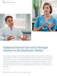
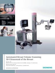

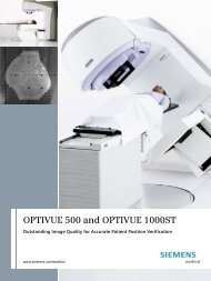
![WalkAway plus Technical Specifications [41 KB] - Siemens Healthcare](https://img.yumpu.com/51018135/1/190x253/walkaway-plus-technical-specifications-41-kb-siemens-healthcare.jpg?quality=85)