Hypopigmented Face Patches; Their Distribution and Relevance to ...
Hypopigmented Face Patches; Their Distribution and Relevance to ...
Hypopigmented Face Patches; Their Distribution and Relevance to ...
You also want an ePaper? Increase the reach of your titles
YUMPU automatically turns print PDFs into web optimized ePapers that Google loves.
INTERNATIONAL JOURNAI.^LEPROSY<br />
Volume 67, Number<br />
i'riuted 1h the<br />
(ISSN 0148-916X)<br />
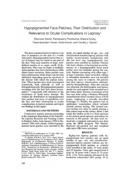
<strong>Hypopigmented</strong> <strong>Face</strong> <strong>Patches</strong>; <strong>Their</strong> <strong>Distribution</strong> <strong>and</strong><br />
<strong>Relevance</strong> <strong>to</strong> Ocular Complications in Leprosyl<br />
Ebenezer Daniel, Ramaswamy Premkumar, Sheena Koshy,<br />
Paraman<strong>and</strong>am Yowan, Nisha Kurian, <strong>and</strong> Timothy J. ffytche''<br />
The most conunon lesion in leprosy is an<br />
area of numbness on the skin or a visible<br />
skin patch. <strong>Hypopigmented</strong> lesions that occur<br />
in leprosy may be found on any part of<br />
the skin. They may manifest as huge, welldefined<br />
patches or as vague, small, ill-defined<br />
ones. They may be single or multiple.<br />
Some patches may have hypoesthesia while<br />
others retain sensation. Some patches may<br />
turn erythema<strong>to</strong>us while others can become<br />
infiltrated, depending upon the spectrum of<br />
the disease with which the patient manifests.<br />
These patches have been investigated<br />
extensively, both clinically as well as<br />
his<strong>to</strong>pathologically. <strong>Hypopigmented</strong> patches<br />
occurring over the face have been investigated<br />
(2.5) especially with reference <strong>to</strong> the<br />
occurrence of facial nerve damage. We<br />
studied the distribution of hypopigmented<br />
face patches <strong>and</strong> arcas of anesthesia over<br />
the face <strong>and</strong> their relationship <strong>to</strong> ocular<br />
complications in leprosy patients <strong>and</strong> report<br />
our findings in this paper.<br />
MATERIALS AND METHODS<br />
Consecutive leprosy patients presenting<br />
at the outpatient department of the Schieffelin<br />
Leprosy Research <strong>and</strong> Training Center<br />
in Karigiri, India, from January 1994 <strong>to</strong> December<br />
1994 with clearly visible hypopigmented<br />
facial patches were enrolled in the<br />
' Received for publication on 27 July 1999. Accepted<br />
for publication on 12 August 1999.<br />
E. Daniel, M.B.B.S., M.S., D.O., Head, Department<br />
of Ophthalmology; R. Premkumar, Ph.D., Head,<br />
Department of Occupational Therapy; S. Koshy,<br />
M.B.B.S., D.O., Ophthalmologist; P. Yowan, Field Coordina<strong>to</strong>r,<br />
Department of Ophthalmology; N. Kurian,<br />
M.Sc., Department of Biostatistics, Schieffelin Leprosy<br />
Research <strong>and</strong> Training Center, Karigiri, Vellore<br />
District, Tamil Nadu, India 632 106. T. J. ffytche,<br />
L.V.O., F.R.C.S., F.R.C.Opth., Consultant Ophthalmologist,<br />
St. Thomas' Hospital, Lambeth Palace<br />
Road, London SEI 7EH, U.K.<br />
study. An equal number of age-, sex-, <strong>and</strong><br />
classification-matched leprosy patients with<br />
similar socioeconomic backgrounds who<br />
did not have any hypopigmented face<br />
patches were enrolled as controls. Patients<br />
who had a his<strong>to</strong>ry of having had an erythema<strong>to</strong>us<br />
or a hypopigmented facial patch<br />
earlier were not included among the controls.<br />
Patients presenting with caber type 1<br />
or type 2 reactions, tinea versicolor, vitiligo<br />
or seborrheic dermatitis were not included<br />
among the cases or controls. Ali patients<br />
had their leprosy classification substantiated<br />
by his<strong>to</strong>pathology. After due consent<br />
was obtained, the demographic <strong>and</strong> leprosy<br />
details for each patient were recorded on a<br />
pro forma sheet. Sensory assessment of the<br />
face was done using a Semmes-Weinstein<br />
monofilament which exerted a force of 0.05<br />
grams, employing a recently described<br />
technique (4). Finally, the patients had an<br />
ophthalmic examination which included<br />
corneal sensory testing. The corneal sensory<br />
testing was done by gently placing a<br />
thin cot<strong>to</strong>n wisp on the cornea, about 2 mm<br />
inside the limbus at the 6 o'clock position<br />
as the patient gazed upward, <strong>and</strong> noting the<br />
reaction of the patient. All data were entered<br />
in<strong>to</strong> a computer <strong>and</strong> analysis was done<br />
using the SPSS program. The Mantel-<br />
Haenszel chi-squared test <strong>and</strong> weighted<br />
odds ratio (OR) were calculated.<br />
RESULTS<br />
Eighty-two leprosy patients with hypopigmented<br />
face patches <strong>and</strong> an equal<br />
number of age-, sex- <strong>and</strong> classificationmatched<br />
leprosy patients without any hypopigmented<br />
facial patches as controls<br />
were enrolled in the study; 53 were males<br />
<strong>and</strong> 29 females. <strong>Their</strong> ages ranged from 6<br />
years <strong>to</strong> 70 years with a mean of 35 years<br />
(S.D. 15.6). Forty patients belonged <strong>to</strong> the<br />
borderline tuberculoid (BT) group, 35 <strong>to</strong><br />
388
67, 4^Daniel, et al.: <strong>Face</strong> <strong>Patches</strong> <strong>and</strong> the Eye^ 389<br />
TABLE 1. <strong>Distribution</strong> of Impopigniented<br />
facial patches <strong>and</strong> anesthesia over<br />
the face in cases <strong>and</strong> controls.<br />
TABLE 3. Anesthesia over the face <strong>and</strong><br />
impaired corneal sensation in cases <strong>and</strong><br />
controls."<br />
Nerve arca<br />
Ophthalmic<br />
Maxillary<br />
M<strong>and</strong>ibular<br />
Greater auricular<br />
No. cases^Cases<br />
with hypo-^with<br />
pigmente('^fadai<br />
patches^anesthesa<br />
Controls<br />
facial<br />
anesthesia<br />
62^5^7<br />
57^9^4<br />
54^4^3<br />
34^ 6<br />
Impaired Facial anesthesia Facial anesthesia<br />
corneal present absent<br />
sensation Cases Controls Cases Controls<br />
Yes 9 3 35 18<br />
No 7 I() 31 51<br />
Total 16 13 66 69<br />
" Mantel-Ilaenszel weighted odds na<strong>to</strong> = 3.36; p<br />
390^ International Journal ofLeprosv^ 1999<br />
Location<br />
TABU. 4. Location offacial patches <strong>and</strong> ocular coniplications.<br />
No.<br />
Decreased vision Madarosis Lagophthalmos Other complications"<br />
No. % No. (X, No. (3. No.<br />
Around right eye 39 7 17.9% 3 7.7e/c 6 15.4% 8 20.5%<br />
Around left eye 38 7 18.4% 4 10.5% 5 13.2% 7 18.4%<br />
Forehead 39 7 17.9% 5 12.8% 6 15.4% 9 23.1%<br />
Temple 30 6 20.0% -) 6.7% 3 10.0% 6 20.0%<br />
Cheek 70 II 15.7% 6 8.6% 7 10.0% 18 25.7%<br />
Chin 25 7 28.0% 3 12.0% -) 8.0% 7 28.0%<br />
Central face IS i 13.3% 1 6.7% 9 60.0% 4 26.7%<br />
'Other coniplications = Cataract, pterygiuni, corneal opacities, iridocyclitis.<br />
lar branches. Most patches overlapped <strong>and</strong><br />
were not contined <strong>to</strong> the arcas supplied by<br />
any one of the three major divisions of the<br />
trigeminal nerve.<br />
The important finding in this study is the<br />
association between the presence of hypopigmented<br />
patches on the face <strong>and</strong> decreased<br />
corneal sensation. Patients with hypopigmentation<br />
over the face were three <strong>to</strong><br />
four times more likely <strong>to</strong> have impaired<br />
colmeal sensation than patients without any<br />
hypopigmented facial patches. The location<br />
of the hypopigmented patches over the face<br />
did not signiticantly alter this association.<br />
<strong>Patches</strong> around the eye <strong>and</strong> over the zygomatic<br />
arca were equally as significant as<br />
those over the nose, forehead <strong>and</strong> chin. Increasing<br />
age can decrease corneal sensation,<br />
but our study showed that even after<br />
adjusting for age, the signiticance of assoeiation<br />
between decreased corneal sensation<br />
<strong>and</strong> hypopigmented patches over the face<br />
remained almost the same. Anesthesia over<br />
the face <strong>to</strong>gether with hypopigmentation<br />
proved <strong>to</strong> be more signiticantly associated<br />
with decreased corneal sensation than anesthesia<br />
over the face without hypopigmentation<br />
(p
67, 4^Daniel, et al.: <strong>Face</strong> <strong>Patches</strong> <strong>and</strong> the Eye^391<br />
were found <strong>to</strong> have more corneal hypoesthesia<br />
than patients who did not have hypopigmented<br />
facial patches. The risk of<br />
having impaired corneal sensation was<br />
three <strong>to</strong> four times higher in patients with<br />
hypopigmented facial patches. This feature<br />
can be used <strong>to</strong> identify decreased corneal<br />
senszttion among leprosy patients under<br />
field conditions where direct estimation of<br />
corneal sensation is not advocated.<br />
RESUMEN<br />
Un grupo de 82 pacientes con lepra, con manchas<br />
hipopigmentadas sobre la cara (casos), y un número<br />
igual de pacientes sin ninguna hipopigmentatición facial<br />
(controles), se examinaron para establecer la distribución<br />
de las áreas de anestesia en la cara y la frecuencia<br />
de complicaciones oculares. Las manchas<br />
hipopigmentadas no siguieron ningún patrón y se sobrepusieron<br />
en las áreas de sensación innervadas por<br />
las tres ramas del nervio trigémino. La anestesia sobre<br />
la cara, evaluada con un monotilamen<strong>to</strong> de Semmes-<br />
Weinstein que ejerce una fuerza de 0.05 gramos, estuvo<br />
presente en el 19.5% de los casos y en el 15.9%<br />
de los controles. Los pacientes con manchas faciales<br />
hipopigmentadas presentaron más hipoanestesia de la<br />
córnea, que los pacientes sin hipopigmentación en la<br />
cara. El riesgo de presentar alguna alteración en la sensación<br />
de la córnea fue 3 a 4 vexes mayor en los pacientes<br />
con manchas hipopigmentadas. Esta caracteristica<br />
puede usarse para identificar aquellos pacientes<br />
con disminución en la sensación corneal bajo condiciones<br />
de campo, donde no es posible hacer medieiones<br />
directas.<br />
RÉSUMÉ<br />
Quatre vingt deux patients atteints de lèpre présentant<br />
des aires hypopigmentées sur la figure (cas) et un<br />
nombre identique de patients hanséniens contrôlés en<br />
ce qui concerne l'âge, le genre et la classification (contrõles)<br />
furent examinés pour la distribution des plaques<br />
hypopigmentées faciales, les aires anesthésiées sur la<br />
figure et les complications oculaires. Les plaques hypopigmentées<br />
ne suivaient pas d'aires &titiles et se<br />
chevatichaient dans les aires de sensibilité des trois<br />
branches du nerf trijumeau. L'anesthésie faciale, évaluée<br />
par le filziment de Semmes-Weinstein qui exerçait<br />
une force de 0,05 grammes, était présente chez 19.5%<br />
des eas et 15.9% des contrôles. Les patients présentant<br />
des plaques hypopigmentées avaient plus d'hypoesthésies<br />
de la cornée que les patients sans plaques<br />
hypopigmentées faciales. Le risque d'avoir une<br />
diminution de lit sensitivité de la cornée étaient trois à<br />
quatre fois plus élevée chez les patients présentant des<br />
plaques hypopigmentées faciales. Cette caractéristique<br />
peut être utilisée pour identifier une baisse de sensitivité<br />
de la cornée parmi les patients hanséniens dans les<br />
conditions du terrain oit l'estimation directe de la sensitivité<br />
cornéenne n'est pas pronée possible.<br />
REFERENCES<br />
1. ANTIA, N. 11., DIVEKAR, S. C. <strong>and</strong> DASTUR, D. K.<br />
The facial nerve in leprosy; clinicai <strong>and</strong> operative<br />
aspects. Int. J. Lepr. 34 (1966) 103-117.<br />
2. Hochwixi, M., KIRAN, K. U. <strong>and</strong> SUNEETHA, S.<br />
The significance of facial patches <strong>and</strong> type I reaction<br />
for the development of facial nerve damage in<br />
leprosy; a rctrospective study among 1226 paucibacillary<br />
leprosy patients. Lepr. Rev. 62 (1991)<br />
143-149.<br />
3. PFALTZGRAFF, R. E. <strong>and</strong> BRvcEsoN, A. Clinicai<br />
leprosy. In: Leprosy. I st edn. Hastings, R. C., ed.<br />
New York: Churchill Livings<strong>to</strong>ne, 1985, pp. 140,<br />
146.<br />
4. PREN1KUMAR, R., DANIEL, E., SUNEUEIA, S. <strong>and</strong><br />
YowAN, P. Quantitative assessment of facial sensation<br />
in leprosy. Int. J. Lepr. 66 (1998) 348-355.<br />
5. REICHART, P. A., SRISUWAN, S. <strong>and</strong> METAH, D. Lesions<br />
of the facial <strong>and</strong> trigeminal nerve in leprosy.<br />
Int. J. Oral Surg. 11 (1982) 14-20.<br />
6. WHO EXPERT Comum: ON LEPROSY. Seventh report.<br />
Geneva: World Health Organization, 1998, p.<br />
25. Tech. Rep. Ser. 874.


