Riding the Waves Handout R6 - Grand County EMS
Riding the Waves Handout R6 - Grand County EMS
Riding the Waves Handout R6 - Grand County EMS

You also want an ePaper? Increase the reach of your titles
YUMPU automatically turns print PDFs into web optimized ePapers that Google loves.
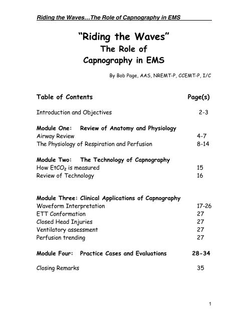
<strong>Riding</strong> <strong>the</strong> <strong>Waves</strong>…The Role of Capnography in <strong>EMS</strong><br />
“<strong>Riding</strong> <strong>the</strong> <strong>Waves</strong>”<br />
The Role of<br />
Capnography in <strong>EMS</strong><br />
By Bob Page, AAS, NREMT-P, CCEMT-P, I/C<br />
Table of Contents<br />
Page(s)<br />
Introduction and Objectives 2-3<br />
Module One: Review of Anatomy and Physiology<br />
Airway Review 4-7<br />
The Physiology of Respiration and Perfusion 8-14<br />
Module Two: The Technology of Capnography<br />
How EtCO 2 is measured 15<br />
Review of Technology 16<br />
Module Three: Clinical Applications of Capnography<br />
Waveform Interpretation 17-26<br />
ETT Conformation 27<br />
Closed Head Injuries 27<br />
Ventilatory assessment 27<br />
Perfusion trending 27<br />
Module Four: Practice Cases and Evaluations 28-34<br />
Closing Remarks 35<br />
1
<strong>Riding</strong> <strong>the</strong> <strong>Waves</strong>…The Role of Capnography in <strong>EMS</strong><br />
INTRODUCTION<br />
Capnography is a noninvasive method for monitoring <strong>the</strong><br />
level of carbon dioxide in exhaled breath (EtCO 2 ), to<br />
assess a patient’s ventilatory status. A true capnogram<br />
produces an EtCO 2 value as well as a waveform, or<br />
capnogram. On Critical Care transports, capnograms are<br />
useful for monitoring ventilator status, warning of airway<br />
leaks and ventilator circuit disconnections. Capnography<br />
is also useful for ensuring proper endotracheal tube<br />
placement. Capnography also helps clinicians diagnose<br />
specific medical conditions, make treatment decisions,<br />
and assess efficacy of code efforts and predict outcome.<br />
Capnography offers numerous clinical uses, but technical<br />
limitations have prevented <strong>EMS</strong> personnel from<br />
embracing its use outside <strong>the</strong> operating room. Today,<br />
technological advances have made it possible for <strong>the</strong>se<br />
devices to be used in <strong>the</strong> demanding setting of <strong>the</strong><br />
prehospital environment.<br />
2
<strong>Riding</strong> <strong>the</strong> <strong>Waves</strong>…The Role of Capnography in <strong>EMS</strong><br />
OBJECTIVES for <strong>the</strong> Session:<br />
By <strong>the</strong> end of this session, you will be able to:<br />
Describe <strong>the</strong> structure and function of <strong>the</strong> upper and lower<br />
airways.<br />
Describe <strong>the</strong> mechanics and science of ventilation and<br />
respiration.<br />
Describe <strong>the</strong> basic physiology of perfusion.<br />
Describe <strong>the</strong> relationship between ventilation and perfusion.<br />
Describe <strong>the</strong> principles behind CO 2 measurement.<br />
Describe <strong>the</strong> various methods of EtCO 2 measurement<br />
including quantitative and qualitative capnometry and<br />
capnography.<br />
Describe <strong>the</strong> technology of EtCO 2 measurement including<br />
mainstream, sidestream and microstream sampling.<br />
Identify <strong>the</strong> components of a normal capnogram waveform.<br />
Identify abnormal capnogram waveforms as related to<br />
various airway, breathing and circulation problems.<br />
Discuss <strong>the</strong> various clinical applications of capnography in<br />
<strong>the</strong> field.<br />
Given various cases, discuss <strong>the</strong> role of capnography in<br />
identifying <strong>the</strong> problem and in <strong>the</strong> management of <strong>the</strong><br />
patient.<br />
3
<strong>Riding</strong> <strong>the</strong> <strong>Waves</strong>…The Role of Capnography in <strong>EMS</strong><br />
MODULE ONE:<br />
REVIEW OF AIRWAY ANATOMY AND PHYSIOLOGY<br />
N a s a l P a s s a g e s<br />
R o o f o f t h e M o u t h<br />
E p ig lo t t is<br />
T ra c h e a ( w in d p ip e )<br />
E s o p h a g u s ( f o o d t u b e )<br />
P u lm o n a ry V e in<br />
B ro n c h io l e<br />
B ro n c h i<br />
A lv e o l i<br />
Two divisions to <strong>the</strong> airway:<br />
1. Upper Airway<br />
2. Lower Airway<br />
4
<strong>Riding</strong> <strong>the</strong> <strong>Waves</strong>…The Role of Capnography in <strong>EMS</strong><br />
Airway Anatomy Review<br />
Upper Airway<br />
Nasopharynx<br />
Oropharnyx<br />
Palates<br />
Hard and soft<br />
Larynx<br />
Epiglottis<br />
Vocal cords<br />
Upper Airway Physiology: PEEP or Positive End Expiratory<br />
Pressure can be defined as <strong>the</strong> pressure against which exhalation<br />
occurs. The purpose of PEEP is to prevent alveolar collapse. The<br />
structure and path way of <strong>the</strong> upper airways provide for a natural<br />
or “physiological PEEP.”<br />
The primary roles of <strong>the</strong> upper airway are to _ _ _ _ _ _ _ _ _ _ _ _ ,<br />
_ _ _ _ _ _ _ _ _ _ _ _ _ _ , and _______________ <strong>the</strong> air entering<br />
<strong>the</strong> lungs.<br />
5
<strong>Riding</strong> <strong>the</strong> <strong>Waves</strong>…The Role of Capnography in <strong>EMS</strong><br />
Lower Airway<br />
Trachea<br />
Bronchi<br />
Bronchioles<br />
Dead Space Air<br />
The lower airway is comprised of <strong>the</strong> trachea, bronchi and <strong>the</strong>n<br />
25 divisions of <strong>the</strong> bronchial tree terminating at <strong>the</strong><br />
respiratory bronchioles and <strong>the</strong> alveoli.<br />
The areas from <strong>the</strong> bronchioles to <strong>the</strong> nose comprise <strong>the</strong> total<br />
dead space air. This is air that is not exchanged.<br />
6
<strong>Riding</strong> <strong>the</strong> <strong>Waves</strong>…The Role of Capnography in <strong>EMS</strong><br />
Bronchioles<br />
Alveoli<br />
The alveoli are tiny air sacs where gas exchange occurs. O 2 and<br />
CO 2 are exchanges at <strong>the</strong> capillary-alveolar membrane.<br />
7
<strong>Riding</strong> <strong>the</strong> <strong>Waves</strong>…The Role of Capnography in <strong>EMS</strong><br />
THE PHYSIOLOGY OF VENTILATION<br />
Rule # 1 of Life<br />
Air must go<br />
in and out<br />
Function of ventilation:<br />
Ventilation is <strong>the</strong> movement of air.<br />
• Designed to eliminate CO 2 and take in O 2<br />
How air moves (and it MUST move)<br />
Chemoreceptors in <strong>the</strong> medulla sense elevated levels of CO 2<br />
or lowered pH, triggering ventilation. Known as hype rcarbic<br />
drive.<br />
Diaphragm contracts and moves downward.<br />
Intercostal muscles spread chest wall out increasing <strong>the</strong><br />
volume inside <strong>the</strong> chest.<br />
Differences in pressure inside <strong>the</strong> chest and outside causes<br />
air to move into <strong>the</strong> lungs.<br />
Hypoxic drive (low O 2 levels) is secondary drive.<br />
8
<strong>Riding</strong> <strong>the</strong> <strong>Waves</strong>…The Role of Capnography in <strong>EMS</strong><br />
VOLUME CAPACITIES<br />
Tidal Volume (Vt): The amount of air moved in one breath<br />
Typically 500 cc in an adult at rest<br />
Anatomical Dead Space (Vd): Air not available for gas<br />
exchange<br />
About 150 cc<br />
Alveolar volume (Va); Air that is available for gas exchange<br />
About 350 cc (Vt – Vd = Va)<br />
Anything that affects <strong>the</strong> tidal volume only affects <strong>the</strong><br />
alveolar volume.<br />
Factors affecting <strong>the</strong> tidal volume<br />
Hyperventilation<br />
Fast breathing (tachypnea) doesn’t necessarily increase<br />
tidal volume<br />
Anxiety, head injuries, diabetic emergencies, PE, AMI,<br />
and o<strong>the</strong>rs<br />
Hypoventilation<br />
Slow breathing (bradypnea) does not necessarily<br />
decrease tidal volume.<br />
Causes include CNS disorders, narcotic use and o<strong>the</strong>rs.<br />
Street Wisdom: An increase or decrease in tidal volume is at<br />
<strong>the</strong> expense or benefit of alveolar air.<br />
9
<strong>Riding</strong> <strong>the</strong> <strong>Waves</strong>…The Role of Capnography in <strong>EMS</strong><br />
Respiration is <strong>the</strong> exchange of gases<br />
Alveolar respiration occurs between <strong>the</strong> _______ and<br />
_________ in <strong>the</strong> lungs.<br />
10
<strong>Riding</strong> <strong>the</strong> <strong>Waves</strong>…The Role of Capnography in <strong>EMS</strong><br />
Cellular respiration occurs between <strong>the</strong> _____ in <strong>the</strong> body<br />
and <strong>the</strong> _____________.<br />
When <strong>the</strong>re is a difference in partial pressure between <strong>the</strong><br />
two containers, gas will move from <strong>the</strong> area of greater<br />
concentrations to <strong>the</strong> area of lower concentration. a.k.a.<br />
diffusion<br />
11
<strong>Riding</strong> <strong>the</strong> <strong>Waves</strong>…The Role of Capnography in <strong>EMS</strong><br />
THE PHYSIOLOGY OF PERFUSION<br />
Rule # 2 of Life<br />
Blood must go<br />
round and round.<br />
Fick principle: Oxygen Transport<br />
In order from adequate cellular perfusion to occur, <strong>the</strong><br />
following must be present:<br />
• Adequate number of Red Blood Cells (RBC’s)<br />
♦ Hemoglobin on <strong>the</strong> RBC’s carry <strong>the</strong> oxygen molecules<br />
• Adequate O 2<br />
♦ Patient must have adequate O 2 coming in. See Rule<br />
of Life #1<br />
• RBC’s must be able to offload and take on O 2<br />
♦ Some conditions such as carbon monoxide poisoning<br />
and cyanide poisoning affect <strong>the</strong> RBC’s ability to<br />
bind and release O 2 molecules.<br />
• Adequate blood pressure to push cells<br />
12
<strong>Riding</strong> <strong>the</strong> <strong>Waves</strong>…The Role of Capnography in <strong>EMS</strong><br />
PHYSIOLOGIC BALANCE<br />
More Air Less Blood<br />
V > Q<br />
Equal Air and Blood<br />
V = Q<br />
More Blood Less Air<br />
V < Q<br />
Pathological Conditions<br />
Normal ventilation, poor perfusion: P.E., Arrest<br />
Abnormal ventilation, good perfusion: obstruction, O.P.D.,<br />
drug OD<br />
Bad ventilation and perfusion: Arrest<br />
Bad exchange area: CHF<br />
13
<strong>Riding</strong> <strong>the</strong> <strong>Waves</strong>…The Role of Capnography in <strong>EMS</strong><br />
Critical Thinking Cases – Designed to illustrate <strong>the</strong><br />
pathophysiology<br />
Normal ventilation/normal perfusion<br />
Normal ventilation/compromised perfusion<br />
Compromised ventilation/normal perfusion<br />
Compromised ventilation and perfusion<br />
1. 26 year-old female patient took an overdose of Valium. She is<br />
UNCONSCIOUS. V/S are 110/70, pulse is 64, RR is 12 and very<br />
shallow, skin is warm and dry.<br />
2. 65 year-old male patient complaining of a sudden onset of right<br />
sided chest pain and dyspnea. He has no medical history except for<br />
a hip replacement surgery about 3 weeks ago. His lung sounds are<br />
clear. VS are B/P 140/78, pulse is 110, and RR is about 20 and<br />
normal depth.<br />
3. 80 year-old man complains of a sudden onset of severe headache.<br />
He has flushed skin, and has obvious facial droop to <strong>the</strong> left side.<br />
He has a history of high blood pressure. V/S are B/P 180/110,<br />
pulse is 100 and RR is 16 and normal depth.<br />
4. 37 year-old female that was involved in a head on collision.<br />
Windshield is starred and <strong>the</strong> steering wheel is broke. Bruising and<br />
crepitus found over <strong>the</strong> left chest. Pt is unconscious, difficult to<br />
bag with absent lungs sounds on <strong>the</strong> left side. Blood pressure is<br />
60/40; pulse is 130 and weak at <strong>the</strong> carotid. There is obvious JVD.<br />
Skin is cool and clammy.<br />
14
<strong>Riding</strong> <strong>the</strong> <strong>Waves</strong>…The Role of Capnography in <strong>EMS</strong><br />
MODULE TWO:<br />
TECHNOLOGY OF CAPNOGRAPHY<br />
The Role of CO 2<br />
CO 2 is <strong>the</strong> “Gas of Life”<br />
Produced as a normal by-product of metabolism.<br />
Measurement of EtCO 2 (Capnometry)<br />
Qualitative<br />
Color change assay<br />
• (CO 2 turns <strong>the</strong> sensor from purple to yellow)<br />
Quantitative<br />
Gives you a value (EtCO 2 )<br />
Respiratory Rate<br />
Waveform Capnography<br />
Features quantitative value and waveform<br />
Capnography includes Capnometry<br />
Street Wisdom: “End Tidal CO 2 reading without a waveform is<br />
like a heart rate without an ECG recording.”<br />
15
<strong>Riding</strong> <strong>the</strong> <strong>Waves</strong>…The Role of Capnography in <strong>EMS</strong><br />
Infrared (IR) Spectroscopy:<br />
Most often used<br />
Infrared light is used to expose <strong>the</strong> sample<br />
IR sensors detect <strong>the</strong> absorbed light and calculate a<br />
value<br />
Broad spectrum IR beams can be absorbed N 2 0 and High<br />
O 2 levels<br />
Side stream sampling<br />
“First generation devices”<br />
Draws large sample into machine from <strong>the</strong> line<br />
Can be used on intubated and non-intubated patients with<br />
a nasal cannula attachment<br />
“Second generation devices”<br />
Airway mounted sensors<br />
Generally for intubated patients<br />
Microstream Technology<br />
Position independent adaptors<br />
Moisture, secretion, and contaminant handling in three<br />
ways<br />
• Samples taken from center of line, and in 1/20 th <strong>the</strong><br />
volume<br />
• Vapor permeable tubing<br />
• Sub micron-multi-surface filters<br />
Expensive parts are protected<br />
Microbeam IR sensor is CO 2 specific<br />
Suitable for adult and pediatric environments.<br />
16
<strong>Riding</strong> <strong>the</strong> <strong>Waves</strong>…The Role of Capnography in <strong>EMS</strong><br />
MODULE THREE:<br />
CLINICAL APPLICATIONS OF CAPNOGRAPHY<br />
THE NORMAL CAPNOGRAM<br />
60<br />
50<br />
40<br />
30<br />
20<br />
10<br />
0<br />
II<br />
I<br />
III<br />
IV<br />
Phase I: Respiratory Baseline<br />
Phase II: Expiratory Upstroke<br />
Phase III: Expiratory Plateau<br />
ETCO 2 : Peak EtCO 2 level<br />
Phase IV: Inspiratory Downstroke<br />
17
<strong>Riding</strong> <strong>the</strong> <strong>Waves</strong>…The Role of Capnography in <strong>EMS</strong><br />
Systematic Approach to Waveform Interpretation<br />
1. Is CO 2 present? (waveform present)<br />
2. Look at <strong>the</strong> respiratory baseline. Is <strong>the</strong>re rebreathing?<br />
3. Expiratory Upstroke: Steep, sloping, or prolonged?<br />
4. Expiratory (alveolar) Plateau: Flat, prolonged, notched, or<br />
sloping?<br />
5. Inspiratory Downstroke: Sleep, sloping, or prolonged?<br />
6. Read <strong>the</strong> EtCO 2<br />
7. If ABG is available, compare <strong>the</strong> EtCO 2 with PACO 2<br />
a. If <strong>the</strong>y are within 5mm/hg of each o<strong>the</strong>r <strong>the</strong>n <strong>the</strong><br />
problem is ventilatory and not perfusion.<br />
b. EtCO 2 can be used in many cases in lieu of ABG’s<br />
The ABC’s of Waveform Interpretation!<br />
A – Airway: Look for signs of obstructed airway (steep,<br />
upsloping expiratory plateau)<br />
B – Breathing: Look at EtCO 2 reading. Look for waveforms,<br />
and elevated respiratory baseline.<br />
C – Circulation: Look at trends, long and short term for<br />
increases or decreases in EtCO 2 readings<br />
18
<strong>Riding</strong> <strong>the</strong> <strong>Waves</strong>…The Role of Capnography in <strong>EMS</strong><br />
Street wisdom: A patient complains of having dif ficulty<br />
breathing. The pulse oximeter shows 98% on 15 lpm O 2 . As<br />
you attempt to listen to lung sounds, <strong>the</strong>y are hard to make<br />
out in <strong>the</strong> back of <strong>the</strong> ambulance. What benefit , if any could<br />
capnography make in <strong>the</strong> diagnosis and management of this<br />
patient?<br />
What is <strong>the</strong> dif ference between pulse oximetry and<br />
capnography?<br />
SpO 2 = Pulse oximetry – measures oxygenation<br />
EtCO 2 = Capnography – measures ventilation<br />
19
<strong>Riding</strong> <strong>the</strong> <strong>Waves</strong>…The Role of Capnography in <strong>EMS</strong><br />
NORMAL CAPNOGRAPHY<br />
5 0<br />
4 0<br />
3 0<br />
2 0<br />
1 0<br />
0<br />
T i m e<br />
This is a normal capnogram that has all of <strong>the</strong> phases that are<br />
easily appreciated. Note <strong>the</strong> gradual upslope and alveolar<br />
“Plateau”<br />
Point for thought:<br />
List <strong>the</strong> things a normal capnogram tells you and <strong>the</strong> things that it<br />
does not tell you.<br />
20
<strong>Riding</strong> <strong>the</strong> <strong>Waves</strong>…The Role of Capnography in <strong>EMS</strong><br />
ABNORMAL CAPNOGRAPHY<br />
Hyperventilation<br />
This capnogram starts slow and has an EtCO 2 reading that is<br />
normal. Notice as <strong>the</strong> rate gets faster, <strong>the</strong> waveform gets<br />
narrower and <strong>the</strong>re is a steady decrease in <strong>the</strong> EtCO 2 to below<br />
30mm/hg. Causes of this type of waveform include:<br />
50<br />
40<br />
30<br />
20<br />
10<br />
0<br />
60<br />
50<br />
40<br />
30<br />
20<br />
10<br />
0<br />
Time<br />
EtCO2 mmHg<br />
Time<br />
Hyperventilation syndrome<br />
Overzealous bagging<br />
Pulmonary embolism<br />
21
<strong>Riding</strong> <strong>the</strong> <strong>Waves</strong>…The Role of Capnography in <strong>EMS</strong><br />
Hypoventilation<br />
5 0<br />
4 0<br />
3 0<br />
2 0<br />
1 0<br />
0<br />
T i m e<br />
In this capnogram, <strong>the</strong>re is a gradual increase in <strong>the</strong> EtCO 2 .<br />
Obstruction is not apparent. Causes of this may include:<br />
Respiratory depression for any reason<br />
Narcotic overdose<br />
CNS dysfunction<br />
Heavy sedation<br />
22
<strong>Riding</strong> <strong>the</strong> <strong>Waves</strong>…The Role of Capnography in <strong>EMS</strong><br />
Apnea<br />
60<br />
50<br />
40<br />
30<br />
20<br />
10<br />
0<br />
EtCOTime<br />
2 mmHg<br />
This capnogram shows a complete loss of waveform indicating<br />
no CO 2 present. Capnography allows for instantaneous<br />
recognition of this potentially fatal condition. Since this<br />
occurred suddenly, consider <strong>the</strong> following causes:<br />
Dislodged ET Tube<br />
Total obstruction of ET Tube<br />
Respiratory arrest in <strong>the</strong> non-intubated patient<br />
Equipment malfunction (If <strong>the</strong> patient is still breathing) Check<br />
all connections and sampling chambers<br />
23
<strong>Riding</strong> <strong>the</strong> <strong>Waves</strong>…The Role of Capnography in <strong>EMS</strong><br />
Loss of Alveolar Plateau<br />
50<br />
40<br />
30<br />
20<br />
10<br />
0<br />
Time<br />
This capnogram displays an abnormal loss of alveolar plateau<br />
meaning incomplete or obstructed exhalation. Note <strong>the</strong><br />
“Shark’s fin” pattern. This pattern is found in <strong>the</strong> following<br />
Bronchoconstriction<br />
Asthma<br />
COPD<br />
Incomplete airway obstruction<br />
Upper airway<br />
Tube kinked or obstructed by mucous<br />
24
<strong>Riding</strong> <strong>the</strong> <strong>Waves</strong>…The Role of Capnography in <strong>EMS</strong><br />
60<br />
50<br />
40<br />
30<br />
20<br />
10<br />
0<br />
Poor perfusion (cardiac arrest)<br />
The capnogram can indicate perfusion during CPR and<br />
effectiveness of resuscitation efforts. Note <strong>the</strong> trough in <strong>the</strong><br />
center of <strong>the</strong> capnogram. During this time, <strong>the</strong>re was a change<br />
in personnel doing CPR. The fatigue of <strong>the</strong> first rescuer was<br />
demonstrated when <strong>the</strong> second rescuer took over<br />
compressions.<br />
EtCO2 mmHg<br />
Time: 30 minute trend<br />
60<br />
50<br />
40<br />
30<br />
20<br />
10<br />
0<br />
EtCO 2 mmHg<br />
Time: trend 30 minutes<br />
This patient was defibrillated successfully with a return of<br />
spontaneous pulse.<br />
Notice <strong>the</strong> dramatic change in <strong>the</strong> EtCO 2 when pulses were<br />
restored.<br />
Studies have shown that consistently low readings (less than<br />
10mm) during resuscitation reflect a poor outcome and futile<br />
resuscitation.<br />
25
<strong>Riding</strong> <strong>the</strong> <strong>Waves</strong>…The Role of Capnography in <strong>EMS</strong><br />
Elevated Baseline<br />
60<br />
50<br />
40<br />
30<br />
20<br />
10<br />
0<br />
EtCO Time 2 mmHg<br />
This capnogram demonstrates an elevation to <strong>the</strong> baseline.<br />
This indicates incomplete inhalation and or exhalation. CO 2<br />
does not get completely washed out on inhalation. Possible<br />
causes for this include:<br />
Air trapping (as in asthma or COPD)<br />
CO 2 rebreathing (ventilator circuit problem)<br />
What o<strong>the</strong>r condition(s) might produce this type of<br />
waveform?<br />
26
<strong>Riding</strong> <strong>the</strong> <strong>Waves</strong>…The Role of Capnography in <strong>EMS</strong><br />
Field Clinical Applications for Capnography<br />
Closed Head Injury<br />
Increased intracranial pressure (ICP) tied to increased<br />
blood flow following injury (swelling)<br />
Hypoxic cells produce CO 2 in <strong>the</strong> brain<br />
CO 2 causes vasodilation and more blood fills <strong>the</strong> cranium,<br />
increasing pressure.<br />
Hyperventilation is no longer recommended<br />
Ventilation should be geared towards controlling CO 2 levels<br />
but not overdoing it.<br />
Obstructive Pulmonary Diseases<br />
Asthma, COPD<br />
Waveform can indicate bronchoconstriction where<br />
wheezes might not have been heard<br />
Monitor <strong>the</strong> effectiveness of bronchodilator <strong>the</strong>rapy<br />
Tube Conformation<br />
Capnography will detect <strong>the</strong> presence of CO 2 in expired<br />
air conforming ETT placement<br />
No longer acceptable to use only lungs sounds to confirm<br />
A dislodged tube will be detected immediately with<br />
capnography<br />
Kinking or clotting tubes can also be detected<br />
In cases of ventilator use, capnography can detect<br />
problems in rebreathing.<br />
Perfusion<br />
Capnography can be set up to trend EtCO 2 to detect <strong>the</strong><br />
presence or absence of perfusion<br />
Is a proven predictor of those who do not survive<br />
resuscitation<br />
When an ABG is available, can detect ventilatory or<br />
perfusion problems<br />
27
<strong>Riding</strong> <strong>the</strong> <strong>Waves</strong>…The Role of Capnography in <strong>EMS</strong><br />
MODULE FOUR:<br />
CASE SIMULATIONS AND EVALUATION<br />
CASE # 1<br />
Presentation<br />
Patient is a 65 year old male complaining of crushing substernal<br />
chest pain. He rates <strong>the</strong> pain as a 10 on a scale of one to ten. He<br />
denies and shortness of breath or any o<strong>the</strong>r complaints. He has a<br />
history of cardiac disease and asthma.<br />
Clinical Situation<br />
V/S: 130/80, Pulse is 100, RR is about 20<br />
SpO 2 is 96%, EtCO 2 is 40<br />
Cardiac Monitor shows Sinus Tachycardia<br />
His capnogram is as follows.<br />
60<br />
50<br />
40<br />
30<br />
20<br />
10<br />
0<br />
EtCO<br />
Time 2 mmHg<br />
Questions:<br />
Is <strong>the</strong> EtCO 2 within normal limits?<br />
Is <strong>the</strong> waveform normal or abnormal? Why or Why Not?<br />
What can you deduce about <strong>the</strong> ventilation status?<br />
A<br />
B<br />
C<br />
28
<strong>Riding</strong> <strong>the</strong> <strong>Waves</strong>…The Role of Capnography in <strong>EMS</strong><br />
CASE #2<br />
Presentation<br />
Patient is a 25-year-old male patient with a history of asthma. He<br />
has been compliant with his medications until he ran out of<br />
albuterol. Today, while at a basketball game, he suddenly gets<br />
short of breath. He does not have his albuterol inhaler with him.<br />
He presents sitting in <strong>the</strong> bleachers, in minor respiratory<br />
distress. It is noisy and hard to hear lung sounds.<br />
Clinical Situation<br />
B/P 120/76<br />
Pulse 100<br />
RR – 14<br />
SpO 2 94<br />
EtCO 2 is 50<br />
60<br />
50<br />
40<br />
30<br />
20<br />
10<br />
0<br />
Questions:<br />
EtCOTime<br />
2 mmHg<br />
Is <strong>the</strong> EtCO 2 within normal limits?<br />
Is <strong>the</strong> waveform normal or abnormal? Why or Why Not?<br />
What can you deduce about <strong>the</strong> ventilation status?<br />
A<br />
B<br />
C<br />
29
<strong>Riding</strong> <strong>the</strong> <strong>Waves</strong>…The Role of Capnography in <strong>EMS</strong><br />
CASE #3<br />
Presentation<br />
You and your partner are working a cardiac arrest and are<br />
successful in resuscitation. The Patient is still unstable and <strong>the</strong><br />
decision is made to load and go because of <strong>the</strong> very short<br />
transport time to <strong>the</strong> ED. He is intubated and EtCO 2 confirmed<br />
with good waveform and an EtCO 2 of about 42mm/hg.<br />
The patient is not breathing on <strong>the</strong>ir own.<br />
Clinical Situation:<br />
60<br />
50<br />
40<br />
30<br />
20<br />
10<br />
0<br />
EtCO Time 2 mmHg<br />
B/P is 100/70<br />
Pulse is 88<br />
RR assisted<br />
SpO 2 is 100% on 15lpm via NRB mask<br />
EtCO 2 is 40-42<br />
After loading him into <strong>the</strong> ambulance, <strong>the</strong> first responders<br />
resume ventilation. The capnography alarm sounds and <strong>the</strong><br />
following waveform is seen:<br />
60<br />
50<br />
40<br />
30<br />
20<br />
10<br />
0<br />
EtCOTime<br />
2 mmHg<br />
30
<strong>Riding</strong> <strong>the</strong> <strong>Waves</strong>…The Role of Capnography in <strong>EMS</strong><br />
CASE # 3 Continued<br />
Questions:<br />
Is <strong>the</strong> EtCO 2 within normal limits?<br />
Is <strong>the</strong> waveform normal or abnormal? Why or Why Not?<br />
What can you deduce about <strong>the</strong> ventilation status?<br />
A<br />
B<br />
C<br />
31
<strong>Riding</strong> <strong>the</strong> <strong>Waves</strong>…The Role of Capnography in <strong>EMS</strong><br />
CASE # 4<br />
Presentation<br />
You have a 30-year-old female who was in status seizures. Your<br />
partner administers Valium to halt <strong>the</strong> seizures. The patient<br />
appears to be post-ictal but is slow to respond fully.<br />
Clinical Situation<br />
B/P is 114/68<br />
Pulse is 96<br />
RR is 12<br />
SpO 2 is 98 on 6 lpm nasal cannula<br />
Glucose is 100<br />
EtCO 2 is as follows<br />
60<br />
50<br />
40<br />
30<br />
20<br />
10<br />
0<br />
Questions:<br />
EtCO Time 2 mmHg<br />
Is <strong>the</strong> EtCO 2 within normal limits?<br />
Is <strong>the</strong> waveform normal or abnormal? Why or Why Not?<br />
What can you deduce about <strong>the</strong> ventilation status?<br />
A<br />
B<br />
C<br />
32
<strong>Riding</strong> <strong>the</strong> <strong>Waves</strong>…The Role of Capnography in <strong>EMS</strong><br />
CASE # 5<br />
Presentation<br />
It’s 3 am and you are called to a residence for a 60 year old man<br />
that is in respiratory distress. You find <strong>the</strong> gentleman sitting up<br />
on his bed with feet dangling off <strong>the</strong> end. He presents in obvious<br />
distress and cannot speak words due to <strong>the</strong> distress. His lung<br />
fields are very diminished with crackles heard. He is pale and<br />
diaphoretic and appears to be getting weaker. Family members<br />
tell you that he has a bad heart and takes a “heart pill”, and a<br />
“water pill”. Pt becomes obtunded with labored breathing. They<br />
still have a gag reflex.<br />
Clinical Presentation<br />
BP is 158/90<br />
HR is 130<br />
RR is labored<br />
SpO 2 is 88%<br />
EtCO 2 as follows<br />
A<br />
B<br />
C<br />
60<br />
50<br />
40<br />
30<br />
20<br />
10<br />
0<br />
EtCO Time 2 mmHg<br />
33
<strong>Riding</strong> <strong>the</strong> <strong>Waves</strong>…The Role of Capnography in <strong>EMS</strong><br />
Case #5 Continued<br />
The decision is made to nasally intubate this patient. The tube is<br />
passed although <strong>the</strong> lung sounds are so diminished <strong>the</strong>y are hard<br />
to hear. The Pulse Ox offers no change, however, <strong>the</strong> capnogram<br />
shows <strong>the</strong> following:<br />
60<br />
50<br />
40<br />
30<br />
20<br />
10<br />
0<br />
EtCO Time 2 mmHg<br />
Questions: Scenario 5<br />
Is <strong>the</strong> EtCO 2 within normal limits?<br />
Is <strong>the</strong> waveform normal or abnormal? Why or Why Not?<br />
What can you deduce about <strong>the</strong> ventilation status?<br />
A<br />
B<br />
C<br />
34
<strong>Riding</strong> <strong>the</strong> <strong>Waves</strong>…The Role of Capnography in <strong>EMS</strong><br />
Closing Remarks,<br />
…From one paramedic to ano<strong>the</strong>r….<br />
Capnography represents ano<strong>the</strong>r great stride in <strong>the</strong> advances in<br />
technology and medicine that have made way into <strong>the</strong> field. Not since <strong>the</strong><br />
cardiac monitor and paramedics manually reading ECG strips has one device<br />
had <strong>the</strong> ability to benefit such a wide variety of patients.<br />
For years, Anes<strong>the</strong>siologists have used waveform capnography as <strong>the</strong>ir<br />
standard for monitoring <strong>the</strong> vital functions of patients. Now, <strong>the</strong> technology<br />
allows a smaller version to be used by paramedics.<br />
And now, YOU are ready to do this! Think of <strong>the</strong> incredible difference<br />
this can make in <strong>the</strong> care of your patients.<br />
To summarize, why do you need waveform capnography?<br />
Ventilation Vital Sign<br />
Confirmation of tube placement<br />
Constant monitoring of airway, ventilation and perfusion<br />
Bronchoconstriction in Obstructive airway disease<br />
Any respiratory patient<br />
Closed head injury to guide <strong>the</strong> careful elimination of CO 2<br />
Progressive monitoring of perfusion and ventilation<br />
Why a color change device isn’t enough<br />
Only confirms <strong>the</strong> presence of CO 2 not <strong>the</strong> amount<br />
Can’t monitor <strong>the</strong> patient<br />
Why a quantitative device is not enough<br />
While a number is better than just a color change,<br />
It can’t detect bronchoconstriction<br />
It can’t trend <strong>the</strong> level of CO 2<br />
There are many different brands and technology out <strong>the</strong>re. I have tried to<br />
present a fair and unbiased account of my extensive research into <strong>the</strong><br />
subject. Take each device for a “test drive” and make your mind up.<br />
35


![EKG Basics.ppt [Read-Only] - Grand County EMS](https://img.yumpu.com/34986415/1/190x146/ekg-basicsppt-read-only-grand-county-ems.jpg?quality=85)