KÑига LXXI - Univerzitet u Beogradu
KÑига LXXI - Univerzitet u Beogradu
KÑига LXXI - Univerzitet u Beogradu

Create successful ePaper yourself
Turn your PDF publications into a flip-book with our unique Google optimized e-Paper software.
Synchrotron radiation X-ray tomographic microscopy (SRXTM) of brachiopod shell interiors for taxonomy: ... 111<br />
20 keV (for “Rhynchonella”<br />
flustracea) and 33 keV (for Terebratulina<br />
imbricata) have<br />
been used. The magnification of<br />
the X-ray microscope was X4.<br />
A scintilator LAG: Ce 20 um<br />
was used. The number of projections<br />
for both specimens is<br />
1501. Reconstruction was performed<br />
on a 32 node Linux PC<br />
farm using highly optimized<br />
filtered back projection routines.<br />
Slice data derived from the<br />
scans were then analyzed and<br />
manipulated using Aviso 5.0<br />
on a Dell Precision T 7400 PC<br />
with 64 GB DDR SDRAM at<br />
the Natural History Museum,<br />
Stockholm.<br />
The specific investigative<br />
parameters for the two samples<br />
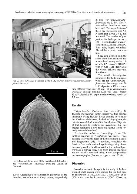
Fig. 2. The TOMCAT Beamline at the SLS, source: http://www.panoramio.com/ were: (i) for “Rhynchonella”<br />
photo/10865922.<br />
flustracea the energy was 20<br />
keV; objective - 4X; exposure<br />
time 300 ms; voxel size 1,85 µm; (ii) for Terebratulina<br />
imbricata on-chip binning (2X) was used; energy<br />
33 keV; objective 4X; exposure time 400 ms; voxel size<br />
3,7 µm.<br />
Results<br />
“Rhynchonella” flustracea SCHLOTHEIM (Fig. 3):<br />
The infilling sediment in this species was fine-grained<br />
limestone. Using SRXTM it was possible to visualise<br />
the 3D shape of the crura, the lack of hinge plates, the<br />
orientation and thickness of the dental plates (Figs. 4a,<br />
b) that helped to confirm our hypothesis that this<br />
species belongs to a new basiliolide genus (to be formally<br />
erected elsewhere).<br />
Terebratulina imbricata OWEN (Figs. 5, 6): The<br />
infilling sediment in T. imbricata was marl. It was<br />
possible to reveal the form of the brachidium in cross<br />
and longitudinal (Fig. 7) sections, especially the<br />
details of the tetebratulide loop forming a ring. Some<br />
traces of growth of shell material in the umbonal part<br />
were also observed (Fig. 5.4). Such growth structures<br />
are not usually revealed using X-ray computerised<br />
microtomography (PAKHNEVICH, pers. com. 2010).<br />
Fig. 3. External dorsal view of the rhynchonellide brachiopod<br />
“Rhynchonella” flustracea from the Danian of<br />
Denmark.<br />
2006). According to the absorption properties of the<br />
samples, monochromatic X-ray beams, respectively<br />
Discussion<br />
Non destructive techniques for the study of the brachiopod<br />
shell interior were applied for the first time<br />
by HAGADORN & NEALSON (2001), HAGADORN et al.<br />
(2001) and later by PAKHNEVICH (2007; 2010a, b),