KÑига LXXI - Univerzitet u Beogradu
KÑига LXXI - Univerzitet u Beogradu
KÑига LXXI - Univerzitet u Beogradu

Create successful ePaper yourself
Turn your PDF publications into a flip-book with our unique Google optimized e-Paper software.
Synchrotron radiation X-ray tomographic microscopy (SRXTM) of brachiopod shell interiors for taxonomy: ... 113<br />
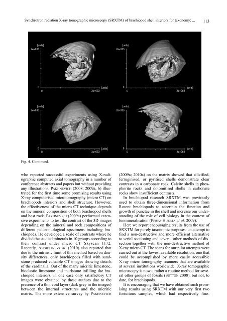
Fig. 4. Continued.<br />
who reported successful experiments using X-radiographic<br />
computed axial tomography in a number of<br />
conference abstracts and papers but without providing<br />
any illustrations. PAKHNEVICH (2008, 2009a, b) illustrated<br />
for the first time some promising results using<br />
X-ray computerised microtomography (micro CT) on<br />
brachiopods interiors and shell structure. However,<br />
the effectiveness of the micro CT technique depends<br />
on the mineral composition of both brachiopod shells<br />
and host rock. PAKHNEVICH (2009a) performed extensive<br />
experiments to test the contrast of the 3D images<br />
depending on the mineral and rock compositions of<br />
different palaeontological specimens including brachiopods.<br />
He developed a scale of contrasts where he<br />
divided the studied minerals in 10 groups according to<br />
their contrast under micro CT Skyscan 1172.<br />
Recently, ANGIOLINI et al. (2010) also reported that<br />
due to the intrinsic limit of this method based on density<br />
differences, only brachiopods filled with sandstone<br />
produced valuable CT images showing details<br />
of the cardinalia. Out of the many micritic limestone,<br />
bioclastic limestone and marlstone infilling the brachiopod<br />
interiors, in one case only satisfactory CT<br />
images were obtained by these authors due to the<br />
presence of a thin void layer (dark grey in the images)<br />
between the internal structures and the micritic<br />
matrix. The more extensive survey by PAKHNEVICH<br />
(2009a; 2010a) on the matrix showed that silicified,<br />
ferruginised, or pyritised shells demonstrate clear<br />
contrasts in a carbonate rock. Calcite shells in phosphorite<br />
rocks and dolomitised shells in carbonate<br />
rocks show insufficient contrasts.<br />
In brachiopod research SRXTM was previously<br />
used to obtain three-dimensional information from<br />
Recent brachiopods to ascertain the function and<br />
growth of punctae in the shell and increase our understanding<br />
of the role of cell biology in the context of<br />
biomineralisation (PÉREZ-HUERTA et al. 2009).<br />
Here we report encouraging results from the use of<br />
SRXTM for purely taxonomic purposes: an attempt to<br />
find a non-destructive and more efficient alternative<br />
to serial sectioning and several other methods of dissection<br />
together with the non-destructive method of<br />
X-ray micro CT. The scans for our pilot attempts were<br />
carried out at the lowest available resolution, one that<br />
could be accomplished by more easily accessible<br />
X-ray micro-tomography scanners that are available<br />
at several institutions worldwide. X-ray tomographic<br />
microscopy is now a rather a routine method for several<br />
other groups of fossils (SUTTON 2008), but not, to<br />
date, for brachiopods.<br />
It is encouraging that we have obtained such promising<br />
results using SRXTM with our very first two<br />
fortuitous samples, which had respectively fine-