Three-dimensional sonographic calculation of the volume of ...
Three-dimensional sonographic calculation of the volume of ...
Three-dimensional sonographic calculation of the volume of ...

You also want an ePaper? Increase the reach of your titles
YUMPU automatically turns print PDFs into web optimized ePapers that Google loves.
Brain <strong>volume</strong> in growth-restricted and appropriate-for-gestational age fetuses 533<br />
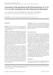
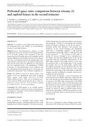
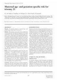
Figure 2 Images obtained with a rotation step <strong>of</strong> 15 ◦ using Virtual Organ Computer-aided AnaLysis (VOCAL) to delineate <strong>the</strong> thalamus for<br />
<strong>volume</strong> <strong>calculation</strong> and reconstruction.<br />
15°<br />
0°<br />
30°<br />
45°<br />
60°<br />
75°<br />
90° 105°<br />
120°<br />
135°<br />
150°<br />
165°<br />
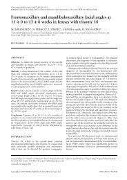
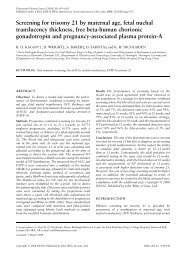
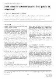
Figure 3 Diagram showing 15 ◦ rotation steps for measurement <strong>of</strong><br />
<strong>the</strong> <strong>volume</strong> <strong>of</strong> <strong>the</strong> frontal lobe starting from <strong>the</strong> axial plane. Note<br />
that <strong>the</strong> starting image at 0 ◦ and <strong>the</strong> final one at 180 ◦ are mirror<br />
images, and so <strong>the</strong> last image (dashed line) is not included in <strong>the</strong><br />
<strong>volume</strong> <strong>calculation</strong>.<br />
180°<br />
index in <strong>the</strong> ductus venosus but present atrial flow.<br />
Both <strong>of</strong> <strong>the</strong>se fetuses developed signs <strong>of</strong> intraventricular<br />
hemorrhage.<br />
Substantial to almost perfect intraobserver reliability<br />
was observed for all regions. Total intracranial, frontal<br />
and cerebellar regions showed similar figures for<br />
interobserver reliability. The only structure showing<br />
moderate interobserver measurement reliability was <strong>the</strong><br />
thalamus (Table 2).<br />
The BPD and HC were significantly smaller in <strong>the</strong> IUGR<br />
group than in AGA fetuses (Table 3). No statistically<br />
significant differences were found in <strong>the</strong> HC/BPD ratio<br />
between <strong>the</strong> two groups. Differences in <strong>the</strong> net <strong>volume</strong>s <strong>of</strong><br />
<strong>the</strong> studied structures are shown in Table 3. All <strong>volume</strong><br />
estimations, except those for <strong>the</strong> thalamic area, were<br />
significantly reduced in <strong>the</strong> IUGR group.<br />
Table 3 also illustrates <strong>the</strong> ratios between <strong>the</strong> different<br />
regions. In IUGR fetuses <strong>the</strong> frontal <strong>volume</strong> was reduced,<br />
and <strong>the</strong> thalamic <strong>volume</strong> was increased, in relation<br />
to <strong>the</strong> total intracranial <strong>volume</strong>. However, statistically<br />
significant differences were found only in ratios including<br />
<strong>the</strong> frontal <strong>volume</strong>.<br />
After adjustment for BPD (Table 4), <strong>the</strong> thalamic<br />
<strong>volume</strong> was found to be significantly larger, and <strong>the</strong><br />
frontal <strong>volume</strong> significantly smaller, in IUGR fetuses,<br />
whereas total intracranial and cerebellar <strong>volume</strong>s did not<br />
differ from those in AGA fetuses.<br />
DISCUSSION<br />
The results <strong>of</strong> this study suggest that fetuses with severe<br />
intrauterine growth restriction have reduced frontal and<br />
increased thalamic <strong>volume</strong>s in relation to <strong>the</strong> total<br />
intracranial <strong>volume</strong>. These differences persisted when <strong>the</strong><br />
<strong>volume</strong>s were adjusted by <strong>the</strong> BPD <strong>of</strong> each fetus. The<br />
Copyright © 2009 ISUOG. Published by John Wiley & Sons, Ltd. Ultrasound Obstet Gynecol 2009; 33: 530–537.