Screening for trisomy 21 by maternal age, fetal nuchal translucency ...
Screening for trisomy 21 by maternal age, fetal nuchal translucency ...
Screening for trisomy 21 by maternal age, fetal nuchal translucency ...

Create successful ePaper yourself
Turn your PDF publications into a flip-book with our unique Google optimized e-Paper software.
Ultrasound Obstet Gynecol 2008; 31: 618–624Published online 6 May 2008 in Wiley InterScience (www.interscience.wiley.com). DOI: 10.1002/uog.5331<strong>Screening</strong> <strong>for</strong> <strong>trisomy</strong> <strong>21</strong> <strong>by</strong> <strong>maternal</strong> <strong>age</strong>, <strong>fetal</strong> <strong>nuchal</strong><strong>translucency</strong> thickness, free beta-human chorionicgonadotropin and pregnancy-associated plasma protein-AK. O. KAGAN*†, D. WRIGHT‡, A. BAKER‡, D. SAHOTA§ and K. H. NICOLAIDES**Harris Birthright Research Centre <strong>for</strong> Fetal Medicine, King’s College Hospital, London and ‡Department of Mathematics and Statistics,University of Plymouth, Plymouth, UK, †Department of Obstetrics and Gynecology, University of Tuebingen, Tuebingen, Germany and§Department of Obstetrics and Gynecology, The Chinese University of Hong Kong, Prince of Wales Hospital, Hong Kong, ChinaKEYWORDS: first-trimester screening; free β-hCG; <strong>nuchal</strong> <strong>translucency</strong>; PAPP-A; <strong>trisomy</strong> <strong>21</strong>ABSTRACTObjectives To derive a model and examine the per<strong>for</strong>manceof first-trimester combined screening <strong>by</strong> <strong>maternal</strong><strong>age</strong>, <strong>fetal</strong> <strong>nuchal</strong> <strong>translucency</strong> (NT) thickness and<strong>maternal</strong> serum free beta-human chorionic gonadotropin(β-hCG) and pregnancy-associated plasma protein-A(PAPP-A).Methods Prospective combined screening <strong>for</strong> <strong>trisomy</strong> <strong>21</strong>was carried out at 11 + 0 to 13+ 6 weeks in 56 771singleton pregnancies, including 56 376 cases with anormal karyotype or delivery of a phenotypically normalba<strong>by</strong> (unaffected group) and 395 cases with <strong>trisomy</strong><strong>21</strong>. The blood test and ultrasound scan were carriedout in the same visit. In each case the <strong>maternal</strong> <strong>age</strong>relatedrisk <strong>for</strong> <strong>trisomy</strong> <strong>21</strong> at term was calculated andadjusted according to the gestational <strong>age</strong> at the time ofscreening to derive the a-priori risk. The measured NTwas trans<strong>for</strong>med into a likelihood ratio using the mixturemodel of NT distributions. The measured free β-hCG andPAPP-A were converted into a multiple of the median(MoM) <strong>for</strong> gestational <strong>age</strong>, adjusted <strong>for</strong> <strong>maternal</strong> weight,ethnicity, smoking status, method of conception andparity, and a likelihood ratio was subsequently calculated.The likelihood ratios <strong>for</strong> NT and <strong>for</strong> the biochemicalmarkers were multiplied <strong>by</strong> the a-priori risk to derivethe patient-specific risk. Detection rates and false-positiverates were calculated <strong>by</strong> taking the proportions withrisks above a given risk threshold after adjustment <strong>for</strong><strong>maternal</strong> <strong>age</strong> according to the distribution of pregnanciesin England and Wales in 2000–2002. These standardizedrates were compared with detection and false-positiverates estimated using Monte Carlo methods to samplefrom the modeled Gaussian distributions.Results The per<strong>for</strong>mance of screening based on themodel was in good agreement with that observed inour population. In a strategy <strong>for</strong> first-trimester combinedscreening where the blood test and scan are carried out inthe same visit it was estimated that, <strong>for</strong> false-positive ratesof 3% and 5%, the detection rates were 92% and 94%,respectively, at 11 weeks, 85% and 90% at 12 weeks, and79% and 83% at 13 weeks. In an alternative strategy,with the blood taken at 10 weeks and the measurement ofNT per<strong>for</strong>med at 12 weeks, the estimated detection rateswere 94% and 96% <strong>for</strong> false-positive rates of 3% and5%, respectively.Conclusions The aim of the first-trimester scan is not justto screen <strong>for</strong> <strong>trisomy</strong> <strong>21</strong> but also to diagnose an increasingnumber of <strong>fetal</strong> mal<strong>for</strong>mations. In this respect the abilityto visualize <strong>fetal</strong> anatomy is better at 12–13 weeksthan at 11 weeks. Consequently, the ideal gestation <strong>for</strong>combined testing in the same visit would be 12 weeks.An alternative strategy, with the blood taken at 10 weeksand the measurement of NT per<strong>for</strong>med at 12 weeks,is associated with higher detection rates of <strong>trisomy</strong> <strong>21</strong>.However, the cost of two-st<strong>age</strong> screening would be higherand, in addition, the potential advant<strong>age</strong> in terms ofdetection rate may be eroded <strong>by</strong> the likely increased noncompliancewith the additional step. Copyright © 2008ISUOG. Published <strong>by</strong> John Wiley & Sons, Ltd.INTRODUCTIONEffective screening <strong>for</strong> <strong>trisomy</strong> <strong>21</strong> is provided <strong>by</strong>assessment of a combination of <strong>maternal</strong> <strong>age</strong>, <strong>fetal</strong><strong>nuchal</strong> <strong>translucency</strong> (NT) thickness, and <strong>maternal</strong> serumCorrespondence to: Prof. K. H. Nicolaides, Harris Birthright Research Centre <strong>for</strong> Fetal Medicine, King’s College Hospital, Denmark Hill,London SE5 8RX, UK (e-mail: fmf@<strong>fetal</strong>medicine.com)Accepted: 3 March 2008Copyright © 2008 ISUOG. Published <strong>by</strong> John Wiley & Sons, Ltd.ORIGINAL PAPER
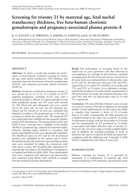
Combined screening 619free beta-human chorionic gonadotropin (β-hCG) andpregnancy-associated plasma protein-A (PAPP-A) at 11to 13 + 6 weeks of gestation 1,2 . Prospective studieshave demonstrated that, <strong>for</strong> a false-positive rate of 5%,combined screening can identify about 90% of affectedfetuses 3,4 . In the assessment of patient-specific risks,the a-priori <strong>maternal</strong> <strong>age</strong>-related risk is multiplied <strong>by</strong>likelihood ratios, determined from the deviation of themeasured NT, free β-hCG and PAPP-A from the respectiveexpected median.The traditional approaches to quantifying the deviationin the measured NT from the normal median <strong>for</strong>crown–rump length (CRL) have been either <strong>by</strong> subtraction(delta NT method) or <strong>by</strong> division (multiples of themedian (MoM) method). However, we have recently proposeda new approach based on the observation thatin both <strong>trisomy</strong> <strong>21</strong> and unaffected pregnancies <strong>fetal</strong> NTfollows two distributions, one which is CRL dependentand another which is CRL independent 5 . In this mixturemodel the proportions of <strong>trisomy</strong> <strong>21</strong> and unaffectedfetuses that follow the CRL-independent distribution are95% and 5%, respectively. The detection rate of a screeningpolicy based on <strong>maternal</strong> <strong>age</strong> and the mixture modelof <strong>fetal</strong> NT was 80% <strong>for</strong> a 5% false-positive rate 5 .In biochemical testing it is necessary to make adjustmentsin the measured <strong>maternal</strong> serum concentration offree β-hCG and PAPP-A to correct <strong>for</strong> certain <strong>maternal</strong>and pregnancy characteristics. Essentially, each measuredlevel is first converted to a multiple of the expected normalmedian (MoM) specific to a pregnancy of the samegestational <strong>age</strong>, <strong>maternal</strong> weight, ethnicity, smoking status,method of conception and parity, as well as themachine and re<strong>age</strong>nts used <strong>for</strong> the assays. In a recentstudy of 491 pregnancies with <strong>trisomy</strong> <strong>21</strong> and 96 803unaffected pregnancies we used multiple regression analysisto define the contribution of <strong>maternal</strong> variables thatinfluence the measured concentration of free β-hCG andPAPP-A, and the interaction between these covariates 6 .The traditional method of sequential adjustment <strong>for</strong> eachindividual parameter fails to take into account the interactionbetween the covariates 7 . The detection rate of ascreening policy based on <strong>maternal</strong> <strong>age</strong> and the two biochemicalmarkers was 65% <strong>for</strong> a 5% false-positive rate 6 .In this study of more than 56 000 normal pregnanciesand 395 cases with <strong>trisomy</strong> <strong>21</strong> we used the mixturemodel <strong>for</strong> <strong>fetal</strong> NT 5 and the new estimates <strong>for</strong> free β-hCGand PAPP-A based on the multiple regression approach 6to examine the per<strong>for</strong>mance of first-trimester combinedscreening <strong>by</strong> <strong>maternal</strong> <strong>age</strong>, <strong>fetal</strong> NT, and <strong>maternal</strong> serumfree β-hCG and PAPP-A.METHODSAt the Fetal Medicine Centre, London, screening <strong>for</strong><strong>trisomy</strong> <strong>21</strong> is carried out <strong>by</strong> a combination of <strong>maternal</strong><strong>age</strong>, <strong>fetal</strong> NT thickness, and <strong>maternal</strong> serum free β-hCGand PAPP-A in a one-stop-clinic <strong>for</strong> first-trimesterassessment of risk at 11 + 0to13+ 6 weeks of gestation 3 .Transabdominal ultrasound examination is per<strong>for</strong>med todiagnose any major <strong>fetal</strong> defects, and <strong>for</strong> measurementof CRL and <strong>fetal</strong> NT thickness 1 . The Kryptor system(Brahms AG, Berlin, Germany) is used to measure PAPP-Aandfreeβ-hCG. Maternal demographic characteristics,ultrasonographic measurements and biochemical resultsare recorded in a computer database. Karyotype resultsand details of pregnancy outcomes are added to thedatabase as soon as they become available. A search of thedatabase was done to identify all singleton pregnancies inwhich first-trimester combined screening was carried outfrom July 1999 to July 2007.Statistical analysisThe following steps were taken. First, the <strong>maternal</strong> <strong>age</strong>relatedrisk <strong>for</strong> <strong>trisomy</strong> <strong>21</strong> at term was calculated andadjusted according to the gestational <strong>age</strong> at the time ofscreening 8,9 . Second, the measured NT was trans<strong>for</strong>medinto a likelihood ratio using the mixture model of NTdistributions 5 . Third, the measured free β-hCG and PAPP-A were converted into a MoM <strong>for</strong> gestational <strong>age</strong>, adjusted<strong>for</strong> <strong>maternal</strong> weight, ethnicity, smoking status, method ofconception and parity, and subsequently a likelihood ratiowas calculated from the fitted bivariate Gaussian distributionsin <strong>trisomy</strong> <strong>21</strong> and unaffected pregnancies. Fourth,the likelihood ratios <strong>for</strong> NT and <strong>for</strong> the biochemical markerswere multiplied <strong>by</strong> the <strong>age</strong>-related risk at the time ofscreening in each case. Fifth, detection rates and falsepositiverates were calculated <strong>by</strong> taking the proportionswith risks above a given risk threshold after adjustment<strong>for</strong> <strong>maternal</strong> <strong>age</strong>, according to the distribution of pregnanciesin England and Wales in 2000–2002 10 .Thesestandardized rates were compared with detection andfalse-positive rates estimated using Monte Carlo methodsto sample from the modeled Gaussian distributions 5,6 .RESULTSThe search of the database identified 60 172 singletonpregnancies. In 3053 (5.1%) cases the outcome or one ofthe covariates were not available and in 348 (0.6%) casesthere was a chromosomal abnormality other than <strong>trisomy</strong><strong>21</strong>. Thus, our study population consisted of 56 376pregnancies with a normal karyotype or delivery of a phenotypicallynormal ba<strong>by</strong> (unaffected group) and 395 caseswith <strong>trisomy</strong> <strong>21</strong>. The characteristics of the study populationare summarized in Table 1. According to the <strong>maternal</strong><strong>age</strong> distribution of our population and the gestational <strong>age</strong>at the time of screening we would have expected 367 (95%prediction interval, 329–405) cases with <strong>trisomy</strong> <strong>21</strong> 9 .The distribution of NT in <strong>trisomy</strong> <strong>21</strong> fetuses is shownin Figure 1. In about 5% of cases the NT followed theCRL-dependent distribution of unaffected pregnancies,whereas in 95% of cases the NT did not change withgestation and in this group the mean (SD) NT was 3.4(1.6) mm. Contour plots <strong>for</strong> free β-hCG and PAPP-Ain <strong>trisomy</strong> <strong>21</strong> and unaffected pregnancies are shown inFigure 2. In the unaffected pregnancies the median freeβ-hCG was 1.0 (range, 0.03–30.4) MoM and the medianCopyright © 2008 ISUOG. Published <strong>by</strong> John Wiley & Sons, Ltd. Ultrasound Obstet Gynecol 2008; 31: 618–624.
620 Kagan et al.Table 1 Characteristics of the study population of 56 771 womenParameterMedian (range)or n (%)Maternal characteristicsAge (years) 35.4 (14.1–52.5)Weight (kg) 63.6 (34.0–165.0)Spontaneous conception 54 135 (95.4)Smoker 2566 (4.5)EthnicityCaucasian 50 708 (89.3)Afro-Caribbean 2430 (4.3)East Asian 640 (1.1)South Asian 2<strong>21</strong>8 (3.9)Mixed 775 (1.4)Gestational <strong>age</strong>11 + 0to11+ 6 weeks 5546 (9.8)12 + 0to12+ 6 weeks 31 883 (56.2)13 + 0to13+ 6 weeks 19 342 (34.1)Crown–rump length (mm) 62.9 (45.0–84.0)KaryotypeNormal 56 376 (99.3)Trisomy <strong>21</strong> 395 (0.7)Nuchal <strong>translucency</strong> thickness (mm)15.08.04.02.01.00.545 50 55 60 65 70 75 80 85Crown−rump length (mm)Figure 1 Distribution of <strong>nuchal</strong> <strong>translucency</strong> thickness with respectto crown–rump length (CRL) in <strong>trisomy</strong> <strong>21</strong> fetuses. In this mixturemodel the <strong>nuchal</strong> <strong>translucency</strong> thickness in 5% of cases follows theCRL-dependent distribution (dark shaded area with median and 5 thand 95 th centiles) and in 95% of cases <strong>nuchal</strong> <strong>translucency</strong> does notchange with gestation and the mean is 3.4 mm (light shaded bells).PAPP-A was 1.0 (range, 0.02–7.9) MoM. In the <strong>trisomy</strong><strong>21</strong> pregnancies the median free β-hCG was 2.0 (range,0.1–11.3) MoM and the median PAPP-A was 0.5 (range,0.05–2.2) MoM. In the <strong>trisomy</strong> <strong>21</strong> pregnancies there wasa significant increase in log MoM PAPP-A (P < 0.0001)and log MoM free β-hCG (P = 0.039) with gestation(Figure 3).Per<strong>for</strong>mance of screening <strong>for</strong> <strong>trisomy</strong> <strong>21</strong> <strong>by</strong> <strong>maternal</strong><strong>age</strong> alone, <strong>maternal</strong> <strong>age</strong> and serum biochemistry, <strong>maternal</strong><strong>age</strong> and <strong>fetal</strong> NT, and combined screening is comparedin Figure 4. For a 5% false-positive rate the respectivedetection rates were 30%, 65%, 80% and 91%. Theper<strong>for</strong>mance of screening <strong>for</strong> different risk cut-offs variedwith <strong>maternal</strong> <strong>age</strong> (Figure 5). For example, at a risk cut-offFree β-hCG MoM10.04.02.01.00.50.250.10.050.05 0.1 0.25 0.5 1 2 4PAPP-A MoMFigure 2 Distribution of multiples of the median (MoM) values <strong>for</strong>free beta-human chorionic gonadotropin (β-hCG) andpregnancy-associated plasma protein-A (PAPP-A) inchromosomally normal pregnancies (90% contour, unshaded) andin <strong>trisomy</strong> <strong>21</strong> pregnancies (90% contour, shaded). Raw data <strong>for</strong>our sample of 395 cases of <strong>trisomy</strong> <strong>21</strong> are plotted (♦).of 1 in 100, the detection rates <strong>for</strong> a 20-, 30- and 40-yearoldwoman were 72%, 77% and 92%, respectively, <strong>for</strong> afalse-positive rate of 1.1%, 1.9% and 9.9%, respectively.In combined screening the detection rates <strong>for</strong> fixedfalse-positive rates observed in our population, afteradjustment <strong>for</strong> <strong>maternal</strong> <strong>age</strong> according to the distributionof pregnancies in England and Wales in 2000–2002,were compared with the values derived from the modeledGaussian distributions (Table 2). These values indicatethat the per<strong>for</strong>mance of screening based on the model isin good agreement with that observed in our population.This is further highlighted <strong>by</strong> the data presented inTable 3, where <strong>for</strong> each range of estimated risks accordingto the model there is good agreement with the number ofobserved cases of <strong>trisomy</strong> <strong>21</strong>. Table 4 shows the modeleddetection and false-positive rates <strong>for</strong> different risk cut-offs.The risk was 1 in 200 or higher in 89% of <strong>trisomy</strong> <strong>21</strong>and in 4.6% of the unaffected pregnancies. Table 5 showsthe estimated detection rates of <strong>trisomy</strong> <strong>21</strong> with a policyof biochemical testing and ultrasound scanning carriedout at two separate visits, with the first done at 10 or11 weeks and the second at 12–13 weeks.DISCUSSIONThis study shows that screening <strong>by</strong> <strong>maternal</strong> <strong>age</strong>, <strong>fetal</strong>NT, free β-hCG and PAPP-A identifies about 90% of allpregnancies with <strong>trisomy</strong> <strong>21</strong> <strong>for</strong> a false-positive rate of5%, and that screening at 11 weeks of gestation per<strong>for</strong>mssubstantially better than at 13 weeks. It also demonstratesthat there is good agreement between the estimated risksderived from the model and the results in our studypopulation.In the calculation of patient-specific risk <strong>for</strong> <strong>trisomy</strong><strong>21</strong> the <strong>maternal</strong> <strong>age</strong>-related a-priori risk is multiplied<strong>by</strong> the likelihood ratios <strong>for</strong> the <strong>fetal</strong> NT and <strong>maternal</strong>serum biochemical markers. Consequently, <strong>for</strong> the samemeasurements of NT, free β-hCG and PAPP-A the finalCopyright © 2008 ISUOG. Published <strong>by</strong> John Wiley & Sons, Ltd. Ultrasound Obstet Gynecol 2008; 31: 618–624.
Combined screening 6<strong>21</strong>(a)1.5(b)0.51.00.50.0Log MoM free β-hCG0.0Log MoM PAPP-A−0.5−0.5−1.0−1.0−1.575808590Gestational <strong>age</strong> (days)95100−1.575808590Gestational <strong>age</strong> (days)95100Figure 3 Individual values and regression lines ( ) of log multiples of the median (MoM) free beta-human chorionic gonadotropin(β-hCG) (a) and pregnancy-associated plasma protein-A (PAPP-A) (b) in <strong>trisomy</strong> <strong>21</strong> pregnancies with respect to gestational <strong>age</strong>. The dashedline is the regression line in normal pregnancies.Table 2 Detection rates of <strong>trisomy</strong> <strong>21</strong> <strong>for</strong> given false-positive rates observed in our population, after adjustment <strong>for</strong> <strong>maternal</strong> <strong>age</strong> accordingto the distribution of pregnancies in England and Wales in 2000–2002, compared with values derived from the modeled GaussiandistributionsFalse-positiveDetection rate (%)Overall (n = 395) 11 weeks (n = 28) 12 weeks (n = 223) 13 + weeks (n = 144)rate (%) Observed Modeled Observed Modeled Observed Modeled Observed Modeled1 78 75 78 83 84 76 77 692 84 81 81 88 88 83 84 753 89 84 90 92 91 85 86 794 90 86 95 93 91 88 89 815 91 89 96 94 92 90 90 83risk will vary with <strong>maternal</strong> <strong>age</strong>, and <strong>for</strong> the samerisk cut-off both the detection and false-positive ratesincrease with <strong>maternal</strong> <strong>age</strong> (Figure 5). This in<strong>for</strong>mation,which is often requested <strong>by</strong> patients, may be useful incounseling. Furthermore, it is essential in comparingoverall detection and false-positive rates between centersor against a national standard because the overallper<strong>for</strong>mance depends on the <strong>maternal</strong> <strong>age</strong> distributionof the screened population.The National <strong>Screening</strong> Committee in the UK recommendedthat a screening test <strong>for</strong> <strong>trisomy</strong> <strong>21</strong> shouldprovide an overall detection rate of at least 75% <strong>for</strong> afalse-positive rate of 3% or less 11 . In our study both themodeled and observed detection rate of 75% was achievedat a false-positive rate of 1%. At a false-positive rate of3% the observed and modeled detection rates were 89%and 84%, respectively. These detection rates are higherthan the respective 53% and 60% achieved <strong>by</strong> the tripleCopyright © 2008 ISUOG. Published <strong>by</strong> John Wiley & Sons, Ltd. Ultrasound Obstet Gynecol 2008; 31: 618–624.
622 Kagan et al.and quadruple second-trimester biochemical tests 12 .Furthermore,the per<strong>for</strong>mance of our combined model issubstantially superior to that in two previous multicenterstudies in the UK and USA 13,14 . The Serum, Urine andUltrasound <strong>Screening</strong> Study (SURUSS) recruited 47 053pregnancies, including 101 with <strong>trisomy</strong> <strong>21</strong>, but NT wasmeasured in only 39 983 cases, including 85 with <strong>trisomy</strong><strong>21</strong>; free β-hCG and PAPP-A were measured retrospectivelyin stored samples from 98 <strong>trisomy</strong> <strong>21</strong> pregnanciesand 1090 matched unaffected controls 13 . The modeleddetection rates from combined screening were 68%, 79%and 84% <strong>for</strong> respective false-positive rates of 1%, 3% and5%. Similarly, in the First- and Second-Trimester Evaluationof Risk (FASTER) study, 38 167 pregnancies wererecruited, including 117 with <strong>trisomy</strong> <strong>21</strong>; free β-hCG andPAPP-A were measured retrospectively in stored samplesfrom 79 <strong>trisomy</strong> <strong>21</strong> pregnancies and 395 matched unaffectedcontrols 14 . The modeled detection rates at 12 weekswere 72% and 85% <strong>for</strong> respective false-positive rates of1% and 5%.The per<strong>for</strong>mance of combined screening <strong>for</strong> <strong>trisomy</strong> <strong>21</strong>decreased with gestation and, <strong>for</strong> a 3% false-positive rate,the estimated detection rate was 92%, 85% and 79% at11, 12 and 13 weeks, respectively. This is a consequenceof both <strong>fetal</strong> NT and serum PAPP-A. In the mixturemodel of NT the overlap between the CRL-dependent andCRL-independent distribution increases with gestation,and there<strong>for</strong>e the separation between <strong>trisomy</strong> <strong>21</strong> and10080unaffected pregnancies decreases. Similarly, the separationin <strong>maternal</strong> serum PAPP-A between <strong>trisomy</strong> <strong>21</strong> andunaffected pregnancies decreases with gestation. Althoughthere is an increase in the separation <strong>for</strong> free β-hCGwith gestation this is considerably less than the decreaseobserved <strong>for</strong> PAPP-A. The implications of these findingsare that, in terms of the per<strong>for</strong>mance of screening <strong>for</strong><strong>trisomy</strong> <strong>21</strong> in a set-up in which biochemical testing and theultrasound scan are carried out in the same visit, the bestgestational <strong>age</strong> is 11 weeks. However, the aim of the firsttrimesterscan is not just to screen <strong>for</strong> <strong>trisomy</strong> <strong>21</strong> but alsoto diagnose an increasing number of <strong>fetal</strong> mal<strong>for</strong>mations,and in this respect the ability to visualize <strong>fetal</strong> anatomy isbetter at 12–13 weeks than at 11 weeks 15 . Consequently,the ideal gestation <strong>for</strong> combined testing in the same visitwould be 12 weeks when, <strong>for</strong> false-positive rates of 3%and 5%, the estimated detection rates of <strong>trisomy</strong> <strong>21</strong> are85% and 90%, respectively.An alternative strategy <strong>for</strong> first-trimester combinedscreening is <strong>for</strong> biochemical testing and ultrasoundscanning to be carried out in two separate visits, with the(a) 100Detection rate (%)9590858075Detection rate (%)60402070(b) 50201053False-positive rate (%)15 20 25 30 35 40 45Maternal <strong>age</strong> (years)020 2 4 6 8 10False-positive rate (%)Figure 4 Receiver–operating characteristics curves <strong>for</strong> theper<strong>for</strong>mance of screening <strong>for</strong> <strong>trisomy</strong> <strong>21</strong> <strong>by</strong> <strong>maternal</strong> <strong>age</strong> alone( ); <strong>maternal</strong> <strong>age</strong>, serum free beta-human chorionicgonadotropin and pregnancy-associated plasma protein-A (- - - - );<strong>maternal</strong> <strong>age</strong> and <strong>fetal</strong> <strong>nuchal</strong> <strong>translucency</strong> thickness ( ......); andcombined screening ( ).115 20 25 30 35 40 45Maternal <strong>age</strong> (years)Figure 5 Per<strong>for</strong>mance of combined first-trimester screening <strong>for</strong><strong>trisomy</strong> <strong>21</strong>. Modeled detection rate (a) and false-positive rate (b)<strong>for</strong> risk cut-offs of 1 in 300 ( ),1in200(......),1in150(- - - - ) and 1 in 100 ( ) in relation to <strong>maternal</strong> <strong>age</strong>.Copyright © 2008 ISUOG. Published <strong>by</strong> John Wiley & Sons, Ltd. Ultrasound Obstet Gynecol 2008; 31: 618–624.
Combined screening 623Table 3 Accuracy of estimated risk of <strong>trisomy</strong> <strong>21</strong> <strong>by</strong> a combination of <strong>maternal</strong> <strong>age</strong>, <strong>fetal</strong> <strong>nuchal</strong> <strong>translucency</strong> thickness, and <strong>maternal</strong> serumfree beta-human chorionic gonadotropin and pregnancy-associated plasma protein-AEstimated risk of <strong>trisomy</strong> <strong>21</strong>Trisomy <strong>21</strong> Unaffected ObservedRange Median (n (%)) (n (%)) risk≥ 1 in 10 1 in 5 273 (69.1) 340 (0.6) 1 in <strong>21</strong> in 11 to 1 in 50 1 in 28 60 (15.2) 909 (1.6) 1 in 161in51to1in100 1in74 16(4.1) 947(1.7) 1in601 in 101 to 1 in 250 1 in 173 15 (3.8) 2305 (4.1) 1 in 1551 in 251 to 1 in 1000 1 in 579 19 (4.8) 7362 (13.1) 1 in 3881 in 1001 to 1 in 5000 1 in 2443 10 (2.5) 16 685 (29.6) 1 in 1 670< 1 in 5000 1 in 16 144 2 (0.5) 27 828 (49.4) 1 in 13 915Table 4 Modeled detection rates (DRs) and false-positive rates(FPRs) <strong>for</strong> given risk cut-offs <strong>for</strong> <strong>trisomy</strong> <strong>21</strong>Risk cut-offDR(%)Total 11 weeks 12 weeks 13 weeksFPR(%)DR(%)FPR(%)DR(%)FPR(%)DR(%)FPR(%)1 in 20 71 0.6 78 0.6 71 0.6 62 0.51 in 50 79 1.4 86 1.4 79 1.4 71 1.31 in 100 84 2.6 90 2.5 85 2.5 77 2.51 in 150 87 3.6 92 3.5 87 3.5 81 3.61 in 200 89 4.6 93 4.3 89 4.5 83 4.71 in 300 91 6.3 95 5.8 91 6.1 86 6.71 in 1000 95 15.9 98 13.3 96 15.2 93 17.9first done at 10–11 weeks and the second at 12–13 weeks.For false-positive rates of 3% and 5%, the estimateddetection rates of combined testing with the blood takenat 10 weeks and the measurement of NT per<strong>for</strong>med at12 weeks are 94% and 96%, respectively. The cost andpatient acceptability of the two alternative policies of firsttrimestertesting will depend on the existing infrastructureof antenatal care. The potential advant<strong>age</strong> of two-st<strong>age</strong>screening in terms of detection rate may be eroded <strong>by</strong> thelikely increased non-compliance with the additional step.Another element to be considered in terms of alternativepolicies of early screening is the contribution of additionalsonographic markers, such as absent nasal bone, widefrontomaxillary facial angle, reversed end-diastolic flowin the ductus venosus and tricuspid regurgitation, whichhave already been incorporated into routine practice insome specialist centers 3,16 –19 .ACKNOWLEDGMENTThis study was supported <strong>by</strong> a grant from The FetalMedicine Foundation (charity number 1037116).REFERENCES1. Snijders RJ, Noble P, Sebire N, Souka A, Nicolaides KH. UKmulticentre project on assessment of risk of <strong>trisomy</strong> <strong>21</strong><strong>by</strong> <strong>maternal</strong> <strong>age</strong> and <strong>fetal</strong> <strong>nuchal</strong>-<strong>translucency</strong> thickness at10–14 weeks of gestation. Lancet 1998; 352: 343–346.2. Spencer K, Souter V, Tul N, Snijders R, Nicolaides KH. Ascreening program <strong>for</strong> <strong>trisomy</strong> <strong>21</strong> at 10–14 weeks using <strong>fetal</strong><strong>nuchal</strong> <strong>translucency</strong>, <strong>maternal</strong> serum free beta-human chorionicgonadotropin and pregnancy-associated plasma protein-A.Ultrasound Obstet Gynecol 1999; 13: 231–237.3. Nicolaides KH, Spencer K, Avgidou K, Faiola S, Falcon O.Multicenter study of first-trimester screening <strong>for</strong> <strong>trisomy</strong> <strong>21</strong>in 75 8<strong>21</strong> pregnancies: results and estimation of the potentialimpact of individual risk-orientated two-st<strong>age</strong> first-trimesterscreening. Ultrasound Obstet Gynecol 2005; 25: 2<strong>21</strong>–226.4. Nicolaides KH. Nuchal <strong>translucency</strong> and other first-trimestersonographic markers of chromosomal abnormalities. Am JObstet Gynecol 2004; 191: 45–67.5. Wright D, Kagan KO, Molina FS, Gazzoni A, Nicolaides KH.A mixture model of <strong>nuchal</strong> <strong>translucency</strong> thickness in screening<strong>for</strong> chromosomal defects. Ultrasound Obstet Gynecol 2008; 31:376–383.6. Kagan KO, Wright D, Spencer K, Molina FS, Nicolaides KH.First-trimester screening <strong>for</strong> <strong>trisomy</strong> <strong>21</strong> <strong>by</strong> free beta-humanchorionic gonadotropin and pregnancy-associated plasmaTable 5 Estimated detection rates of <strong>trisomy</strong> <strong>21</strong> from different policies on screening according to the timing of biochemical testing andultrasound scanningFalse-positiveDetection rate (%)Scan and blood at same visit Scan 12 weeks Scan 13 weeksrate (%) 11 weeks 12 weeks 13 weeks Blood 10 weeks Blood 11 weeks Blood 10 weeks Blood 11 weeks1 83 76 69 88 81 85 782 88 83 75 92 87 90 833 92 85 79 94 89 92 864 93 88 81 95 91 94 885 94 90 83 96 92 95 9010 97 94 89 98 96 97 94Copyright © 2008 ISUOG. Published <strong>by</strong> John Wiley & Sons, Ltd. Ultrasound Obstet Gynecol 2008; 31: 618–624.
624 Kagan et al.protein-A: impact of <strong>maternal</strong> and pregnancy characteristics.Ultrasound Obstet Gynecol 2008; 31: 493–502.7. Draper N, Smith R. Applied Regression Analysis (3 rd edn).Wiley: New York, NY, 1998.8. Cuckle HS, Wald NJ, Thompson SG. Estimating a woman’s riskof having a pregnancy associated with Down’s syndrome usingher <strong>age</strong> and serum alpha-fetoprotein level. Br J Obstet Gynaecol1987; 94: 387–402.9. Snijders RJ, Sundberg K, Holzgreve W, Henry G, NicolaidesKH. Maternal <strong>age</strong>- and gestation-specific risk <strong>for</strong> <strong>trisomy</strong> <strong>21</strong>.Ultrasound Obstet Gynecol 1999; 13: 167–170.10. Office <strong>for</strong> National Statistics. Birth Statistics. Review of theRegistrar General on births and patterns of family building inEngland and Wales. Series FM1, number 29–31. StationeryOffice: London, 2000–2002.11. National <strong>Screening</strong> Committee. National <strong>Screening</strong> CommitteePolicy – Down’s Syndrome <strong>Screening</strong> (compiled July 2006).http://www.library.nhs.uk/screening/ViewResource.aspx?resID=35689 [Accessed 23 December 2007].12. Cuckle H, Benn P, Wright D. Down syndrome screening in thefirst and/or second trimester: model predicted per<strong>for</strong>manceusing meta-analysis parameters. Semin Perinatol 2005; 29:252–257.13. Wald NJ, Rodeck C, Hackshaw AK, Walters J, Chitty L, MackinsonAM; SURUSS Research Group. First and second trimesterantenatal screening <strong>for</strong> Down’s syndrome: the results of theSerum, Urine and Ultrasound <strong>Screening</strong> Study (SURUSS). HealthTechnol Assess 2003; 7: 1–77.14. Malone FD, Canick JA, Ball RH, Nyberg DA, Comstock CH,Bukowski R, Berkowitz RL, Gross SJ, Dugoff L, Craigo SD,Timor-Tritsch IE, Carr SR, Wolfe HM, Dukes K, Bianchi DW,Rudnicka AR, Hackshaw AK, Lambert-Messerlian G, Wald NJ,D’Alton ME; First- and Second-Trimester Evaluation of Risk(FASTER) Research Consortium. First-trimester or secondtrimesterscreening, or both, <strong>for</strong> Down’s syndrome. NEnglJMed2005; 353: 2001–2011.15. Souka AP, Pilalis A, Kavalakis Y, Kosmas Y, Antsaklis P,Antsaklis A. Assessment of <strong>fetal</strong> anatomy at the 11–14-weekultrasound examination. Ultrasound Obstet Gynecol 2004; 24:730–734.16. Cicero S, Curcio P, Pap<strong>age</strong>orghiou A, Sonek J, Nicolaides K.Absence of nasal bone in fetuses with <strong>trisomy</strong> <strong>21</strong> at 11–14 weeksof gestation: an observational study. Lancet 2001; 358:1665–1667.17. Sonek J, Borenstein M, Dagklis T, Persico N, Nicolaides KH.Frontomaxillary facial angle in fetuses with <strong>trisomy</strong> <strong>21</strong>at 11–13(6) weeks. Am J Obstet Gynecol 2007; 196:271.e1–271.e4.18. Matias A, Gomes C, Flack N, Montenegro N, Nicolaides KH.<strong>Screening</strong> <strong>for</strong> chromosomal abnormalities at 11–14 weeks: therole of ductus venosus blood flow. Ultrasound Obstet Gynecol1998; 2: 380–384.19. Faiola S, Tsoi E, Huggon IC, Allan LD, Nicolaides KH. Likelihoodratio <strong>for</strong> <strong>trisomy</strong> <strong>21</strong> in fetuses with tricuspid regurgitationat the 11 to 13 + 6-week scan. Ultrasound Obstet Gynecol2005; 26: 22–27.Copyright © 2008 ISUOG. Published <strong>by</strong> John Wiley & Sons, Ltd. Ultrasound Obstet Gynecol 2008; 31: 618–624.