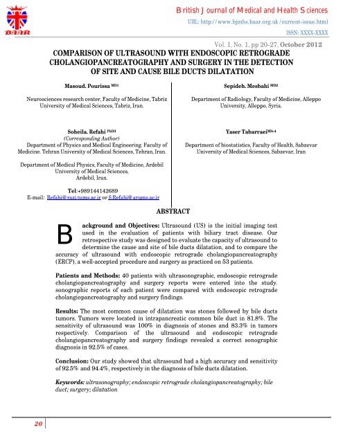
comparison of ultrasound with endoscopic retrograde ...
comparison of ultrasound with endoscopic retrograde ...
comparison of ultrasound with endoscopic retrograde ...

Create successful ePaper yourself
Turn your PDF publications into a flip-book with our unique Google optimized e-Paper software.
British Journal <strong>of</strong> Medical and Health Sciences<br />
URL: http://www.bjmhs.baar.org.uk /current-issue.html<br />
ISSN: XXXX-XXXX<br />
Vol. 1, No. 1, pp 20-27, October 2012<br />
COMPARISON OF ULTRASOUND WITH ENDOSCOPIC RETROGRADE<br />
CHOLANGIOPANCREATOGRAPHY AND SURGERY IN THE DETECTION<br />
OF SITE AND CAUSE BILE DUCTS DILATATION<br />
Masoud. Pourissa MD1<br />
Neurosciences research center, Faculty <strong>of</strong> Medicine, Tabriz<br />
University <strong>of</strong> Medical Sciences, Tabriz, Iran.<br />
Sepideh. Mesbahi MD2<br />
Department <strong>of</strong> Radiology, Faculty <strong>of</strong> Medicine, Alleppo<br />
University, Alleppo, Syria.<br />
Soheila. Refahi PhD3<br />
(Corresponding Author)<br />
Department <strong>of</strong> Physics and Medical Engineering. Faculty <strong>of</strong><br />
Medicine. Tehran University <strong>of</strong> Medical Sciences, Tehran, Iran.<br />
Yaser Tabarraei MSc4<br />
Department <strong>of</strong> biostatistics, Faculty <strong>of</strong> Health, Sabzevar<br />
University <strong>of</strong> Medical Sciences, Sabzevar, Iran<br />
Department <strong>of</strong> Medical Physics, Faculty <strong>of</strong> Medicine, Ardebil<br />
University <strong>of</strong> Medical Sciences,<br />
Ardebil, Iran.<br />
Tel:+989144142689<br />
E-mail: Refahi@razi.tums.ac.ir or S.Refahi@arums.ac.ir<br />
ABSTRACT<br />
B<br />
ackground and Objectives: Ultrasound (US) is the initial imaging test<br />
used in the evaluation <strong>of</strong> patients <strong>with</strong> biliary tract disease. Our<br />
retrospective study was designed to evaluate the capacity <strong>of</strong> <strong>ultrasound</strong> to<br />
determine the cause and site <strong>of</strong> bile ducts dilatation, and to compare the<br />
accuracy <strong>of</strong> <strong>ultrasound</strong> <strong>with</strong> <strong>endoscopic</strong> <strong>retrograde</strong> cholangiopancreatography<br />
(ERCP), a well-accepted procedure and surgery as practiced on 53 patients.<br />
Patients and Methods: 40 patients <strong>with</strong> ultrasonographic, <strong>endoscopic</strong> <strong>retrograde</strong><br />
cholangiopancreatography and surgery reports were entered into the study.<br />
sonographic reports <strong>of</strong> each patient were compared <strong>with</strong> <strong>endoscopic</strong> <strong>retrograde</strong><br />
cholangiopancreatography and surgery findings.<br />
Results: The most common cause <strong>of</strong> dilatation was stones followed by bile ducts<br />
tumors. Tumors were located in intrapancreatic common bile duct in 81.8%. The<br />
sensitivity <strong>of</strong> <strong>ultrasound</strong> was 100% in diagnosis <strong>of</strong> stones and 83.3% in tumors<br />
respectively. Comparison <strong>of</strong> the <strong>ultrasound</strong> and <strong>endoscopic</strong> <strong>retrograde</strong><br />
cholangiopancreatography and surgery findings revealed a correct sonographic<br />
diagnosis in 92.5% <strong>of</strong> cases.<br />
Conclusion: Our study showed that <strong>ultrasound</strong> had a high accuracy and sensitivity<br />
<strong>of</strong> 92.5% and 94.4%, respectively in the diagnosis <strong>of</strong> bile ducts dilatation.<br />
Keywords: ultrasonography; <strong>endoscopic</strong> <strong>retrograde</strong> cholangiopancreatography; bile<br />
duct; surgery; dilatation
Introduction<br />
British Journal <strong>of</strong> Medical and Health Sciences<br />
URL: http://www.bjmhs.baar.org.uk /current-issue.html<br />
ISSN: XXXX-XXXX<br />
Vol. 1, No. 1, pp 20-27, October 2012<br />
Ultrasound (US) is the initial imaging test used in the evaluation <strong>of</strong> patients <strong>with</strong> biliary tract disease such as<br />
bile duct stones (Varghese et al., 1999). Endoscopic <strong>retrograde</strong> cholangiopancreatography (ERCP) is a wellestablished<br />
method <strong>of</strong> diagnosing and treating disorders <strong>of</strong> the bile and pancreatic ducts (Loperfido et al., 1998). In<br />
the past ten years, there have been major developments in imaging <strong>of</strong> the pancreatic and bile ducts which give the<br />
same information as diagnostic cholangiography and pancreatography in all but a few cases. In the future, ERCP<br />
will be reserved for therapy (Sheridan, 2002). A major benefit <strong>of</strong> ERCP in the evaluation <strong>of</strong> choledocholithiasis is<br />
that ERCP provides a means <strong>of</strong> diagnosis and therapeutic intervention in the same setting (Ann and Fulche, 2002).<br />
This study was designed to evaluate the sites and causes <strong>of</strong> bile ducts dilatation. An additional aim <strong>of</strong> the present<br />
approach was to compare the accuracy <strong>of</strong> <strong>ultrasound</strong> <strong>with</strong> <strong>endoscopic</strong> <strong>retrograde</strong> cholangiopancreatography<br />
(ERCP), a well-accepted procedure and surgery.<br />
Patients and Methods<br />
From September 2005 through July 2007, in a retrospective study we reviewed reports <strong>of</strong> fifty-three patients<br />
who referred for <strong>ultrasound</strong> diagnosis <strong>of</strong> bile duct <strong>with</strong>in the Department <strong>of</strong> <strong>ultrasound</strong> at teaching Imam Hospital,<br />
Tabriz, Iran. The patients were fasted overnight and examined in the supine and right anterior oblique positions to<br />
optimally visualize the biliary ducts. Special maneuvers such as compression were routinely performed and probe<br />
was placed as axial and sagittal on the lower portion <strong>of</strong> pancreas to visualize the lower bile duct (Figuers 1, 2).<br />
.<br />
Fig1. A 41 years old female <strong>with</strong> history <strong>of</strong> pain at RUQ. and mild jaundice.Echogene lesion is noted in CBD <strong>with</strong> acoustic shaddow
British Journal <strong>of</strong> Medical and Health Sciences<br />
URL: http://www.bjmhs.baar.org.uk /current-issue.html<br />
ISSN: XXXX-XXXX<br />
Vol. 1, No. 1, pp 20-27, October 2012<br />
Fig 2. A 67 years old man <strong>with</strong> history <strong>of</strong> severe jaundice.Sagittal section is performed along common bile duct which shows a<br />
hypoechoi.<br />
All <strong>ultrasound</strong> examinations were performed by an experienced sonologist using a General Electronic Model X200<br />
<strong>ultrasound</strong> unit <strong>with</strong> 3.5 MHz curvilinear probe. Diameter <strong>of</strong> >7 mm were considered as dilated-caliber ducts. The<br />
ERCP examination was performed by two endoscopists and the taking <strong>of</strong> images supervised by one radiologist. For<br />
the purposes <strong>of</strong> this study the hard copy images <strong>of</strong> ERCP and <strong>ultrasound</strong> examinations and surgery findings <strong>of</strong> each<br />
patient were collected and the patient details masked. Patients who had not exact US and ERCP and surgery reports<br />
excluded from the study. The final diagnosis was based on ERCP and surgery. The extrahepatic biliary system was<br />
divided into three regions: the porta hepatic, the suprapancreatic common bile duct, and the intrapancreatic or<br />
ampullary portion <strong>of</strong> the common bile duct (Figure 3).<br />
Fig3. Schematic drawing <strong>of</strong> the three anatomic divisions <strong>of</strong> the extrahepatic common bile duct.
British Journal <strong>of</strong> Medical and Health Sciences<br />
URL: http://www.bjmhs.baar.org.uk /current-issue.html<br />
ISSN: XXXX-XXXX<br />
Vol. 1, No. 1, pp 20-27, October 2012<br />
When possible the site <strong>of</strong> obstruction was determined from the <strong>ultrasound</strong> study to be one <strong>of</strong> these three regions.<br />
Data were analyzed in SPSS version 11.5 s<strong>of</strong>tware for descriptive assessment and <strong>comparison</strong> <strong>of</strong> the US and ERCP<br />
and surgery diagnoses were performed manually by related formulas. The sites and causes <strong>of</strong> dilatation <strong>of</strong> bile<br />
ducts, accuracy, sensitivity, specificity, and positive and negative predictive values were determined for US<br />
assessment.<br />
Results<br />
Thirteen patients were excluded from this study for the following reason: ERCP hard- copy images and surgery<br />
findings were not available. 40 patients (F: M 25/15; mean age, 54.9±9.58 years; range 38-75 years) were entered<br />
into the study. Table 1 summarized the ERCP and surgery findings in patients.<br />
Table 1. Causes <strong>of</strong> dilatation based on ERCP and surgery (n=40)<br />
Cause No. <strong>of</strong> patients %<br />
Stone 20 50<br />
Tumor 12 30<br />
Cyst 2 5<br />
Pancreatitis 1 2.5<br />
Stricture 1 2.5<br />
No organic lesion 4 10<br />
Total 40 100<br />
The most common diagnosis made by ERCP and surgery was bile duct stone (BDS) followed by bile duct<br />
tumor. Findings at ERCP and surgery showed bile duct stone in 20 <strong>of</strong> 40 patients. In all these cases US was able to<br />
detect BDS by sensitivity <strong>of</strong> 100%.<br />
In four <strong>of</strong> patients was not a specific cause for bile duct dilatation found. According to ERCP and surgery<br />
reports, the rate <strong>of</strong> a bile duct tumor was 12 in 40 patients. Ten <strong>of</strong> bile duct tumors were correctly identified by<br />
<strong>ultrasound</strong> by sensitivity <strong>of</strong> 83/3%. The sensitivity <strong>of</strong> US for detection <strong>of</strong> cysts, pancreatitis and stricture was 100%.<br />
Comparison <strong>of</strong> results as assessed by ERCP and surgery <strong>with</strong> US illustrated in Table 2.
British Journal <strong>of</strong> Medical and Health Sciences<br />
URL: http://www.bjmhs.baar.org.uk /current-issue.html<br />
ISSN: XXXX-XXXX<br />
Vol. 1, No. 1, pp 20-27, October 2012<br />
Table 2. Comparison <strong>of</strong> ERCP and surgery findings <strong>with</strong> US reports<br />
ERCP Positive Negative Total<br />
Sonography<br />
Positive 34 1 35<br />
Negative 2 3 5<br />
Total 36 4 40<br />
Note. Sensitivity (TP/TP+FN) = 94.4%%. Specificity (TN/TN+FP) = 75%. Positive predictive value (TP/TP+FP) = 97%. Negative predictive value<br />
(TN/TN+FN) =60%. Accuracy (TP+TN)/ (P+N) =92.5%<br />
There were 34 true-positive and two false-negative reading by sonograms yielding a sensitivity <strong>of</strong> 94.4%,<br />
specificity <strong>of</strong> 75%, positive predictive (PPV) <strong>of</strong> 97%, and negative predictive value (NPV) <strong>of</strong> 60%. A correct US<br />
diagnosis was obtained in 37 <strong>of</strong> 40 patients yielding an accuracy <strong>of</strong> 92.5%.<br />
Table 3 demonstrates the results <strong>of</strong> the assessment <strong>of</strong> the 40 cases <strong>with</strong> respect to site <strong>of</strong> obstruction.<br />
Table 3. Site <strong>of</strong> obstruction<br />
Site<br />
Determination<br />
Correct<br />
Incorrect<br />
Intrapancreatic common duct 26 1<br />
Port hepatis 3 1<br />
Suprapancreatic common duct 1 1<br />
The common site <strong>of</strong> obstruction was in the intrapancreatic common duct followed by porta hepatis. Of the 40<br />
cases, we were unable to determine the site <strong>of</strong> obstruction in 7(17.5%). We determined the site <strong>of</strong> obstruction in 33<br />
(82.5%) cases and we were correct in 30 (90.9%) <strong>of</strong> these cases, or 75% <strong>of</strong> the total patient group. None <strong>of</strong> the three<br />
sites appeared to be more easily determined than the others.
Discussion<br />
British Journal <strong>of</strong> Medical and Health Sciences<br />
URL: http://www.bjmhs.baar.org.uk /current-issue.html<br />
ISSN: XXXX-XXXX<br />
Vol. 1, No. 1, pp 20-27, October 2012<br />
The goal <strong>of</strong> biliary tract imaging is tw<strong>of</strong>old: (1) to detect the presence <strong>of</strong> biliary duct dilatation and (2) to<br />
identify common duct stones (Joseph and Ferrucci, 2001). At present, US is <strong>of</strong>ten the first imaging modality used<br />
for evaluation <strong>of</strong> biliary disease (Khan et al., 2002). In the detection <strong>of</strong> bile duct dilatation, <strong>ultrasound</strong> reveals up to<br />
100% sensitivity for experienced examiners (Rosch et al., 2002; Bloom et al., 1999). The stone detection rate is also<br />
influenced by patient factors such as the number, size and site <strong>of</strong> stones, patient body habitus and the presence <strong>of</strong><br />
overlying bowel gas (Prat., et al 1996). The accurate diagnosis <strong>of</strong> choledocholithiasis is difficult and <strong>of</strong>ten relies on<br />
direct cholangiographic techniques such as ERCP. In clinical practice the non-invasive diagnosis <strong>of</strong><br />
choledocholithiasis is based on a combination <strong>of</strong> clinical suspicion, biochemical analysis and imaging findings<br />
(Rieger and Wayand, 1995; Jensen et al., 1985; Barkun et al., 1994; Abboud et al., 1996). Unfortunately, all <strong>of</strong><br />
these tests have varying diagnostic accuracies and there is no one reliable method <strong>of</strong> uniformly identifying patients<br />
<strong>with</strong> BDS (Guiband et al., 1995). US is less effective when choledochal stones are located far distally in the head <strong>of</strong><br />
the pancreas, when there is overlying bowel gas, or when obese patient habitus degrades image quality. Moreover,<br />
US remain a highly operator-dependent method, and the results are always influenced by the skill <strong>of</strong> the examiner<br />
(Joseph and Ferrucci, 2001). In our study sensitivity US for detection <strong>of</strong> BDS was 100%.Varghese et al in their<br />
study, showed that US detected BDS in 13 patients and missed stones in 21 patients, 6 <strong>of</strong> whom had non-dilated<br />
bile ducts. They reported sensitivity, specificity and diagnostic accuracy <strong>of</strong> 38%, 100% and 89%, in the US<br />
diagnosis <strong>of</strong> choledocholithiasis (Varghese et al., 1999). The results <strong>of</strong> previous studies indicate that sensitivity and<br />
specificity <strong>of</strong> US for detection <strong>of</strong> BDS is at 25-80%, 80-100%, respectively (Sheridan, 2002). In another study by<br />
David indicate sensitivity <strong>of</strong> US in the detection choledocholithiasis is rather poor, <strong>with</strong> only 22% <strong>of</strong> cases<br />
interpreted as positive (Einstein et al., 1984). Bile duct cyst (BDC) are rare and <strong>of</strong> unknown origin and may<br />
represent a precursor for biliary tract cancer development (Adkins et al., 2000; Söreide et al., 2004; Tashiro et al.,<br />
2003). BDC are <strong>of</strong>ten first suspected or diagnosed by the patobiliary imaging studies initiated for evaluation <strong>of</strong><br />
upper abdominal complications (Søreide and Søreide 2007). A BDC may be visualized by any <strong>of</strong> the modalities<br />
available such as US or by direct (invasive) ductal imaging using ERCP (Levy and Rohrmann, 2003). In our<br />
approach, there were two cases <strong>with</strong> BDC which detect by US <strong>with</strong> sensitivity <strong>of</strong> 100%. The most common biliary<br />
tumor is cholangiocarcinoma, an adenocarcinoma arising from the ductal epithelium <strong>of</strong> the biliary system (Zech et<br />
al., 2004). In this study the value <strong>of</strong> US in demonstrating the tumor itself was compared <strong>with</strong> ERCP and surgery in<br />
patients <strong>with</strong> biliary duct tumor. The sensitivity <strong>of</strong> US in the diagnosis <strong>of</strong> bile ducts tumor was 83.3%. In Horing's<br />
et al study correct visualization by US <strong>of</strong> the tumor itself was possible in 68% <strong>of</strong> proximal tumors and 36% <strong>with</strong><br />
distal tumors (Horing et al., 1993). In another study by Rigants et al US correctly defined the cause <strong>of</strong> obstruction<br />
in 90% <strong>of</strong> the patients <strong>with</strong> tumoral bile duct obstruction (Rigauts et al., 1992). The results <strong>of</strong> previous studies<br />
indicate that US compares well <strong>with</strong> direct cholangiography in accurately determining the site and causes <strong>of</strong> biliary<br />
obstruction. We visualized 91% <strong>of</strong> sites <strong>of</strong> obstruction. Failure to determine the site <strong>of</strong> obstruction in 17.5% <strong>of</strong> the<br />
patients was primarily due to the inability to visualize the common duct in its entirety. One must see the dilated<br />
common duct terminate at a specific point or merge into a normal-sized common duct to accurately determine the<br />
site <strong>of</strong> obstruction. Thus even duct could not always be seen (Honickman et al., 1983).<br />
In conclusion, our results show ultrasonography is an effective method in assessment <strong>of</strong> bile ducts diseases and<br />
90% <strong>of</strong> patients <strong>with</strong> bile ducts obstrauction can diagnose using US.
References<br />
British Journal <strong>of</strong> Medical and Health Sciences<br />
URL: http://www.bjmhs.baar.org.uk /current-issue.html<br />
ISSN: XXXX-XXXX<br />
Vol. 1, No. 1, pp 20-27, October 2012<br />
Abboud PC, Malet PF, Berlin JA et al., Predictors <strong>of</strong> common bile duct stones prior to cholecystectomy: a mesaanalysis.<br />
Gastrointest Endoscopy 44 (1996), 450–459.<br />
Adkins RB Jr, Chapman WC, Reddy VS. Embryology, anatomy, and surgical applications <strong>of</strong> the extrahepatic<br />
biliary system. Surg Clin North Am 2000; 80:363–79.<br />
Ann S. Fulcher. MRCP and ERCP in the diagnosis <strong>of</strong> common bile duct stones. Gastrointestinal Endoscopy,<br />
Volume 56, Issue 6, Supplement 1, December 2002, S178-S182.<br />
Barkun AN, Barkun JS, Fried GM et al., Useful predictors <strong>of</strong> bile duct stones in patients undergoing laparoscopic<br />
cholecystectomy. Annals Surg 220 (1994), 32–39.<br />
Bloom CM, Langer B, Wilson SR (1999) Role <strong>of</strong> US in the detection, characterization, and staging <strong>of</strong><br />
cholangiocarcinoma. Radiographics 19:1199–1218.<br />
Einstein DM, Lapin SA, Ralls PW, Halls JM. The insensitivity <strong>of</strong> sonography in the detection <strong>of</strong><br />
choledocholithiasis. AJR Am J Roentgenol. 1984 Apr; 142(4):725-8.<br />
Guiband L, Bret PM, Reinhold C et al., Bile duct obstruction and choledocholithiasis: Diagnosis <strong>with</strong> MR<br />
cholangiography. Radiology 197 (1995), 109–115.<br />
Honickman SP, Mueller PR, Wittenberg J, Simeone JF, Ferrucci JT Jr, Cronan JJ, vanSonnenberg E. Ultrasound in<br />
obstructive jaundice: prospective evaluation <strong>of</strong> site and cause. Radiology. 1983 May; 147(2):511-5.<br />
Höring E, Künzig B, von Gaisberg U. Value <strong>of</strong> <strong>ultrasound</strong> in diagnosis <strong>of</strong> bile duct tumors. Ultraschall Med. 1993<br />
Dec; 14(6):269-71.<br />
Jensen-Hauer M, Karesen R, Nygaard K et al., Predictive ability <strong>of</strong> choledocholithiasis indicators - a prospective<br />
evaluation. Annals Surg 202 (1985), 64–68.<br />
Joseph T. Ferrucci. Noninvasive imaging <strong>of</strong> the biliary ducts. Journal <strong>of</strong> Gastrointestinal Surgery, Volume 5, Issue<br />
3, May-June 2001, 232-234.<br />
Khan SA, Davidson BR, Goldin R et al (2002) Guidelines for the diagnosis and treatment <strong>of</strong> cholangiocarcinoma:<br />
consensus document. Gut 51[Suppl 6]: VI 1–9.<br />
Söreide K, Körner H, Havnen J, Söreide JA.. Bile duct cysts in adults. Br J Surg 2004; 91:1538–48.
British Journal <strong>of</strong> Medical and Health Sciences<br />
URL: http://www.bjmhs.baar.org.uk /current-issue.html<br />
ISSN: XXXX-XXXX<br />
Vol. 1, No. 1, pp 20-27, October 2012<br />
Søreide K, Søreide JA. Bile Duct Cyst as Precursor to Biliary Tract Cancer. Annals <strong>of</strong> Surgical Oncology, Volume<br />
14, Number 3 / March, 2007. 1200-1211.<br />
Levy AD, Rohrmann CA Jr. Biliary cystic disease. Curr Probl Diagn Radiol 2003; 32:233–63.<br />
Loperfido S, Angelini G, Benedetti G, Chilovi F, Costan F, Berardinis FDe et al., Major early complications from<br />
diagnostic and therapeutic ERCP: a prospective multicenter study. Gastrointest Endosc 48 (1998), 1–10.<br />
Prat F, Amouyal G, Amouyal P et al., Prospective controlled study <strong>of</strong> <strong>endoscopic</strong> ultrasonography and <strong>endoscopic</strong><br />
<strong>retrograde</strong> cholangiography in patients <strong>with</strong> suspected common-bile duct lithiasis. Lancet 347 (1996), 75–79.<br />
Rieger R and Wayand W, Yield <strong>of</strong> prospective, non-invasive evaluation <strong>of</strong> the common bile duct combined <strong>with</strong><br />
selective ERCP/sphincterotomy in 1,930 consecutive laparoscopic cholecystectomy patients. Gastrointestinal<br />
Endoscopy 42 (1995), 6–12.<br />
Rigauts H, Marchal G, Van Steenbergen W, Ponette E. Comparison <strong>of</strong> <strong>ultrasound</strong> and E.R.C.P. in the detection <strong>of</strong><br />
the cause <strong>of</strong> obstructive biliary disease. R<strong>of</strong>o. 1992 Mar; 156(3):252-7.<br />
Rosch T, Meining A, Fruhmorgen S et al (2002) A prospective <strong>comparison</strong> <strong>of</strong> the diagnostic accuracy <strong>of</strong> ERCP,<br />
MRCP, CT, and EUS in biliary strictures. Gastrointest Endosc 55:870–876.<br />
Sheridan MB. Endoscopic <strong>retrograde</strong> cholangiopancreatography should no longer be used as a diagnostic test: the<br />
case in favour. Dig Liver Dis. 2002 May; 34(5):370-4.<br />
Tashiro S, Imaizumi T, Ohkawa H, et al. Pancreaticobiliary maljunction: retrospective and nationwide survey in<br />
Japan. J Hepatobiliary Pancreat Surg 2003; 10:345–51.<br />
Varghese JC, Liddell RP, Farrell MA, Murray FE, Osborne H, Lee MJ. The diagnostic accuracy <strong>of</strong> magnetic<br />
resonance cholangiopancreatography and <strong>ultrasound</strong> compared <strong>with</strong> direct cholangiography in the detection <strong>of</strong><br />
choledocholithiasis. Clin Radiol. 1999 Sep; 54(9):604-14.<br />
Zech CJ, Schoenberg SO, Reiser M, Helmberger T. Cross-sectional imaging <strong>of</strong> biliary tumors: current clinical<br />
status and future developments. Eur Radiol. 2004 Jul; 14(7):1174-87.