comparison of ultrasound with endoscopic retrograde ...
comparison of ultrasound with endoscopic retrograde ...
comparison of ultrasound with endoscopic retrograde ...

You also want an ePaper? Increase the reach of your titles
YUMPU automatically turns print PDFs into web optimized ePapers that Google loves.
British Journal <strong>of</strong> Medical and Health Sciences<br />
URL: http://www.bjmhs.baar.org.uk /current-issue.html<br />
ISSN: XXXX-XXXX<br />
Vol. 1, No. 1, pp 20-27, October 2012<br />
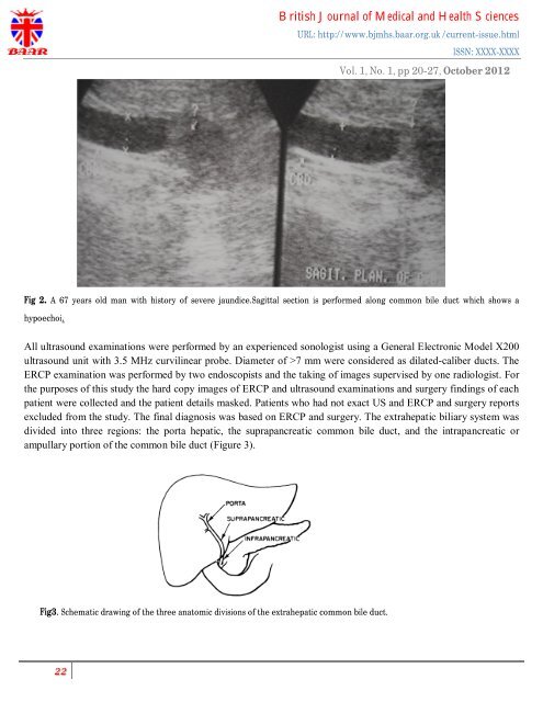
Fig 2. A 67 years old man <strong>with</strong> history <strong>of</strong> severe jaundice.Sagittal section is performed along common bile duct which shows a<br />
hypoechoi.<br />
All <strong>ultrasound</strong> examinations were performed by an experienced sonologist using a General Electronic Model X200<br />
<strong>ultrasound</strong> unit <strong>with</strong> 3.5 MHz curvilinear probe. Diameter <strong>of</strong> >7 mm were considered as dilated-caliber ducts. The<br />
ERCP examination was performed by two endoscopists and the taking <strong>of</strong> images supervised by one radiologist. For<br />
the purposes <strong>of</strong> this study the hard copy images <strong>of</strong> ERCP and <strong>ultrasound</strong> examinations and surgery findings <strong>of</strong> each<br />
patient were collected and the patient details masked. Patients who had not exact US and ERCP and surgery reports<br />
excluded from the study. The final diagnosis was based on ERCP and surgery. The extrahepatic biliary system was<br />
divided into three regions: the porta hepatic, the suprapancreatic common bile duct, and the intrapancreatic or<br />
ampullary portion <strong>of</strong> the common bile duct (Figure 3).<br />
Fig3. Schematic drawing <strong>of</strong> the three anatomic divisions <strong>of</strong> the extrahepatic common bile duct.