Joint Annual Research Report 2004 - The Royal Marsden
Joint Annual Research Report 2004 - The Royal Marsden
Joint Annual Research Report 2004 - The Royal Marsden

You also want an ePaper? Increase the reach of your titles
YUMPU automatically turns print PDFs into web optimized ePapers that Google loves.
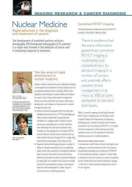
IMAGING RESEARCH & CANCER DIAGNOSIS<br />
Nuclear Medicine<br />
Rapid advances in the diagnosis<br />
and treatment of cancers<br />
<strong>The</strong> development of combined positron emission<br />
tomography (PET)/computed tomography (CT) scanners<br />
is a major step forward in the detection of cancer and<br />
in evaluating response to treatment.<br />
Gary Cook<br />
MSc MD FRCP FRCR<br />
Dr Gary Cook and Dr Val<br />
Lewington are Consultants<br />
in Nuclear Medicine and<br />
PET at <strong>The</strong> <strong>Royal</strong> <strong>Marsden</strong><br />
NHS Foundation Trust<br />
Val Lewington<br />
MSc FRCP<br />
Two key areas of rapid<br />
development in<br />
nuclear medicine<br />
Nuclear medicine involves the use of radioactive isotopes<br />
in the diagnosis and treatment of many diseases but has<br />
an especially important role in oncology. Whilst many<br />
diagnostic and therapeutic nuclear medicine techniques<br />
are used in day-to-day routine patient management<br />
there are two key areas that have shown rapid recent<br />
development, with evidence of improvement in patient<br />
management and care.<br />
Positron emission tomography (PET) scanning uses<br />
radiopharmaceuticals such as 18 F-fluorodeoxyglucose<br />
(FDG) to detect abnormally increased glucose<br />
metabolism in malignant cells to help to pinpoint<br />
cancer more sensitively and to monitor treatment<br />
more effectively. <strong>The</strong> most recent advance in this<br />
modality is in the development of combined PET/CT<br />
scanners that can monitor tumour function (PET) as<br />
well as detect structural changes (CT) in a single scan,<br />
maximising the advantages of each type of scan.<br />
Targeted radionuclide therapy employs a number of<br />
different radiopharmaceuticals that can specifically<br />
target cancer cells, resulting in a therapeutic radiation<br />
dose to tumour tissue rather than normal tissues.<br />
Whilst this technique has been successfully employed<br />
for many years in a number of rare cancers including<br />
thyroid and neuroendocrine tumours, novel agents<br />
have now been developed for a wider range of<br />
common tumours, including lymphoma.<br />
Combined PET/CT imaging<br />
<strong>The</strong> <strong>Royal</strong> <strong>Marsden</strong> installed one of the first PET/CT<br />
scanners in the NHS in February <strong>2004</strong>.<br />
<strong>The</strong>re is evidence that<br />
the extra information<br />
gained from combined<br />
PET/CT imaging is<br />
incremental and<br />
complementary to<br />
standard imaging in a<br />
number of cancers<br />
and positively affects<br />
patients’ clinical<br />
management in as<br />
many as 30% of cases<br />
compared to standard<br />
techniques.<br />
Work is currently underway at the <strong>Royal</strong> <strong>Marsden</strong><br />
PET/CT Unit in collaboration with Professor Janet<br />
Husband (Head of the Department of Diagnostic<br />
Radiology) and a number of clinical units to further<br />
evaluate the role and efficacy of FDG-PET in a number<br />
of clinical situations, in order that the use of this<br />
valuable resource can be optimised to help tailor<br />
treatment protocols in individual patients.<br />
Recurrent colorectal cancer<br />
In collaboration with Professor David Cunningham and<br />
colleagues in the Gastrointestinal Unit, this project is<br />
prospectively evaluating the contribution of FDG-PET<br />
in clinical decision-making in patients in whom there<br />
is suspicion of recurrent cancer as a result of a positive<br />
tumour marker blood test (carcino-embryonic antigen<br />
– CEA), but in whom standard imaging tests such as<br />
CT are negative.