35â4 The Senses - Downtown Magnets High School
35â4 The Senses - Downtown Magnets High School
35â4 The Senses - Downtown Magnets High School

You also want an ePaper? Increase the reach of your titles
YUMPU automatically turns print PDFs into web optimized ePapers that Google loves.
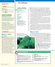
Fovea<br />
Optic nerve<br />
Blood vessels<br />
Retina<br />
Inner layer of eye<br />
that contains<br />
photoreceptors.<br />
Choroid<br />
Middle layer of<br />
eye that is rich in<br />
blood vessels.<br />
Vitreous humor<br />
Just behind the iris is the lens. Small muscles attached to<br />
the lens change its shape to help you adjust your eyes’ focus to<br />
see near or distant objects. Behind the lens is a large chamber<br />
filled with a transparent, jellylike fluid called vitreous (VIHtree-uhs)<br />
humor.<br />
<strong>The</strong> lens focuses light onto the retina. Photoreceptors are<br />
arranged in a layer in the retina. <strong>The</strong> photoreceptors convert<br />
light energy into nerve impulses that are carried to the central<br />
nervous system. <strong>The</strong>re are two types of photoreceptors: rods and<br />
cones. Rods are extremely sensitive to light, but they do not<br />
distinguish different colors. Cones are less sensitive than rods,<br />
but they do respond to light of different colors, producing color<br />
vision. Cones are concentrated in the fovea. <strong>The</strong> fovea is the site<br />
of sharpest vision. <strong>The</strong>re are no photoreceptors where the optic<br />
nerve passes through the back of the eye. This place is called the<br />
blind spot.<br />
<strong>The</strong> impulses assembled by this complicated layer of interconnected<br />
cells leave each eye by way of an optic nerve. <strong>The</strong><br />
optic nerves then carry the impulses to the appropriate regions<br />
of the brain. <strong>The</strong> brain interprets them as visual images and<br />
provides information about the external world.<br />
Where are the photoreceptors located in the eye<br />
Sclera<br />
Outer layer of eye that maintains its<br />
shape. Serves as point of attachment<br />
for muscles that move the eye.<br />
Muscle<br />
Lens<br />
Aqueous humor<br />
Cornea<br />
Pupil<br />
Iris<br />
Ligaments<br />
Figure 35–13 <strong>The</strong> eye is a<br />
complicated sense organ. <strong>The</strong><br />
sclera, choroid, and retina are three<br />
layers of tissue that form the inner<br />
wall of the eyeball. Interpreting<br />
Graphics What is the function of<br />
the sclera<br />
NSTA<br />
For: Links on the<br />
senses<br />
Visit: www.SciLinks.org<br />
Web Code: cbn-0354<br />
Use Visuals<br />
Figure 35–13 Ask students to locate<br />
the three layers of tissue (sclera,<br />
choroid, and retina) that form the<br />
inner wall of the eyeball. <strong>The</strong>n, have<br />
students trace the path of light<br />
through the eye. Call on students to<br />
identify each of the structures the<br />
light passes through. Finally, ask:<br />
What purpose do the muscles<br />
around the lens serve (<strong>The</strong>y change<br />
the shape of the lens to help the eye<br />
focus to see near or distant objects.)<br />
Where does the lens focus the<br />
light (On the retina) What happens<br />
to the impulses after they leave the<br />
eye by way of the optic nerve<br />
(<strong>The</strong>y go to the appropriate regions of<br />
the brain, where the visual images are<br />
interpreted.)<br />
Build Science Skills<br />
Using Analogies Ask: If the lens of<br />
the eye is analogous to a projector,<br />
what part of the eye is analogous<br />
to the screen (<strong>The</strong> retina) How is<br />
the image projected on the screen<br />
different from the image projected<br />
on the retina (<strong>The</strong>re are no receptors<br />
on the screen to convert the image into<br />
electrical impulses.)<br />
NSTA<br />
Download a worksheet<br />
on the senses for students to complete,<br />
and find additional teacher<br />
support from NSTA SciLinks.<br />
UNIVERSAL ACCESS<br />
Inclusion/Special Needs<br />
Help students understand the terms for sensory<br />
receptors by writing the following prefixes on the<br />
board: thermo-, mechano-, chemo-, and photo-.<br />
Challenge students to think of words that begin<br />
with these or similar prefixes, such as thermometer<br />
and mechanic. List the words on the board.<br />
<strong>The</strong>n, ask students to define the prefixes based<br />
on the meanings of those words.<br />
Advanced Learners<br />
Give students who need an extra challenge an<br />
opportunity to investigate surgical methods for<br />
correcting vision problems, including photorefractive<br />
keratectomy (PRK) and laser in situ<br />
keratomileusis (LASIK). Encourage students to<br />
present their findings to the class in an oral<br />
report, with diagrams showing how the procedures<br />
correct specific vision problems.<br />
Answers to . . .<br />
<strong>The</strong> retina<br />
Figure 35–13 <strong>The</strong> function of the<br />
sclera is to maintain the shape of the<br />
eye and serve as a point of attachment<br />
for muscles that move the eye.<br />
Nervous System 907