35â4 The Senses - Downtown Magnets High School
35â4 The Senses - Downtown Magnets High School
35â4 The Senses - Downtown Magnets High School

You also want an ePaper? Increase the reach of your titles
YUMPU automatically turns print PDFs into web optimized ePapers that Google loves.
Section 35–4<br />
1 FOCUS<br />
Objectives<br />
35.4.1 Name the five types of sensory<br />
receptors.<br />
35.4.2 Identify the five sense<br />
organs.<br />
Vocabulary Preview<br />
Ask: Which Vocabulary terms refer<br />
to parts of the eye (Pupil, lens,<br />
retina, rod, and cone) Which terms<br />
refer to parts of the ear (Cochlea,<br />
semicircular canal)<br />
Reading Strategy<br />
Have students preview the section by<br />
studying the figures and reading the<br />
captions.<br />
2 INSTRUCT<br />
Build Science Skills<br />
Applying Concepts Ask students<br />
to imagine they are at a picnic on a<br />
beautiful summer day with a picnic<br />
basket full of their favorite foods.<br />
<strong>The</strong>n, ask: How might the different<br />
categories of your sensory receptors<br />
be stimulated at the picnic<br />
(Students may say, for example, that<br />
their thermoreceptors might be stimulated<br />
by holding a cold drink and their<br />
chemoreceptors by smelling and tasting<br />
food.)<br />
Vision<br />
Make Connections<br />
Physics Explain that light is part of<br />
the electromagnetic spectrum, which<br />
includes electromagnetic waves of<br />
different wavelengths. Add that<br />
humans can see only light that falls<br />
within a very limited range of wavelengths<br />
and that light in this range is<br />
called visible light.<br />
Print:<br />
35– 4 <strong>The</strong> <strong>Senses</strong><br />
BI 9.e. Students know the roles of sensory neurons, interneurons, and motor neurons in sensation, thought, and<br />
response.<br />
Key Concept<br />
• What are the five types of<br />
sensory receptors<br />
Vocabulary<br />
sensory receptor<br />
pupil<br />
lens<br />
retina<br />
rod<br />
cone<br />
cochlea<br />
semicircular canal<br />
taste bud<br />
Reading Strategy:<br />
Outlining Before you read,<br />
use the headings of the section<br />
to make an outline about the<br />
five sense organs. As you read,<br />
fill in the subtopics and smaller<br />
topics. <strong>The</strong>n, add phrases or a<br />
sentence after each to provide<br />
key information.<br />
(magnification: 2000)<br />
SECTION RESOURCES<br />
<strong>The</strong> body contains millions of neurons that react directly to<br />
stimuli from the environment, including light, sound,<br />
motion, chemicals, pressure, and changes in temperature. <strong>The</strong>se<br />
neurons, known as sensory receptors, react to a specific<br />
stimulus such as light or sound by sending impulses to other<br />
neurons, and eventually to the central nervous system. Sensory<br />
receptors are located throughout the body but are concentrated<br />
in the sense organs. <strong>The</strong>se sense organs include the eyes, the<br />
inner ears, the nose, the mouth, and the skin. Sensory receptors<br />
within each organ enable it to respond to a particular stimulus.<br />
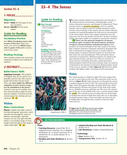
<strong>The</strong>re are five general categories of sensory<br />
receptors: pain receptors, thermoreceptors, mechanoreceptors,<br />
chemoreceptors, and photoreceptors. Pain<br />
receptors are located throughout the body except in the brain.<br />
Pain receptors respond to chemicals released by damaged cells.<br />
Pain is important to recognize because it usually indicates<br />
danger, injury, or disease. <strong>The</strong>rmoreceptors are located in the<br />
skin, body core, and hypothalamus. <strong>The</strong>rmoreceptors detect<br />
variations in temperature. Mechanoreceptors are found in the<br />
skin, skeletal muscles, and inner ears. <strong>The</strong>y are sensitive to<br />
touch, pressure, stretching of muscles, sound, and motion.<br />
Chemoreceptors, located in the nose and taste buds, are sensitive<br />
to chemicals in the external environment. Photoreceptors, found<br />
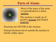
in the eyes, are sensitive to light. Figure 35–12 shows how photoreceptor<br />
cells appear under a scanning electron microscope.<br />
Vision<br />
• Teaching Resources, Lesson Plan 35–4,<br />
Adapted Section Summary 35–4, Adapted<br />
Worksheets 35–4, Section Summary 35–4,<br />
Worksheets 35–4, Section Review 35–4,<br />
Enrichment<br />
• Reading and Study Workbook A, Section<br />
35–4<br />
Time<br />
Saver<br />
<strong>The</strong> world around us is bathed in light. <strong>The</strong> sense organs that<br />
we use to sense light are the eyes. <strong>The</strong> structures of the eye are<br />
shown in Figure 35–13. Light enters the eye through the cornea,<br />
a tough transparent layer of cells. <strong>The</strong> cornea helps to focus the<br />
light, which then passes through a chamber filled with a fluid<br />
called aqueous (AY-kwee-uhs) humor. At the back of the chamber<br />
is a disklike structure called the iris. <strong>The</strong> iris is the colored<br />
part of the eye. In the middle of the iris is a small opening called<br />
the pupil. Tiny muscles in the iris adjust the size of the pupil<br />
to regulate the amount of light that enters the eye. In dim light,<br />
the pupil becomes larger so that more light can enter the eye.<br />
In bright light, the pupil becomes smaller so that less light<br />
enters the eye.<br />
Figure 35–12 <strong>The</strong>re are two types of lightsensitive<br />
photoreceptor cells in the retina—rods<br />
and cones. This color-enhanced scanning electron<br />
micrograph shows the rod cells of an eye.<br />
• Adapted Reading and Study Workbook B,<br />
Section 35–4<br />
• Lab Worksheets, Chapter 35 Real-World Lab<br />
Technology:<br />
• iText, Section 35–4<br />
• Transparencies Plus, Section 35–4<br />
906 Chapter 35
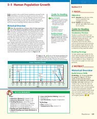
Fovea<br />
Optic nerve<br />
Blood vessels<br />
Retina<br />
Inner layer of eye<br />
that contains<br />
photoreceptors.<br />
Choroid<br />
Middle layer of<br />
eye that is rich in<br />
blood vessels.<br />
Vitreous humor<br />
Just behind the iris is the lens. Small muscles attached to<br />
the lens change its shape to help you adjust your eyes’ focus to<br />
see near or distant objects. Behind the lens is a large chamber<br />
filled with a transparent, jellylike fluid called vitreous (VIHtree-uhs)<br />
humor.<br />
<strong>The</strong> lens focuses light onto the retina. Photoreceptors are<br />
arranged in a layer in the retina. <strong>The</strong> photoreceptors convert<br />
light energy into nerve impulses that are carried to the central<br />
nervous system. <strong>The</strong>re are two types of photoreceptors: rods and<br />
cones. Rods are extremely sensitive to light, but they do not<br />
distinguish different colors. Cones are less sensitive than rods,<br />
but they do respond to light of different colors, producing color<br />
vision. Cones are concentrated in the fovea. <strong>The</strong> fovea is the site<br />
of sharpest vision. <strong>The</strong>re are no photoreceptors where the optic<br />
nerve passes through the back of the eye. This place is called the<br />
blind spot.<br />
<strong>The</strong> impulses assembled by this complicated layer of interconnected<br />
cells leave each eye by way of an optic nerve. <strong>The</strong><br />
optic nerves then carry the impulses to the appropriate regions<br />
of the brain. <strong>The</strong> brain interprets them as visual images and<br />
provides information about the external world.<br />
Where are the photoreceptors located in the eye<br />
Sclera<br />
Outer layer of eye that maintains its<br />
shape. Serves as point of attachment<br />
for muscles that move the eye.<br />
Muscle<br />
Lens<br />
Aqueous humor<br />
Cornea<br />
Pupil<br />
Iris<br />
Ligaments<br />
Figure 35–13 <strong>The</strong> eye is a<br />
complicated sense organ. <strong>The</strong><br />
sclera, choroid, and retina are three<br />
layers of tissue that form the inner<br />
wall of the eyeball. Interpreting<br />
Graphics What is the function of<br />
the sclera<br />
NSTA<br />
For: Links on the<br />
senses<br />
Visit: www.SciLinks.org<br />
Web Code: cbn-0354<br />
Use Visuals<br />
Figure 35–13 Ask students to locate<br />
the three layers of tissue (sclera,<br />
choroid, and retina) that form the<br />
inner wall of the eyeball. <strong>The</strong>n, have<br />
students trace the path of light<br />
through the eye. Call on students to<br />
identify each of the structures the<br />
light passes through. Finally, ask:<br />
What purpose do the muscles<br />
around the lens serve (<strong>The</strong>y change<br />
the shape of the lens to help the eye<br />
focus to see near or distant objects.)<br />
Where does the lens focus the<br />
light (On the retina) What happens<br />
to the impulses after they leave the<br />
eye by way of the optic nerve<br />
(<strong>The</strong>y go to the appropriate regions of<br />
the brain, where the visual images are<br />
interpreted.)<br />
Build Science Skills<br />
Using Analogies Ask: If the lens of<br />
the eye is analogous to a projector,<br />
what part of the eye is analogous<br />
to the screen (<strong>The</strong> retina) How is<br />
the image projected on the screen<br />
different from the image projected<br />
on the retina (<strong>The</strong>re are no receptors<br />
on the screen to convert the image into<br />
electrical impulses.)<br />
NSTA<br />
Download a worksheet<br />
on the senses for students to complete,<br />
and find additional teacher<br />
support from NSTA SciLinks.<br />
UNIVERSAL ACCESS<br />
Inclusion/Special Needs<br />
Help students understand the terms for sensory<br />
receptors by writing the following prefixes on the<br />
board: thermo-, mechano-, chemo-, and photo-.<br />
Challenge students to think of words that begin<br />
with these or similar prefixes, such as thermometer<br />
and mechanic. List the words on the board.<br />
<strong>The</strong>n, ask students to define the prefixes based<br />
on the meanings of those words.<br />
Advanced Learners<br />
Give students who need an extra challenge an<br />
opportunity to investigate surgical methods for<br />
correcting vision problems, including photorefractive<br />
keratectomy (PRK) and laser in situ<br />
keratomileusis (LASIK). Encourage students to<br />
present their findings to the class in an oral<br />
report, with diagrams showing how the procedures<br />
correct specific vision problems.<br />
Answers to . . .<br />
<strong>The</strong> retina<br />
Figure 35–13 <strong>The</strong> function of the<br />
sclera is to maintain the shape of the<br />
eye and serve as a point of attachment<br />
for muscles that move the eye.<br />
Nervous System 907
35–4 (continued)<br />
Hearing and Balance<br />
Use Visuals<br />
Figure 35–14 Name the structures<br />
that sound waves pass through after<br />
they enter the ear. As you name each<br />
structure, ask: What role does this<br />
structure play in hearing (Students<br />
might say, for example, that the auditory<br />
canal channels the sound waves to<br />
the tympanum and that the tympanum<br />
vibrates in response to the sound<br />
waves.)<br />
Make Connections<br />
Health Science Explain that the<br />
region of the ear called the middle<br />
ear, which is the area between the<br />
tympanum and the semicircular<br />
canals, is prone to infections. This is<br />
because of the close connection<br />
between the middle ear and the<br />
eustachian tube, which originates in<br />
the throat. Viruses and bacteria in the<br />
throat can easily travel to the middle<br />
ear through the eustachian tube and<br />
cause infections, inflammation, and<br />
pain.<br />
Build Science Skills<br />
Inferring Tell students that middleear<br />
infections often block the<br />
transmission of sounds from the outside<br />
world but not sounds, such as<br />
chewing sounds, that originate within<br />
the head. Ask: Why can people<br />
hear “head” sounds even when<br />
their ears are blocked because of<br />
an infection (Because the sound<br />
waves are transmitted directly to the<br />
inner ear through the bones of the<br />
head)<br />
Demonstration<br />
Half fill a glass container with water<br />
and, as students watch, slowly tilt<br />
the container from side to side.<br />
Because the water always stays<br />
parallel to the floor due to gravity,<br />
students will observe it move up and<br />
down the sides of the glass as the<br />
glass tilts. Explain that this is also<br />
how the fluid inside the semicircular<br />
canals moves as the head changes<br />
position.<br />
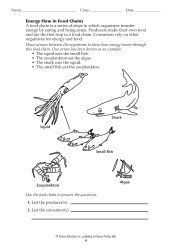
Auditory canal<br />
Hammer<br />
(magnification: about 3500)<br />
Figure 35 –14 <strong>The</strong> diagram (top)<br />
shows the structures in the ear that<br />
transmit sounds. <strong>The</strong> scanning electron<br />
micrograph shows hair cells (yellow) in<br />
the inner ear. <strong>The</strong> motion of these hairs<br />
produces nerve impulses that travel to<br />
the brain through the cochlear nerve.<br />
Predicting How would frequent<br />
exposure to loud noise affect a person’s<br />
threshold for detecting sound<br />
TEACHER TO TEACHER<br />
Tympanum<br />
Anvil<br />
When I teach about the senses, I give students a<br />
chance to experience sensory “fatigue.” I have<br />
students rest a penny on the inside of the forearm<br />
against the skin and measure how long it<br />
takes until they can no longer sense the presence<br />
of the coin. When everyone is finished, we discuss<br />
why sensory fatigue occurs and when it is<br />
useful (for example, when you are wearing clothing).<br />
I also demonstrate sensory fatigue with the<br />
sense of smell. I ask a volunteer to come to the<br />
Oval window<br />
Stirrup<br />
Semicircular canals<br />
Round window<br />
Hearing and Balance<br />
Cochlear nerve<br />
Cochlea<br />
Bone<br />
Eustachian tube<br />
<strong>The</strong> human ear has two sensory functions. One of these functions<br />
is hearing. <strong>The</strong> other function is detecting positional<br />
changes associated with movement.<br />
Hearing Sound is nothing more than vibrations in the air<br />
around us. <strong>The</strong> ears are the sensory organs that can distinguish<br />
both the pitch and loudness of those vibrations. <strong>The</strong><br />
structure of the ear is shown in Figure 35–14.<br />
Vibrations enter the ear through the auditory canal. <strong>The</strong><br />
vibrations cause the tympanum (TIM-puh-num), or eardrum,<br />
to vibrate. <strong>The</strong>se vibrations are picked up by three tiny bones,<br />
commonly called the hammer, anvil, and stirrup. <strong>The</strong> last of<br />
these bones, the stirrup, transmits the vibrations to the oval<br />
window. Vibrations of the oval window create pressure waves<br />
in the fluid-filled cochlea (KAHK-lee-uh) of the inner ear.<br />
<strong>The</strong> cochlea is lined with tiny hair cells that are pushed<br />
back and forth by these pressure waves. In response to these<br />
movements, the hair cells produce nerve impulses that are<br />
sent to the brain through the cochlear nerve.<br />
Balance Your ears contain structures that help your central<br />
nervous system maintain your balance, or equilibrium. Within<br />
the inner ear just above the cochlea are three tiny canals at right<br />
angles to one another. <strong>The</strong>y are called semicircular canals<br />
because each forms a half circle. <strong>The</strong> semicircular canals and<br />
the two tiny sacs located behind them monitor the position of<br />
your body, especially your head, in relation to gravity.<br />
front of the room and hold one nostril closed<br />
while I hold a bottle of oil of wintergreen (available<br />
at pharmacies) under the other nostril. <strong>The</strong><br />
class measures the time it takes until the volunteer<br />
can no longer distinguish the smell of<br />
wintergreen.<br />
—Duane Nichols<br />
Biology Teacher<br />
Alhambra <strong>High</strong> <strong>School</strong><br />
Alhambra, CA<br />
908 Chapter 35
<strong>The</strong> semicircular canals and the sacs are filled with fluid<br />
and lined with hair cells. As the head changes position, the fluid<br />
in the canals also changes position. This causes the hair on the<br />
hair cells to bend. This action, in turn, sends impulses to the<br />
brain that enable it to determine body motion and position.<br />
Smell and Taste<br />
You may never have thought of it this way, but your sense of<br />
smell is actually an ability to detect chemicals. Chemoreceptors<br />
in the lining of the nasal passageway respond to specific chemicals<br />
and send impulses to the brain through sensory nerves.<br />
Your sense of smell is capable of producing thousands of<br />
different sensations. In fact, much of what we commonly call the<br />
“taste” of food and drink is actually smell. To prove this to<br />
yourself, eat a few bites of food while holding your nose. You’ll<br />
discover that much of the taste of food disappears until you open<br />
your nose and breathe freely.<br />
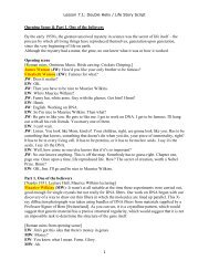
Like the sense of smell, the sense of taste is a chemical sense.<br />
<strong>The</strong> sense organs that detect taste are the taste buds. Most of<br />
the taste buds are on the tongue, but a few are found at other<br />
locations in the mouth. <strong>The</strong> surface of the tongue is shown in<br />
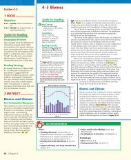
Figure 35–15. <strong>The</strong> tastes detected by the taste buds are classified<br />
as salty, bitter, sweet, and sour. Sensitivity to these different<br />
categories varies on different parts of the tongue.<br />
Touch and Related <strong>Senses</strong><br />
<strong>The</strong> sense of touch, unlike the other senses you have just read<br />
about, is not found in one particular place. All of the regions of<br />
the skin are sensitive to touch. In this respect, your largest<br />
sense organ is your skin. Skin contains sensory receptors that<br />
respond to temperature, touch, and pain. Not all parts of the<br />
body are equally sensitive to touch, because not all parts have<br />
the same number of receptors. <strong>The</strong> greatest density of touch<br />
receptors is found on your fingers, toes, and face.<br />
35 – 4 Section Assessment<br />
1. Key Concept Name the<br />
five types of sensory receptors<br />
and list where they are found in<br />
the body.<br />
2. Identify the functions of the<br />
cornea, pupil, lens, retina, and<br />
optic nerve.<br />
3. What are the four basic tastes<br />
detected by the tongue<br />
4. Explain why you can’t “taste”<br />
food when you have a bad cold.<br />
5. Critical Thinking Applying<br />
Concepts If you spin around for<br />
a time, the fluid in your semicircular<br />
canals also moves. When you<br />
stop suddenly, you feel as though<br />
you are still moving. Why do you<br />
think you might feel dizzy<br />
(magnification: 86)<br />
Figure 35–15 This colorenhanced<br />
scanning electron micrograph<br />
shows the surface of the<br />
tongue. <strong>The</strong> large pink objects are the<br />
taste buds. Chemoreceptors<br />
found in the taste buds are sensitive<br />
to chemicals in food.<br />
Creative Writing<br />
Imagine that you have to do<br />
without your sense of taste<br />
for one day. How would this<br />
influence your food choices<br />
Write a 3- to 4-paragraph<br />
essay describing how the<br />
absence of this sense organ<br />
would affect your day.<br />
Smell and Taste<br />
Demonstration<br />
Have volunteers taste and try to identify<br />
a variety of different fruit juices<br />
while wearing blindfolds and pinching<br />
their noses shut. (Without sight<br />
cues and the sense of smell, students<br />
will find it difficult to distinguish the<br />
tastes.)<br />
Touch and Related<br />
<strong>Senses</strong><br />
Build Science Skills<br />
Designing Experiments Challenge<br />
students to design an experiment to<br />
determine the distribution of heat<br />
and cold receptors in a small area of<br />
skin on the back of the hand.<br />
3 ASSESS<br />
Evaluate Understanding<br />
Provide students with copies of<br />
Figure 35–13 without the labels.<br />
Have students label each part of the<br />
eye shown in the figure.<br />
Reteach<br />
On the chalkboard or an overhead<br />
transparency, list five general categories<br />
of sensory receptors. Have<br />
students give examples of each category<br />
of receptors at work. (For<br />
chemoreceptors, for example, students<br />
might say smelling a flower or tasting<br />
food.)<br />
Answers will vary. For example,<br />
students might describe how their<br />
sense of taste is affected and how<br />
this, in turn, affects their enjoyment<br />
of food and their appetite.<br />
35 – 4 Section Assessment<br />
1. Pain receptors: everywhere except the brain;<br />
thermoreceptors: skin, body core, hypothalamus;<br />
mechanoreceptors: skin, skeletal<br />
muscles, inner ears; chemoreceptors: nose,<br />
taste buds; photoreceptors: eyes<br />
2. Cornea: helps to focus light; pupil: controls<br />
the amount of light that enters the eye; lens:<br />
adjusts focus for near or far distances; retina:<br />
rod and cone photoreceptors convert light<br />
into electrical impulses; optic nerve: carries<br />
the electrical impulses to the brain<br />
3. <strong>The</strong> four basic taste receptors are receptors<br />
for salty, bitter, sweet, and sour tastes.<br />
4. Because much of the sense of taste is actually<br />
due to interaction with the sense of smell<br />
5. Sensory receptors lag behind the rapid<br />
changes in position.<br />
If your class subscribes to the iText,<br />
use it to review the Key Concepts in<br />
Section 35–4.<br />
Answer to . . .<br />
Figure 35 –14 Loud noises can<br />
damage tiny hair cells and raise the<br />
threshold for detecting sounds.<br />
Nervous System 909