Pyrexia - PACT - ESICM
Pyrexia - PACT - ESICM
Pyrexia - PACT - ESICM

Create successful ePaper yourself
Turn your PDF publications into a flip-book with our unique Google optimized e-Paper software.
Task 2. Determining the cause of fever in the critically ill patient<br />
Tracheal aspirates: their diagnostic significance is greater if (semi-)<br />
quantitative rather than qualitative cultures are performed; this helps to obviate<br />
false-positive results by colonisation of upper airways in the absence of lower<br />
respiratory tract infection by the bacteria. Usually, a cut-off point of 10 5 cfu/mL<br />
is taken. Indeed, some micro-organisms are obligatory pathogens while the lowgrade<br />
presence of others, such as Gram-negative bacilli, may merely represent<br />
colonisation. Microscopy of the aspirates is necessary to exclude saliva with<br />
many epithelial cells, and elastin staining may confirm a lower (versus upper)<br />
respiratory tract origin of the aspirate. In the case of VAP, the aspirate typically<br />
contains numerous neutrophils.<br />
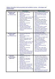
Distal bronchial specimens: tracheal aspirate results are less specific than<br />
those of Gram stains, microscopy and cultures of lower (distal) pulmonary<br />
secretions obtained by bronchoscopy and bronchoalveolar lavage (BAL) or<br />
protected specimen brush (PSB). It remains unclear, however, whether<br />
antibiotic guidance based on the latter is associated with lower morbidity and<br />
mortality for suspected VAP than antibiotic treatment guided by tracheal<br />
aspirates. Nevertheless, utilisation of these invasive tools may prevent<br />
overtreatment by antibiotics and reduce antibiotic pressure. This may be<br />
increasingly relevant because of the increase in multiresistant pathogens<br />
causing VAP.<br />
Finally, positive blood or, when present, pleural fluid cultures with the same<br />
organism as recovered from the airway can be found in VAP.<br />
Before proceeding to the next section, consider searching the Web for<br />
evidence that treatment guided by BAL/PSB specimens is superior to that guided by<br />
conventional, less invasive techniques and also for more specific clinical indications for<br />
the use of the invasive technique. Then assess the views expressed below.<br />
The table below outlines a diagnostic approach to VAP when invasive<br />
procedures are performed.<br />
Criteria for the diagnosis of VAP<br />
I Three or more of the following:<br />
a) Rectal temperature >38.0 °C or 103 cfu/L) or >5% of leukocytes containing phagocytosed bacteria.<br />
b) Positive blood culture with the same micro-organism as that present in the airway<br />
c) Positive culture of pleural fluid<br />
[16]