Dermatologic Manifestations of HIV in Africa - International AIDS ...
Dermatologic Manifestations of HIV in Africa - International AIDS ...
Dermatologic Manifestations of HIV in Africa - International AIDS ...

You also want an ePaper? Increase the reach of your titles
YUMPU automatically turns print PDFs into web optimized ePapers that Google loves.
<strong>HIV</strong> Dermatology <strong>in</strong> <strong>Africa</strong> Volume 18 Issue 1 February/March 2010<br />
ticularly pediatric patients, <strong>in</strong> <strong>Africa</strong><br />
(Figure 16). MC is caused by a poxvirus.<br />
It presents with dome-shaped,<br />
umbilicated papules, typically 3 mm to<br />
8 mm, although giant lesions can occur<br />
<strong>in</strong> advanced <strong>HIV</strong> disease. Antiretroviral<br />
therapy is the ma<strong>in</strong>stay <strong>of</strong> treatment,<br />
but curettage or silver nitrate treatment<br />
may also be used.<br />
Differential Diagnosis to Consider<br />
Cryptococcosis. Dissem<strong>in</strong>ated cryptococcal<br />
disease can present <strong>in</strong> the<br />
sk<strong>in</strong>, <strong>of</strong>ten preced<strong>in</strong>g or simultaneous<br />
with the onset <strong>of</strong> men<strong>in</strong>gitis symptoms.<br />
Cl<strong>in</strong>ically, the lesions resemble<br />
MC, although the central umbilication<br />
<strong>of</strong>ten has a hemorrhagic crust. The<br />
sk<strong>in</strong> lesions <strong>of</strong> cryptococcosis are <strong>of</strong>ten<br />
eruptive, as opposed to the slower,<br />
<strong>in</strong>sidious onset <strong>of</strong> MC (Figure 17). Cl<strong>in</strong>icians<br />
should ma<strong>in</strong>ta<strong>in</strong> a high level <strong>of</strong><br />
suspicion for cutaneous cryptococcosis<br />
<strong>in</strong> <strong>Africa</strong>, where cryptococcal disease<br />
is very prevalent, particularly if the lesions<br />
appear over a short time course.<br />
Warts<br />
Viral warts, both genital and nongenital,<br />
are very common <strong>in</strong> <strong>Africa</strong> (Figure<br />
16). Particularly <strong>in</strong> pediatric patients,<br />
hundreds <strong>of</strong> flat warts are a frequent,<br />
and stigmatiz<strong>in</strong>g, f<strong>in</strong>d<strong>in</strong>g that may or<br />
may not improve with antiretroviral<br />
therapy. Cryotherapy, the standard<br />
wart treatment <strong>in</strong> developed countries,<br />
is generally not available. Large genital<br />
lesions may be treated with 25% podophyll<strong>in</strong>,<br />
which is pa<strong>in</strong>ted on the warts<br />
and washed <strong>of</strong>f <strong>in</strong> 4 hours to 6 hours,<br />
and nongenital warts may be treated<br />
with salicylic acid preparations. Genital<br />
warts should be monitored for rapidly<br />
grow<strong>in</strong>g firm nodules or ulcers that<br />
may <strong>in</strong>dicate human papillomavirus<strong>in</strong>duced<br />
squamous cell carc<strong>in</strong>oma.<br />
Herpes Simplex Virus and<br />
Varicella-Zoster Virus Infections<br />
Infections with herpes simplex virus<br />
and varicella-zoster virus are quite<br />
common <strong>in</strong> <strong>Africa</strong> and most <strong>of</strong>ten<br />
present <strong>in</strong> the typical fashion. Large,<br />
chronic ulcerations <strong>of</strong> the face, genitals,<br />
or buttocks due to herpes simplex<br />
virus <strong>in</strong>fection are frequent <strong>in</strong> patients<br />
with advanced <strong>HIV</strong> disease (Figure 18).<br />
A “scalloped” border to a chronic ulceration<br />
can be a clue to the diagnosis.<br />
Oral acyclovir treatment is generally<br />
available, whereas <strong>in</strong>travenous acyclovir<br />
can be difficult to obta<strong>in</strong>, and alternative<br />
antiherpetic drugs are not widely<br />
available.<br />
Varicella-zoster virus <strong>in</strong>fection <strong>in</strong> the<br />
V1 distribution (<strong>in</strong>volv<strong>in</strong>g the eye) may<br />
eventuate <strong>in</strong> bl<strong>in</strong>dness. Intravenous<br />
acyclovir treatment is recommended<br />
<strong>in</strong> this case, as the absorption is superior<br />
to the oral route. Postherpetic<br />
neuralgia is less <strong>of</strong> a problem <strong>in</strong> patients<br />
with <strong>HIV</strong>, with the exception <strong>of</strong><br />
varicella-zoster virus <strong>in</strong>fection <strong>in</strong> the V1<br />
region (Figure 19).<br />
Figure 19. Varicella-zoster virus <strong>in</strong>fection affect<strong>in</strong>g<br />
the V1 dermatome.<br />
T<strong>in</strong>ea<br />
T<strong>in</strong>ea is a universally common sk<strong>in</strong><br />
problem <strong>in</strong> <strong>HIV</strong>-<strong>in</strong>fected patients. Typical<br />
t<strong>in</strong>ea has an <strong>in</strong>flammatory, scaly<br />
border; <strong>in</strong> darkly pigmented patients,<br />
the area <strong>of</strong> central “clear<strong>in</strong>g” is usually<br />
hyperpigmented (Figure 20). Common<br />
locations <strong>in</strong>clude the hands, feet, and<br />
lower back or buttocks. Special considerations<br />
<strong>in</strong> <strong>Africa</strong> <strong>in</strong>clude t<strong>in</strong>ea <strong>in</strong>cogni-<br />
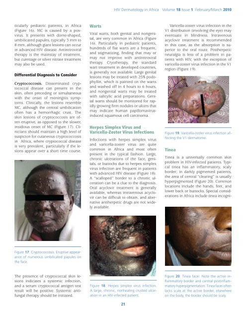
Figure 17. Cryptococcosis. Eruptive appearance<br />
<strong>of</strong> numerous umbilicated papules on<br />
the face.<br />
The presence <strong>of</strong> cryptococcal sk<strong>in</strong> lesions<br />
<strong>in</strong>dicates a systemic <strong>in</strong>fection,<br />
and a serum cryptococcal antigen test<br />
result will be positive. Systemic antifungal<br />
therapy should be <strong>in</strong>itiated.<br />
Figure 18. Herpes simplex virus <strong>in</strong>fection.<br />
A large, chronic, nonheal<strong>in</strong>g crusted ulceration<br />
<strong>in</strong> an <strong>HIV</strong>-<strong>in</strong>fected patient.<br />
21<br />
Figure 20. T<strong>in</strong>ea facei. Note the active <strong>in</strong>flammatory<br />
border and central post<strong>in</strong>flammatory<br />
hyperpigmentation. T<strong>in</strong>ea facei <strong>of</strong>ten<br />
lacks scale at the active border; elsewhere<br />
on the body, the border should be scaly.