Synthetic-lethal Interactions Identify Two Novel Genes, SLA/and ...
Synthetic-lethal Interactions Identify Two Novel Genes, SLA/and ...
Synthetic-lethal Interactions Identify Two Novel Genes, SLA/and ...

You also want an ePaper? Increase the reach of your titles
YUMPU automatically turns print PDFs into web optimized ePapers that Google loves.
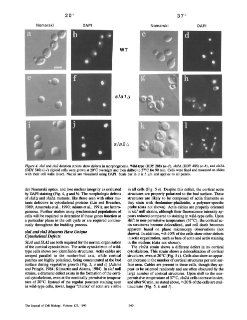
Figure 4. sial <strong>and</strong> sla2 deletion strains show defects in morphogenesis. Wild-type (DDY 288) (a-d), s/a/A (DDY 485) (e-h), <strong>and</strong> sla2A<br />
(DDY 540) (i-l) diploid cells were grown at 20°C overnight <strong>and</strong> then shifted to 37°C for 90 min. Cells were fixed <strong>and</strong> mounted on slides<br />
with their cell walls intact. Nuclei are visualized using DAPI. Scale bar in a is 5 #m <strong>and</strong> applies to all panels.<br />
der Nomarski optics, <strong>and</strong> lose nuclear integrity as evaluated<br />
by DAPI staining (Fig. 4, g <strong>and</strong> h). The morphologic defects<br />
of s/a/A <strong>and</strong> sla2A mutants, like those seen with other mutants<br />
defective in cytoskeletal proteins (Liu <strong>and</strong> Brescher,<br />
1989; Amatruda et al., 1990; Adams et al., 1991), are heterogeneous.<br />
Further studies using synchronized populations of<br />
cells will be required to determine if these genes function at<br />
a particular phase in the cell cycle or are required continuously<br />
throughout the budding process.<br />
sial <strong>and</strong> sla2 Mutants Have Unique<br />
Cytoskeletal Defects<br />
<strong>SLA</strong>/<strong>and</strong> <strong>SLA</strong>2 are both required for the normal organization<br />
of the cortical cytoskeleton. The actin cytoskeleton of wildtype<br />
cells shows two identifiable structures. Actin cables are<br />
arrayed parallel to the mother-bud axis, while cortical<br />
patches are highly polarized, being concentrated at the bud<br />
surface during vegetative growth (Fig. 5, a <strong>and</strong> c) (Adams<br />
<strong>and</strong> Pringle, 1984; Kilmartin <strong>and</strong> Adams, 1984). In sial null<br />
strains, a dramatic defect exists in the formation of the cortical<br />
cytoskeleton, even at the nominally permissive temperature<br />
of 20°C. Instead of the regular punctate staining seen<br />
in wild-type cells, fewer, larger "chunks" of actin are visible<br />
in all cells (Fig. 5 e). Despite this defect, the cortical actin<br />
structures are properly polarized to the bud surface. These<br />
structures are likely to be composed of actin filaments as<br />
they stain with rhodamine-phalloidin, a polymer-specific<br />
probe (data not shown). Actin cables are properly oriented<br />
in slal null strains, although their fluorescence intensity appears<br />
reduced compared to staining in wild-type cells. Upon<br />
shift to non-permissive temperature (37°C), the cortical actin<br />
structures become delocalized, <strong>and</strong> cell death becomes<br />
apparent based on phase microscopy observations (not<br />
shown). In addition, •5-10% of the cells show other defects<br />
in actin organization, such as bars of actin <strong>and</strong> actin staining<br />
in the nucleus (data not shown).<br />
The sla2A strain shows a different defect in its cortical<br />
cytoskeleton. This strain shows a delocalization of cortical<br />
structures, even at 20°C (Fig. 5 i). Cells also show an apparent<br />
increase in the number of cortical structures per unit surface<br />
area. Cables are present in these cells, though they appear<br />
to be oriented r<strong>and</strong>omly <strong>and</strong> are often obscured by the<br />
large number of cortical structures. Upon shift to the nonpermissive<br />
temperature of 37°C, sla2A cells increase in size,<br />
<strong>and</strong> after 90 min, as stated above, ~20% of the ceils are multinucleate<br />
(Fig. 5, k <strong>and</strong> 1).<br />
The Journal of Cell Biology, Volume 122, 1993 640