Cell-based Screening Assays - Ozyme
Cell-based Screening Assays - Ozyme
Cell-based Screening Assays - Ozyme

You also want an ePaper? Increase the reach of your titles
YUMPU automatically turns print PDFs into web optimized ePapers that Google loves.
0<br />
0<br />
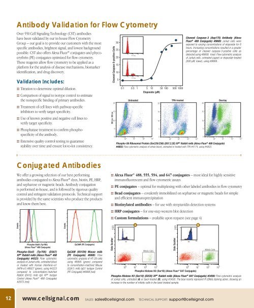
Antibody Validation for Flow Cytometry<br />
Over 550 <strong>Cell</strong> Signaling Technology (CST) antibodies<br />
have been validated by our in-house Flow Cytometry<br />
Group – our goal is to provide our customers with the most<br />
specific antibodies, brightest signal, and lowest background<br />
possible. CST also offers Alexa Fluor ® conjugates and phycoerythrin<br />
(PE) conjugates optimized for flow cytometry.<br />
These reagents allow flow cytometry to be applied as a<br />
platform for the analysis of disease mechanisms, biomarker<br />
identification, and drug discovery.<br />
Validation Includes:<br />
:: Titration to determine optimal dilution.<br />
:: Comparison of signal to isotype control to estimate<br />
the nonspecific binding of primary antibodies.<br />
:: Treatment of cell lines with pathway-specific<br />
inhibitors to verify target specificity.<br />
:: Use of known positive and negative cell lines to<br />
verify target specificity.<br />
:: Phosphatase treatment to confirm phosphospecificity<br />
of the antibody.<br />
:: Extensive quality control testing to guarantee<br />
stability over time and ensure lot-to-lot consistency.<br />
Cleaved Caspase-3-positive <strong>Cell</strong>s<br />
SS Lin: SS Lin<br />
20<br />
15<br />
10<br />
5<br />
0<br />
30<br />
25<br />
20<br />
15<br />
10<br />
5<br />
63<br />
48<br />
10 0 101<br />
FL1 Log<br />
10 0<br />
10 1<br />
FL1 Log<br />
10 2 10z<br />
10 2 10z<br />
0.1 0.5 1 5 10 50 100 500 1000<br />
Etoposide (µM)<br />
Untreated<br />
10 0 10 1 10 2 10 3<br />
20<br />
15<br />
10<br />
5<br />
0<br />
TPA-treated<br />
10 0 10 1 10 2 10 3<br />
Cleaved Caspase-3 (Asp175) Antibody (Alexa<br />
Fluor ® 488 Conjugate) #9669: Jurkat cells were<br />
exposed to varying concentrations of etoposide for 6<br />
hours. Increasing concentrations resulted in a greater<br />
percentage of cleaved caspase-3-positive cells as<br />
detected using #9669. Inset: Flow cytometric analysis<br />
of Jurkat cells, untreated (upper) or etoposide-treated<br />
(500 µM, lower), using #9669.<br />
20<br />
15<br />
10<br />
5<br />
0<br />
Overlay<br />
Phospho-S6 Ribosomal Protein (Ser235/236) (D57.2.2E) XP ® Rabbit mAb (Alexa Fluor ® 488 Conjugate)<br />
#4803: Flow cytometric analysis of whole blood, untreated or treated with TPA #4174, using #4803.<br />
10 0 10 1 10 2 10 3<br />
Conjugated Antibodies<br />
We offer a growing selection of our best performing<br />
antibodies conjugated to Alexa Fluor ® dyes, biotin, PE, HRP,<br />
and sepharose or magnetic beads. Antibody conjugation<br />
is performed in-house, and is followed by rigorous quality<br />
control and stringent validation protocols. Technical support<br />
is provided by the same scientists who produce the products<br />
and know them best.<br />
:: Alexa Fluor ® 488, 555, 594, and 647 conjugates – most ideal for highly sensitive<br />
immunofluorescent and flow cytometric assays<br />
:: PE conjugates – optimal for multiplexing with other labeled antibodies in flow cytometry<br />
:: Bead conjugates – covalently immobilized on sepharose or magnetic beads for simple<br />
and efficient immunoprecipitation<br />
:: Biotinylated antibodies – for use with streptavidin detection systems<br />
:: HRP conjugates – for one-step western blot detection<br />
:: Custom formulations – available upon request (see page 4)<br />
Events<br />
Events<br />
1023<br />
73<br />
A<br />
1023<br />
76<br />
B<br />
Phospho-Stat3 (Tyr705)<br />
(Alexa Fluor ® 488 Conjugate)<br />
Phospho-Stat3 (Tyr705) (D3A7)<br />
XP ® Rabbit mAb (Alexa Fluor ® 488<br />
Conjugate) #4323: Flow cytometric<br />
analysis of Jurkat cells, untreated (blue)<br />
or treated with Human Interferon-α1<br />
(hIFN-α1) #8927 (green), using #4323<br />
compared to concentration-matched<br />
Rabbit (DA1E) mAb IgG XP ® Isotype<br />
Control (Alexa Fluor ® 488 Conjugate)<br />
#2975 (red).<br />
EpCAM (PE Conjugate)<br />
EpCAM (VU1D9) Mouse mAb<br />
(PE Conjugate) #8995: Flow<br />
cytometric analysis of HT-29 cells<br />
using #8995 (green) compared<br />
to concentration-matched Mouse<br />
(G3A1) mAb IgG1 Isotype Control<br />
(PE Conjugate) #6899 (red).<br />
Side Scatter<br />
0<br />
0<br />
DNA<br />
1023<br />
Mitotic <strong>Cell</strong>s<br />
Side Scatter<br />
0<br />
10 0 10 1 10 2 10 3<br />
10 0 10 1 10 2 10 3<br />
Phospho-Histone H3 (Ser10) (Alexa Fluor ® 647 Conjugate)<br />
Phospho-Histone H3 (Ser10) (D2C8) XP ® Rabbit mAb (Alexa Fluor ® 647 Conjugate) #3458: Flow cytometric analysis<br />
of Jurkat cells, untreated (A) or taxol-treated (B), using #3458. The blue inserts represent PI (DNA) staining alone, showing an<br />
increase in the number of mitotic cells in the taxol-treated sample.<br />
0<br />
DNA<br />
1023<br />
Mitotic <strong>Cell</strong>s<br />
12 www.cellsignal.com Sales: sales@cellsignal.com Technical Support: support@cellsignal.com