Cell-based Screening Assays - Ozyme
Cell-based Screening Assays - Ozyme
Cell-based Screening Assays - Ozyme

You also want an ePaper? Increase the reach of your titles
YUMPU automatically turns print PDFs into web optimized ePapers that Google loves.
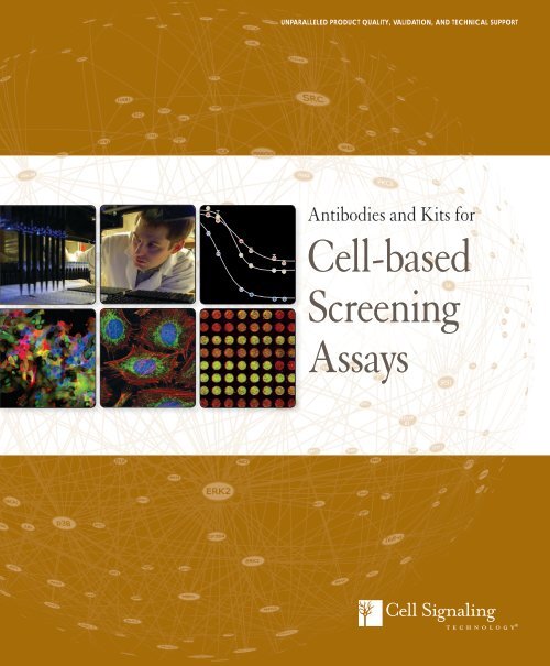
UNPARALLELED PRODUCT QUALITY, VALIDATION, AND TECHNICAL SUPPORT<br />
Antibodies and Kits for<br />
<strong>Cell</strong>-<strong>based</strong><br />
<strong>Screening</strong><br />
<strong>Assays</strong>
XP® Monoclonal Antibodies<br />
for <strong>Cell</strong>-<strong>based</strong> <strong>Screening</strong> <strong>Assays</strong><br />
XP ® monoclonal antibodies are a line of high quality<br />
rabbit monoclonal antibodies exclusively available<br />
from <strong>Cell</strong> Signaling Technology (CST). Any product<br />
labeled with XP has been carefully selected <strong>based</strong> on<br />
superior performance in all approved applications.<br />
XP monoclonal antibodies are generated using<br />
XMT ® technology, a proprietary monoclonal<br />
method developed at CST. This technology provides<br />
access to a broad range of antibody-producing<br />
B cells unattainable with traditional monoclonal<br />
technologies, allowing more comprehensive<br />
screening and the identification of XP monoclonal<br />
antibodies with:<br />
Autophagy<br />
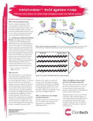
LC3B (D11) XP ® Rabbit mAb #3868: Confocal IF analysis of HeLa cells, untreated (left) or<br />
chloroquine-treated (right), using #3868 (green). Actin filaments were labeled using DY-554<br />
phalloidin (red). Blue pseudocolor = DRAQ5 ® #4084 (fluorescent DNA dye).<br />
Epigenetics<br />
Jak/Stat Signaling<br />
Events<br />
eXceptional specificity<br />
As with all CST antibodies, the antibody is specific to your<br />
target of interest, saving you valuable time and resources.<br />
+ eXceptional sensitivity<br />
The antibody will provide a stronger signal for your target protein<br />
in cells and tissues, allowing you to monitor expression of low<br />
levels of endogenous proteins, saving you valuable materials.<br />
+ eXceptional stability and reproducibility<br />
XMT technology combined with our stringent quality control ensures<br />
maximum lot-to-lot consistency and the most reproducible results.<br />
= eXceptional Performance<br />
XMT technology coupled with our extensive antibody validation<br />
and stringent quality control delivers XP monoclonal antibodies<br />
with eXceptional Performance in the widest range of applications.<br />
SUZ12 (D39F6) XP ® Rabbit mAb #3737: Confocal<br />
iF analysis of mouse embryonic stem cells growing<br />
on mouse embryonic fibroblast (MEF) feeder cells<br />
using #3737 (green). Actin filaments were labeled<br />
with DY-554 phalloidin (red).<br />
RTK Signaling<br />
Phospho-Stat5 (Tyr694)<br />
Phospho-Stat5 (Tyr694) (D47E7)<br />
XP ® Rabbit mAb #4322: Flow cytometric<br />
analysis of TF-1 cells, untreated<br />
(blue) or treated with Human Granulocyte<br />
Macrophage Colony Stimulating<br />
Factor (hGM-CSF) #8922 (green),<br />
using #4322.<br />
Phospho-Met (Tyr1234/1235) (D26) XP ® Rabbit mAb #3077: IHC analysis of paraffinembedded<br />
HCC827 xenograft (left) and paraffin-embedded papillary renal cell carcinoma (right)<br />
using #3077.<br />
Visit our website for more experimental details, additional information,<br />
and a complete list of available XP ® monoclonal antibodies.
Winner! Life Science Industry Awards ®<br />
:: Best Antibodies :: Best Breakthrough Products for Cancer Research<br />
<strong>Cell</strong> Signaling Technology is the leading provider of signal transduction reagents<br />
for the academic and drug discovery communities. Phosphorylation-specific<br />
antibodies from CST are the most highly cited in PubMed and are core reagents<br />
for multiple drug discovery platforms. We offer a complete line of products to<br />
support your cell-<strong>based</strong> assays at any stage of the drug discovery process. Our<br />
high quality reagents have been validated on a large number of popular screening<br />
and assay platforms and can be used in most biochemical or cell-<strong>based</strong> assays.<br />
Moreover, our exclusive line of XP ® monoclonal antibodies are especially suited<br />
for drug discovery applications where exceptional specificity, sensitivity, and<br />
reproducibility are of utmost importance.<br />
Find out more:<br />
All CST reagents are readily available for testing, lot reservation,<br />
custom lot production, and bulk purchases for high throughput<br />
screening campaigns.<br />
:: Please visit www.cellsignal.com/ddt/custom_reagents.html for further<br />
information.<br />
:: Contact sales@cellsignal.com for a list of carrier-free formulation antibodies.<br />
:: Custom Product Scientists are available to answer technical questions relating<br />
to your assay. For assistance, contact support@cellsignal.com.<br />
Sales Department<br />
:: Phone: 978-867-2396<br />
:: Toll-free phone: 866-310-9776<br />
:: U.S. E-mail: sales@cellsignal.com<br />
:: International E-mail:<br />
cst_intsales@cellsignal.com<br />
Table of Contents<br />
Custom Reagents<br />
PathScan ® Sandwich ELISA<br />
Kits and Antibody Pairs<br />
PathScan ® Antibody Array Kits<br />
<strong>Cell</strong>ular Analysis Tools<br />
Whole <strong>Cell</strong> <strong>Assays</strong>: Immunofluorescence<br />
PathScan ® Multiplex IF Kits<br />
Whole <strong>Cell</strong> <strong>Assays</strong>: Flow Cytometry<br />
Complementary Reagents<br />
for Whole <strong>Cell</strong> <strong>Assays</strong><br />
PTMscan ® Proteomics Services<br />
Cytokines and Growth Factors<br />
Environmental commitment: eco.cellsignal.com
Custom Reagents<br />
For custom reagents, requests, and services contact sales@cellsignal.com<br />
Custom Formulations<br />
:: Carrier-free formulation is available for a broad portfolio of products<br />
and is useful for conjugations, custom ELISA development, and other<br />
types of customized assay development.<br />
Direct Conjugation Services<br />
:: Many of our high quality antibodies are available off-the-shelf already<br />
conjugated to Alexa Fluor ® 488, 555, 594, and 647 fluorescent dyes,<br />
PE, biotin, HRP, or magnetic or sepharose beads.<br />
:: Price-competitive and high quality custom antibody conjugation<br />
of our catalog antibodies to Alexa Fluor dyes, PE, biotin, HRP, or<br />
sepharose or magnetic beads performed upon request.<br />
:: Recently revised sizing options available.<br />
Contact sales@cellsignal.com for sizing and pricing.<br />
Custom ELISA Production<br />
:: Custom ELISA products provided in convenient bulk packaging.<br />
:: Chemiluminescent and fluorescent detection options available.<br />
Bulk Reservations<br />
:: Single lots of any of our reagents can be reserved for medium or<br />
high-throughput assays, such as high content screening,<br />
HTRF ® -FRET, AlphaScreen ® , Meso Scale Discovery ® , or other assays.<br />
:: Custom bulk quantities are immediately available for most of our<br />
antibodies, including all of our high performance XP ® monoclonal<br />
antibodies.<br />
:: Volume-<strong>based</strong> discounts for carrier-free and all standard glycerol<br />
formulations are available upon request.<br />
Custom Product Quality Control<br />
Custom products are held to the same unparalleled standards<br />
of quality as off-the-shelf CST antibodies. Antibody purity<br />
and integrity are assessed by HPLC-SEC, confirming a single<br />
population of protein of the correct size, and excluding protein<br />
aggregation or degradation. SEC analysis helps ensure the most<br />
efficient antibody labeling or spotting. Lot-to-lot consistency<br />
is verified in a peptide immunofluorometric assay (DELFIA ® )<br />
comparing each lot to a previous lot.<br />
0.075<br />
0.070<br />
0.065<br />
0.060<br />
0.055<br />
0.050<br />
0.045<br />
0.040<br />
0.035<br />
0.030<br />
0.025<br />
0.020<br />
0.015<br />
0.010<br />
0.005<br />
0.000<br />
0.00 5.00 10.00 15.00 20.00 25.00 30.00 35.00 40.00<br />
Minutes<br />
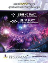
Phospho-Histone H3 (Ser10) (D2C8) XP ® Rabbit mAb #3377:<br />
HPLC-SEC analysis of a carrier-free lot of #3377 confirms product purity.<br />
Absorbance 280 nm<br />
Fluorescent Intensity 615 nm<br />
(x106)<br />
12<br />
10<br />
8<br />
6<br />
2<br />
4<br />
0<br />
-6 -5 -4 -3<br />
Lot 6 phosphopeptide<br />
Lot 6 non-phosphopeptide<br />
Lot 8 phosphopeptide<br />
Lot 6 non-phosphopeptide<br />
Antibody Concentration (log µM)<br />
Phospho-S6 Ribosomal Protein (Ser235/236) (D57.2.2E) XP ® Rabbit<br />
mAb #4858: Time-resolved immunofluorometric peptide assay was<br />
performed with increasing concentrations of two independent lots of #4858<br />
(carrier-free formulation) in the presence of phosphopeptide antigen (blue<br />
and green) or the corresponding non-phosphopeptide (red and purple). The<br />
assay verifies the phospho-specificity of the antibody and demonstrates<br />
consistent performance and minimal lot-to-lot variation.<br />
4 www.cellsignal.com Sales: sales@cellsignal.com Technical Support: support@cellsignal.com
PathScan ®<br />
Sandwich ELISA Kits<br />
Sandwich ELISA<br />
CST has applied its antibody expertise to<br />
identify antibody pairs with optimal activity<br />
in solid phase sandwich enzyme-linked<br />
immunosorbent assays (ELISA). These<br />
assays enable the detection of low amounts<br />
of target protein from cell lysates. Over<br />
170 PathScan ® Sandwich ELISA Kits are<br />
available for signaling proteins that serve as<br />
assay endpoints in drug discovery screening<br />
campaigns. Kits and pairs are developed,<br />
produced, and validated in-house, ensuring<br />
the highest kit quality.<br />
:: PathScan ® Sandwich ELISA Kits contain all necessary<br />
components for detection of endogenous levels of key signaling<br />
molecules. Matched phospho-specific and total protein ELISA kits<br />
are available for many targets. Colorimetric and chemiluminescent<br />
detection options available.<br />
:: PathScan ® Sandwich ELISA Antibody Pairs provide scientists<br />
with an economical alternative to our complete ELISA kits.<br />
:: PathScan ® Multi-Target ELISA Kits examine several important<br />
and well-characterized signaling events in a single assay.<br />
:: PathScan ® ELISA Control <strong>Cell</strong> Extracts provide the appropriate<br />
positive and negative controls and allow the standardization of<br />
signal obtained from different plates.<br />
:: Custom ELISA products allow researchers the option of different<br />
detection methods and plate formats. Convenient bulk packaging is<br />
available upon request.<br />
Colorimetric<br />
Sandwich<br />
ELISA Kits<br />
Target<br />
Phospho-4E-BP1 (Thr37/<br />
Thr46)<br />
4E-BP1 #7179<br />
Chemiluminescent<br />
Sandwich ELISA<br />
Kits<br />
Antibody<br />
Pairs<br />
#7216 #7854<br />
Multi-Target Kits<br />
Phospho-Acetyl-CoA<br />
#7986<br />
Carboxylase (Ser79)<br />
Acetyl-CoA Carboxlyase #7996<br />
β-Actin #7880 #7881<br />
Phospho-Akt (Thr308) #7252 #7135 #7144 <strong>Cell</strong> Growth #7239<br />
Phospho-Akt1 (Ser473) #7160 #7134 #7143 <strong>Cell</strong> Growth #7239<br />
Signaling Nodes #7272<br />
Akt1 #7170 #7132 #7142 <strong>Cell</strong> Growth #7239<br />
Signaling Nodes #7272<br />
Phospho-Akt2 (Ser474) #7048<br />
Phospho-Akt2 (Ser474)<br />
#7932<br />
(mouse preferred)<br />
Akt2 #7046<br />
Akt2 (mouse preferred) NEW #7930<br />
Phospho-Akt3 (Ser472)<br />
#7942<br />
(mouse preferred)<br />
Akt3 (mouse preferred) #7934<br />
Phospho-ALK (Tyr1586) #7159<br />
Phospho-ALK (Tyr1604) #7324 #7020<br />
ALK #7322 #7084<br />
Phospho-AMPKα (Thr172) #7959 #7955<br />
Phospho-ATF-2 (Thr71) #7185<br />
Phospho-Aurora A (Thr288) #7114 #7115<br />
Aurora A #7116 #7117<br />
Phospho-Axl (panTyr) #7042<br />
Axl #7040<br />
Phospho-Bad (Ser112) #7182 #7842 Apoptosis #7105<br />
Bad #7162 #7840 Apoptosis #7105<br />
E-Cadherin #7886 #7887<br />
Cleaved Caspase-3 (Asp175) #7190 Apoptosis #7105<br />
β-Catenin #7308 #7309<br />
Target<br />
Colorimetric<br />
Sandwich<br />
ELISA Kits<br />
Chemiluminescent<br />
Sandwich ELISA<br />
Kits<br />
Antibody<br />
Pairs<br />
Phospho-cdc2 (Tyr15) #7176 #7838<br />
Phospho-Chk1 (Ser317) #7870<br />
Chk1 #7872 #7873<br />
Phospho-Chk2 (Thr68) #7037<br />
Chk2 #7045 #7090<br />
Phospho-CREB (Ser133) NEW #7385<br />
CREB NEW #7390<br />
Cox2 NEW #7291<br />
Phospho-DDR1 (panTyr) #7863<br />
DDR1 #7845<br />
Phospho-EGFR (Tyr845) #7189<br />
Phospho-EGFR (Tyr1068) #7240 NEW #7036<br />
Phospho-EGFR (Tyr1173) #7187<br />
EGFR (E746-A750del NEW #8950<br />
Specific)<br />
EGFR (L858R Mutant Specific) NEW #8951<br />
EGFR #7250 NEW #7297<br />
Phospho-eIF2α (Ser51) #7948<br />
eIF2α #7952<br />
Phospho-eIF4E (Ser209) #7938<br />
eIF4E #7940<br />
Phospho-eNOS (Ser1177) #7980<br />
Phospho-Erk1 (p44 MAPK)<br />
(Thr202/Tyr204)<br />
#7315 #7278<br />
Phospho-Erk1/2 (p44/42<br />
MAPK) (Thr202/Tyr204)<br />
Erk1/2 (p44/42 MAPK) #7050<br />
Fatty Acid Synthase NEW #7689<br />
Phospho-FGFR2 (panTyr) NEW #7954<br />
FGFR2 NEW #7939<br />
Phospho-FLT3 (panTyr) #7761<br />
Multi-Target Kits<br />
#7177 #7246 <strong>Cell</strong> Growth #7239<br />
MAP Kinase #7274<br />
continued><br />
UNPARALLELED PRODUCT QUALITY, VALIDATION, AND TECHNICAL SUPPORT<br />
5
Normalized Absorbanc<br />
PathScan<br />
IC ® Sandwich ELISA Kits 50<br />
:<br />
continued<br />
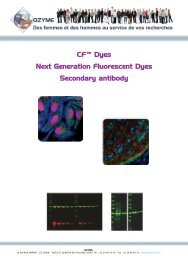
Normalized Absorbance 450 nm<br />
A-431 cells were exposed to varying concentrations of gefitinib (2 hr) or Wortmannin #9951 (1 hr),<br />
then treated with 100 ng/ml Human Epidermal Growth Factor (hEGF) #8916 (5 min). With increasing<br />
concentrations of gefitinib, a significant decrease (~25 fold and ~6.25 fold, respectively) in phospho-<br />
EGFR and phospho-Akt1 signals were detected using the PathScan ® Phospho-EGF Receptor (Tyr1068)<br />
Sandwich ELISA Kit #7240 and the PathScan ® Phospho-Akt1 (Ser473) Sandwich ELISA Kit<br />
#7160. With increasing concentrations of wortmannin, a significant decrease (~5 fold) in phospho-Akt1<br />
signal was detected. Wortmannin had no effect on phospho-EGFR levels.<br />
Normalized Absorbance 450 nm<br />
A-431 120 cells were exposed to varying concentrations of gefitinib for 2 hr, then treated with a combination<br />
of 100 ng/ml Human Epidermal Growth Factor (hEGF) #8916 and 100 ng/ml HGF (5 min). With increasing<br />
concentrations of gefitinib, a significant decrease (~20 fold and ~3.5 fold, respectively) in phospho-EGFR<br />
and phospho-p44/42<br />
100<br />
MAPK signals were detected using the PathScan ® Phospho-EGF Receptor<br />
(Tyr1068) Sandwich ELISA Kit #7240 and the PathScan ® Phospho-p44/42 MAPK (Thr202/Tyr204)<br />
Sandwich ELISA Kit #7177. In contrast, there was no decrease in phospho-Met signal as detected by<br />
the PathScan ® Phospho-Met (Tyr1234/1235) Sandwich ELISA Kit #7227.<br />
80<br />
malized Absorbance 450 nm<br />
60<br />
40<br />
20<br />
0<br />
PathScan 0 ® 0.0001 Sandwich 0.001 0.01 ELISA 0.1 1 Kits 10<br />
Gefitinib (µM)<br />
120<br />
100<br />
80<br />
Phospho-Met (Tyr1234/1235)<br />
Phospho-EGFR 60<br />
(Tyr1068)<br />
Phospho-p44/42 MAPK<br />
(Thr202/Tyr204)<br />
40 IC 50<br />
:<br />
20<br />
0<br />
120<br />
100<br />
80<br />
60<br />
40<br />
20<br />
0<br />
60<br />
www.cellsignal.com<br />
IC 50<br />
:<br />
6 Sales: sales@cellsignal.com Technical Support: support@cellsignal.com<br />
40<br />
0 0.0001 0.001 0.01 0.1 1 10<br />
Treatment (µM)<br />
0<br />
IC 50<br />
:<br />
26 nM<br />
290 nM<br />
Compound titration curves obtained using<br />
6 nM<br />
9 nM<br />
14 nM<br />
26 nM<br />
290 nM<br />
0.0001 0.001 0.01 0.1 1 10<br />
Gefitinib (µM)<br />
6 nM<br />
Phospho-EGFR (Tyr1068)<br />
Wortmannin<br />
Phospho-Akt1 (Ser473)<br />
Phospho-EGFR (Tyr1068)<br />
Gefitinib<br />
Phospho-Akt1 (Ser473)<br />
Phospho-Met (Tyr1234/1235)<br />
Phospho-EGFR (Tyr1068)<br />
Phospho-p44/42 MAPK<br />
(Thr202/Tyr204)<br />
Target<br />
Colorimetric<br />
Sandwich<br />
ELISA Kits<br />
Chemiluminescent<br />
Sandwich ELISA<br />
Kits<br />
Phospho-FLT3 (Tyr591) #7206 #7021<br />
FLT3 #7202<br />
Antibody<br />
Pairs<br />
GFP #7878 #7879<br />
Multi-Target Kits<br />
Phospho-HER2 (panTyr) #7968<br />
Phospho-HER2<br />
#7148 #7817<br />
(Tyr1221/1222)<br />
HER2 #7310<br />
Phospho-HER3 (panTyr) #7890<br />
HER3 #7888<br />
Acetyl-Histone H2A #7233<br />
Acetyl-Histone H2B #7178<br />
Acetyl-Histone H2B (Lys5) #7218<br />
Acetyl-Histone H2B (Lys20) #7222<br />
Acetyl-Histone H3 #7232 #7209<br />
Acetyl-Histone H3 (Lys9) #7121<br />
Acetyl-Histone H3 (Lys18) #7122<br />
Mono-Methyl Histone H3 #7123<br />
(Lys4)<br />
Di-Methyl-Histone H3 (Lys4) #7124<br />
Tri-Methyl-Histone H3 (Lys4) #7125<br />
Pan-Methyl-Histone H3 (Lys9) #7864<br />
Di-Methyl-Histone H3 (Lys9) #7862<br />
Tri-Methyl-Histone H3 (Lys27) #7866<br />
Di-Methyl-Histone H3 (Lys36) #7868<br />
Phospho-Histone H3 (Ser10) #7155 NEW #7303 #7207<br />
Histone H3 #7253<br />
Acetyl-Histone H4 #7238<br />
Acetyl-Histone H4 (Lys8) #7224<br />
Acetyl-Histone H4 (Lys12) #7228<br />
Phospho-HSP27 (Ser78) #7290<br />
Phospho-HSP27 (Ser82) #7152<br />
HSP27 #7295<br />
Phospho-IGF-I Receptor β #7302 #7820<br />
(Tyr1131)<br />
Phospho-IκBα (Ser32) #7355 #7343 Inflammation #7276<br />
IκBα #7360 #7831<br />
Phospho-IKKα (Ser176/180) Phospho-EGFR (Tyr1068) #7073<br />
Total-IKKα<br />
Wortmannin<br />
Phospho-Akt1 (Ser473) #7078<br />
Phospho-IKKβ (Ser177/181) Phospho-EGFR (Tyr1068) #7080<br />
iNOS<br />
Gefitinib<br />
Phospho-Akt1 (Ser473) #7097<br />
Phospho-Insulin Receptor β #7082<br />
(panTyr)<br />
Phospho-Insulin Receptor β #7254 #7827<br />
(Tyr1146)<br />
Phospho-Insulin Receptor β #7258 #7828<br />
(Tyr1150/1151)<br />
Phospho-Insulin Receptor β #7326 #7823<br />
(Tyr1345)<br />
Insulin Receptor β #7069<br />
Phospho-IRS-1 (panTyr) #7133 #7347<br />
Phospho-IRS-1 (Ser302) #7283 #7284<br />
Phospho-IRS-1 (Ser307) #7287 #7288<br />
Phospho-IRS-1 (Ser612) #7332<br />
IRS-1 #7328<br />
Phospho-IRS-2 (panTyr) #7860 #7861
Target<br />
Colorimetric<br />
Sandwich<br />
ELISA Kits<br />
Chemiluminescent<br />
Sandwich ELISA<br />
Kits<br />
Antibody<br />
Pairs<br />
IRS-2 #7884 #7885<br />
Phospho-c-Jun (Ser63) #7145 #7027 #7141<br />
c-Jun #7150 #7028 #7314<br />
Phospho-c-Kit (panTyr) #7231 #7294<br />
Phospho-c-Kit (Tyr719) #7298 #7299<br />
c-Kit #7197<br />
Phospho-LAT (Tyr191) #7936 #7937<br />
Multi-Target Kits<br />
Phospho-Lck (Tyr505) #7941 #7993<br />
Phospho-MARCKS<br />
NEW #7251<br />
(Ser152/156)<br />
MARCKS NEW #7188<br />
Phospho-MEK1 (Ser217/221) #7175 #7029 #7211 Signaling Nodes #7272<br />
MAP Kinase #7274<br />
MEK1 #7165 #7030 #7215 MAP Kinase #7274<br />
Phospho-Met (panTyr) #7333 #7334<br />
Phospho-Met (Tyr1003) #7241<br />
Phospho-Met (Tyr1234/1235) #7227 #7229<br />
Phospho-Met (Tyr1349) #7896<br />
Met #7242<br />
Phospho-NF-κB p65 (Ser536) #7173 #7834 Inflammation #7276<br />
Signaling Nodes #7272<br />
NF-κB p65 #7174 #7836 Inflammation #7276<br />
Cleaved Notch1 (Val1744) NEW #7194<br />
Notch1 NEW #7245<br />
p21 Waf1/Cip1 #7167 #7856<br />
Phospho-p38 (Thr180/Tyr182) #7946 MAP Kinase #7274<br />
Inflammation #7276<br />
Signaling Nodes #7272<br />
Acetyl-p53 #7236 #7848<br />
Phospho-p53 (Ser15) #7365 #7846 Apoptosis #7105<br />
p53 #7370 #7844 Apoptosis #7105<br />
Phospho-p70 S6 Kinase #7063 #7153 #7053<br />
(Thr389)<br />
p70 S6 Kinase #7038 #7039<br />
Phospho-p90 RSK1 (Ser380) #7965<br />
p90 RSK1 #7966<br />
Cleaved PARP (Asp214) #7262 #7858 Apoptosis #7105<br />
Phospho-PDGFR α/β (panTyr) NEW #7235<br />
Phospho-PDGFR α (Tyr849) #7296 #7317<br />
PDGFR α #7318 #7264<br />
Phospho-PDGFR β (Tyr751) #7345 #7826<br />
Phospho-PRAS40 (Thr246) #7327<br />
PRAS40 NEW #7331<br />
Phospho-PTEN (Ser380) #7285<br />
PTEN #7882 #7883<br />
Phospho-Ret (panTyr) #7034<br />
Ret #7032<br />
Phospho-Ros (panTyr) #7093<br />
Ros #7091<br />
Phospho-S6 Ribosomal #7205 NEW #7316 #7201 <strong>Cell</strong> Growth #7239<br />
Protein (Ser235/236)<br />
S6 Ribosomal Protein #7225 NEW #7337 #7203 <strong>Cell</strong> Growth #7239<br />
Phospho-SAPK/JNK1/2/3<br />
(Thr183/Tyr185)<br />
#7325 #7849 #7217 MAP Kinase #7274<br />
Inflammation #7276<br />
SAPK/JNK1/2/3 #7330 #7869 #7219 MAP Kinase #7274<br />
Colorimetric<br />
Sandwich<br />
ELISA Kits<br />
Target<br />
Phospho-Smad2<br />
#7348<br />
(Ser465/467)<br />
Smad2 #7244<br />
Chemiluminescent<br />
Sandwich ELISA<br />
Kits<br />
Antibody<br />
Pairs<br />
Multi-Target Kits<br />
Phospho-Src (Tyr416) #7953 #7963<br />
Src #7984 #7992<br />
Phospho-Stat1 (Tyr701) #7234<br />
Phospho-Stat3 (Tyr705) #7300 #7149 #7146 Signaling Nodes #7272<br />
Inflammation #7276<br />
Phospho-Stat3 (Ser727) #7995 NEW #8026<br />
Stat3 #7305<br />
Phospho-Stat5 (Tyr694) #7113 #7281<br />
Phospho-Stat6 (Tyr641) NEW #7304<br />
Phospho-Survivin (Thr34) NEW #7193<br />
Survivin #7169<br />
Phospho-Syk (panTyr) #7928 #7929<br />
Phospho-Syk (Tyr525/526) #7970<br />
Phospho-mTOR (Ser2448) NEW #7976<br />
mTOR NEW #7974<br />
Phospho-TrkA (Tyr490) #7210<br />
Phospho-TrkA (Tyr674/675) #7212<br />
TrkA #7208<br />
Phospho-TrkB (panTyr) #7108<br />
Phospho-TrkB (Tyr516) #7111<br />
Phospho-TrkB (Tyr706/707) #7118<br />
TrkB #7106<br />
α-Tubulin #7944 #7945<br />
Acetyl-α-Tubulin #7204<br />
Phospho-VEGFR-2 (Tyr1175) #7335 #7824<br />
VEGFR-2 #7340 #7825<br />
Phospho-Zap-70 (Tyr319) #7171 #7852<br />
Zap-70 #7172 #7850<br />
Complementary Reagents for<br />
PathScan® Sandwich ELISA<br />
<strong>Cell</strong> Signaling Technology offers a full set of<br />
complementary reagents for ELISA sample<br />
preparation and detection.<br />
#9998 BSA<br />
#9803 <strong>Cell</strong> Lysis Buffer (10X)<br />
#7018 PathScan ® Sandwich ELISA<br />
Lysis Buffer (1X)<br />
#9808 Phosphate Buffered Saline<br />
(PBS-20X)<br />
#9809 Phosphate Buffered Saline<br />
with Tween 20 (PBST-20X)<br />
#7002 STOP Solution<br />
#7004 TMB Substrate<br />
UNPARALLELED PRODUCT QUALITY, VALIDATION, AND TECHNICAL SUPPORT<br />
7
PathScan ®<br />
Chemiluminescent<br />
Sandwich ELISA Kits<br />
PathScan ® Chemiluminescent Sandwich ELISA Kits<br />
provide the broadest dynamic range and assay<br />
sensitivity while requiring half the sample size.<br />
:: Chemiluminescent detection system offers broader dynamic<br />
range and greater assay sensitivity.<br />
:: Low volume microplates retain high signal and sensitivity while<br />
using half the sample size of traditional colorimetric ELISA.<br />
:: Any standard plate-<strong>based</strong> luminometer can be used for<br />
detection, making specialized equipment unnecessary.<br />
:: Technical support is provided by the same scientists that<br />
develop and produce the kits, ensuring a thorough, fast, and<br />
accurate response.<br />
RLU (x10 5 )<br />
25<br />
20<br />
15<br />
10<br />
5<br />
phospho<br />
nonphospho<br />
0<br />
0.00 0.25 0.50 0.75 1.00 1.25 1.50<br />
Protein conc. of lysate (mg/ml)<br />
6<br />
4<br />
2<br />
0<br />
0.00 0.03 0.05 0.08 0.10 0.13 0.15<br />
PathScan ® Phospho-ALK (Tyr1604) Chemiluminescent Sandwich ELISA Kit #7020: Relationship<br />
between protein concentrations of lysates from NCI-H2228 cells, lysed with (phospho) and without<br />
(nonphospho) the addition of phosphatase inhibitors to the lysis buffer, and immediate light generation<br />
with chemiluminescent substrate. Graph inset corresponding to the shaded area shows high sensitivity<br />
and a linear response at the low protein concentration range.<br />
PathScan ®<br />
Antibody Array Kits<br />
PathScan ® Antibody Array Kits provide the<br />
opportunity to simultaneously analyze numerous<br />
targets with minimal sample requirements.<br />
The kits are slide-<strong>based</strong> antibody arrays founded on the sandwich<br />
immunoassay principle and are designed to investigate signal transduction<br />
occuring through key pathway nodes in a multiplex format. These kits include<br />
all necessary reagents for either fluorescent or chemiluminescent detection.<br />
:: Arrays are developed and optimized in-house, incorporating the highest<br />
quality antibodies and ensuring results you can trust.<br />
:: Arrays allow the analysis of phosphorylation levels of a large number of<br />
proteins per assay, saving valuable time and reagents.<br />
:: Arrays are designed to detect RTKs and/or key intracellular molecules,<br />
allowing the most comprehensive readout of downstream signaling events.<br />
:: Technical support is provided by our in-house Molecular Assay Group, the<br />
same scientists who developed and produce the kits.<br />
:: The option of chemiluminescent readout allows convenient and easy<br />
detection without specialized instrumentation.<br />
PathScan ® Antibody Array Kits<br />
#7982 PathScan ® RTK Signaling Antibody Array Kit<br />
(Chemiluminescent Readout)<br />
#7949 PathScan ® RTK Signaling Antibody Array Kit<br />
(Fluorescent Readout)<br />
NEW #7323 PathScan ® Intracellular Signaling Antibody Array Kit<br />
(Chemiluminescent Readout)<br />
NEW #7744 PathScan ® Intracellular Signaling Antibody Array Kit<br />
(Fluorescent Readout)<br />
Reactivity<br />
H<br />
H<br />
H, M<br />
H, M<br />
A<br />
RFU<br />
B<br />
20000<br />
16000<br />
12000<br />
8000<br />
4000<br />
0<br />
HT-29<br />
HT-29 + UV<br />
HT-29<br />
Erk1/2 (Thr202/Tyr204)<br />
Stat1 (Tyr701)<br />
Stat3 (Tyr705)<br />
Akt (Thr308)<br />
Akt (Ser473)<br />
AMPKα (Thr172)<br />
S6 (Ser235/236)<br />
mTOR (Ser2448)<br />
HSP27 (Ser78)<br />
Bad (Ser112)<br />
p70S6K (Thr389)<br />
PRAS40 (Ser246)<br />
p53 (Ser15)<br />
SAPK/JNK (Thr183/Tyr185)<br />
p38α (Thr180/Tyr182)<br />
PARP (Cleavaed Asp214)<br />
Phospho-HSP27 (Ser78)<br />
Phospho-SAPK/JNK<br />
(Thr183/Tyr185)<br />
HT-29 + UV<br />
Phospho-p53 (Ser15)<br />
Caspase-3 (Cleaved Asp175)<br />
GSK-3β (Ser9)<br />
Phospho-p38 (Thr180/Tyr182)<br />
PathScan ® Intracellular Signaling Antibody Array Kit (Fluorescent Readout) #7744: HT-29 cells<br />
were grown to 80% confluency, then either left untreated or irradiated with UV for 60 minutes. Lysates<br />
were prepared and analyzed using the PathScan ® Intracellular Signaling Antibody Array Kit (Fluorescent<br />
Readout) #7744. Panel A shows quantification of results. Panel B shows images that were acquired using<br />
the LI-COR ® Odyssey ® system.<br />
8 www.cellsignal.com Sales: sales@cellsignal.com Technical Support: support@cellsignal.com
<strong>Cell</strong>ular Analysis Tools<br />
Cyclic AMP and Cyclic GMP XP ® Assay Kits<br />
Cyclic AMP and Cyclic GMP XP ® Assay Kits (#4339 and<br />
#4360) are <strong>based</strong> on the competition immunoassay principle and<br />
can be used to measure the activation of many G protein-coupled<br />
receptors (GPCRs). Kits using a chemiluminescent option are also<br />
available (#8019 and #8020). In these kits, cyclic nucleotide<br />
in the sample of interest competes with a fixed amount of cyclic<br />
nucleotide-HRP conjugate provided in the kit for binding to<br />
cyclic nucleotide-specific XP ® rabbit monoclonal antibody that is<br />
precoated on the assay plate. Because of the competitive nature of<br />
this assay, the magnitude of the absorbance is inversely proportional<br />
to the quantity of cyclic nucleotide in the sample.<br />
:: Highest quality XP monoclonal antibodies employed in the assay<br />
ensure the greatest possible sensitivity and specificity.<br />
:: Technical support is provided by the scientists who designed and<br />
produce these products and know them best.<br />
RLU (x10 6 )<br />
5<br />
4<br />
3<br />
2<br />
1<br />
0<br />
-5<br />
-4 -3 -2 -1 0 1 2 3<br />
Log (FSK, µM)<br />
110<br />
90<br />
70<br />
50<br />
30<br />
10<br />
-5<br />
-4 -3 -2 -1 0 1 2 3<br />
Log (FSK, µM)<br />
Cyclic AMP XP ® Chemiluminescent Assay Kit #8019: Treatment of CHO cells with Forskolin (FSK)<br />
#3828 increases cAMP concentration as detected by #8019. CHO cells were seeded at 4x10 4 cells/<br />
well in a 96-well plate and incubated overnight. <strong>Cell</strong>s were pretreated with 0.5 mM IBMX (30 min)<br />
prior to FSK treatment (15 min) and lysed with 1X <strong>Cell</strong> Lysis Buffer #9803. The light emission values<br />
(left) and percentage of activity (right) are shown. The percentage of activity is calculated as follows:<br />
% activity =100X[(RLU-RLU basal<br />
)/(RLU max<br />
-RLU basal<br />
)], where RLU is the sample relative light unit, RLU max<br />
is the light emission at maximum stimulation (i.e. high FSK concentration), and RLU basal<br />
is the light<br />
emission at basal level (no FSK). FSK directly activates adenylyl cyclases and increases cellular cAMP<br />
concentration. IBMX is a non-specific inhibitor of cAMP and cGMP phosphodiesterases and promotes<br />
accumulation of cAMP and cGMP in cells.<br />
% Activity<br />
NEW XTT <strong>Cell</strong> Viability Kit<br />
The XTT <strong>Cell</strong> Viability Kit #9095 offers a simple means of<br />
performing drug discovery compound screens and toxicity studies.<br />
In this kit, cellular metabolism is monitored through cellular<br />
dehydrogenase-mediated enzymatic reduction of the tetrazolium salt<br />
XTT to a colored formazan dye. Metabolically active cells are capable<br />
of reducing XTT to a colored dye, and hence, the proportion of dye<br />
quantified by measuring absorbance at 450 nm is proportional to the<br />
number of viable cells in a sample.<br />
Absorbance<br />
450nm<br />
3.5<br />
3.0<br />
2.5<br />
2.0<br />
1.5<br />
1.0<br />
0.5<br />
BrdU<br />
XTT<br />
XTT <strong>Cell</strong> Viability Kit #9095: HeLa cells were seeded at 1x10 4 cells/well in a 96-well plate and incubated<br />
overnight. <strong>Cell</strong>s were then treated with various concentrations of Staurosporine #9953 overnight. The<br />
cytotoxicity was measured using #9095 (red) and BrdU <strong>Cell</strong> Proliferation Assay Kit #6813 (blue) as<br />
shown in the left panel. The percent inhibition in each assay was calculated and plotted in the right panel.<br />
Staurosporine is a nonspecific kinase inhibitor and induces cellular apoptosis.<br />
% Inhibition<br />
0<br />
0<br />
-3 -2 -1 0 1 2 -3 -2 -1 0 1 2<br />
Log (Staurosporine, µM)<br />
Log (Staurosporine, µM)<br />
120<br />
100<br />
80<br />
60<br />
40<br />
20<br />
BrdU<br />
XTT<br />
BrdU <strong>Cell</strong> Proliferation Assay Kit<br />
The BrdU <strong>Cell</strong> Proliferation Assay Kit #6813 is a plate-<strong>based</strong><br />
immunoassay that provides a straight forward means of assaying<br />
fundamental cellular activity. This kit offers an accurate, sensitive,<br />
and direct readout of cell division unattainable with viability dyes.<br />
:: Ability to interface with microplate environment, allowing higher<br />
throughput.<br />
:: Elimination of the need for microscopy, yielding results without<br />
specialized equipment.<br />
:: Elimination of the need for radioactive isotope labeling, providing<br />
a safer and simpler protocol.<br />
Absorbance<br />
450nm<br />
4<br />
3<br />
2<br />
1<br />
0<br />
0.001 0.01 0.1 1 10 100<br />
hEGF (ng/ml)<br />
BrdU <strong>Cell</strong> Proliferation Assay<br />
Kit #6813: Treatment of MCF 10A<br />
cells with Human Epidermal Growth<br />
Factor (hEGF) #8916 increases cell<br />
proliferation as detected by #6813.<br />
MCF 10A cells were seeded at<br />
1x10 4 cells/well in a 96-well plate<br />
and incubated overnight. <strong>Cell</strong>s were<br />
then starved in serum-free medium<br />
overnight. hEGF was added to the<br />
plate and cells were incubated for 24<br />
hr. Finally, 10 μM BrdU was added to<br />
the plate and cells were incubated<br />
for 4 hr.<br />
UNPARALLELED PRODUCT QUALITY, VALIDATION, AND TECHNICAL SUPPORT<br />
9
Whole <strong>Cell</strong> <strong>Assays</strong><br />
Antibody Validation for <strong>Cell</strong>-<strong>based</strong> Immunofluorescent <strong>Assays</strong><br />
Scientists at <strong>Cell</strong> Signaling Technology (CST) have validated over 800<br />
activation state-specific (e.g., phosphorylation-specific) and total protein<br />
antibodies for immunofluorescence (IF) applications such as automated<br />
cell-<strong>based</strong> assays.<br />
All CST antibodies approved for use in fluorescent cell-<strong>based</strong> assays have<br />
undergone a rigorous validation process to ensure optimal performance,<br />
including in-house testing on various high content platforms, such as the<br />
TTP ® LabTech Acumen ® e X3 and <strong>Cell</strong>omics ® ArrayScan ® V TI . Additionally,<br />
our exclusive line of XP ® monoclonal antibodies display the exceptional<br />
specificity, sensitivity, and reproducibility needed for the most robust<br />
automated fluorescent assays.<br />
Please contact our High Content <strong>Screening</strong> Group at HCS@cellsignal.com<br />
for more information or assistance with your HCS assays.<br />
Validation Includes:<br />
:: Antibody titration to determine optimal antibody concentration.<br />
:: Treatment of cell lines with pathway-specific inhibitors to verify<br />
treatment-induced translocation.<br />
:: Phosphatase treatment to verify phospho-specificity.<br />
:: Requirement of threshold signal-to-noise ratio in antibody:isotype<br />
comparison to ensure greatest possible sensitivity.<br />
:: Stringent testing to ensure lot-to-lot consistency.<br />
:: Antibody performance demonstrated in-house on multiple high content<br />
platforms.<br />
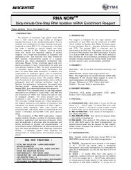
MAP Kinase Signaling<br />
Normalized Mean Fluorescence Intensity<br />
100<br />
80<br />
60<br />
40<br />
20<br />
Phospho-CREB<br />
Phospho-p44/42<br />
Phospho-p90RSK<br />
0 0.1 0.5 1 5 10 50 100<br />
U0126 (µM)<br />
Akt/PI3K and MAP Kinase Signaling<br />
Normalized Mean Fluorescence Intensity<br />
140<br />
120<br />
100<br />
80<br />
60<br />
40<br />
20<br />
0<br />
A<br />
Phospho-p44/42 MAPK (Erk1/2)<br />
Phospho-Akt<br />
0 0.05 0.1 0.5 1 5 10 50 100<br />
LY294002 (µM)<br />
-U0126/+TPA +U0126/+TPA<br />
Untreated LY294002-treated<br />
Normalized Mean Fluorescence Intensity<br />
100<br />
80<br />
60<br />
40<br />
20<br />
0<br />
B<br />
Phospho-p44/42 MAPK (Erk1/2)<br />
Phospho-Akt<br />
0 0.05 0.1 0.5 1 5 10 50 100<br />
U0126 (µM)<br />
Untreated<br />
U0126-treated<br />
phospho-p44/42 MAPK (Erk1/2) (green)<br />
red = phospho-Akt; green = phospho-p44/42 MAPK (Erk1/2); blue = nuclei<br />
red = phospho-Akt; green = phospho-p44/42 MAPK (Erk1/2); blue = nuclei<br />
A549 cells were concurrently exposed to U0126 (MEK1/2 Inhibitor)<br />
#9903 and the phorbol ester TPA #4174 to assess the effect on<br />
MAPK signaling. Phospho-p44/42 MAPK (Erk1/2), detected<br />
with Phospho-p44/42 MAPK (Erk1/2) (Thr202/Tyr204)<br />
(D13.14.4E) XP ® Rabbit mAb #4370, and two downstream<br />
signaling proteins, phospho-p90RSK and phospho-CREB,<br />
detected with Phospho-p90RSK (Thr573) Antibody #9346 and<br />
Phospho-CREB (Ser133) (87G3) Rabbit mAb #9198, were<br />
used as readouts. U0126 was titrated as an 8-point dose curve<br />
in triplicate to monitor the reproducibility and validity of the assay.<br />
The signal for each antibody was analyzed using an Acumen ®<br />
e X3 and images were acquired with a <strong>Cell</strong>omics ® ArrayScan ® V TI .<br />
With increasing concentrations of U0126, a significant decrease<br />
in phospho-p44/42 MAPK (Erk1/2) signal (~10-fold), as well as<br />
phospho-p90RSK and phospho-CREB (>2-fold), was observed as<br />
compared to the TPA-stimulated control.<br />
Normalized Mean Fluorescence Intensity<br />
120<br />
100<br />
80<br />
60<br />
40<br />
20<br />
0<br />
C<br />
Phospho-Akt<br />
Phospho-p44/42 MAPK (Erk1/2)<br />
Phospho-S6 Ribosomal Protein<br />
0 0.05 0.1 0.5 1 5 10 50 100<br />
LY294002 (µM) / U0126 (µM) / Rapamycin (nM)<br />
MCF7 cells were grown under normal conditions in 96-well plates and<br />
treated for 2 hr at 37 ˚C with vehicle or increasing concentrations of<br />
LY294002 (PI3 Kinase Inhibitor) #9901 (A), U0126 (MEK1/2 Inhibitor)<br />
#9903 (B), or a combination of LY294002, U0126, and Rapamycin<br />
(FRAP/mTOR Inhibitor) #9904 (C). <strong>Cell</strong>s were fixed, incubated<br />
overnight with the PathScan ® Signaling Nodes Multiplex IF Kit<br />
#8999 primary antibody cocktail, and subsequently labeled with a<br />
detection cocktail of Alexa Fluor ® conjugated secondary antibodies.<br />
Nuclei were labeled with Hoechst 33342 #4082. Fluorescence<br />
quantification was performed using a TTP ® LabTech Acumen ® e X3<br />
high content screening platform, and images were acquired using a<br />
<strong>Cell</strong>omics ® ArrayScan ® V TI . Fluorescence intensities per well were<br />
normalized to 100% of vehicle control, and inhibition curves were<br />
generated using Spotfire ® .<br />
10 www.cellsignal.com Sales: sales@cellsignal.com Technical Support: support@cellsignal.com
Autophagy Markers<br />
LC3B #3868<br />
LC3A #4599<br />
Rab7 #9367<br />
Average spot count per cell<br />
25<br />
20<br />
15<br />
10<br />
5<br />
0<br />
untreated<br />
starved<br />
chloroquine<br />
Untreated<br />
loperamide<br />
nicardipine<br />
pimozide<br />
fluspirilene<br />
35<br />
30<br />
25<br />
20<br />
15<br />
10<br />
5<br />
0<br />
Average spot count per cell<br />
40<br />
untreated<br />
Nicardipine<br />
starved<br />
chloroquine<br />
loperamide<br />
nicardipine<br />
pimozide<br />
fluspirilene<br />
Loperamide<br />
Average of mean total count<br />
30<br />
25<br />
20<br />
15<br />
10<br />
5<br />
0<br />
untreated<br />
starved<br />
chloroquine<br />
loperamide<br />
nicardipine<br />
Chloroquine<br />
pimozide<br />
fluspirilene<br />
LC3B<br />
HeLa cells were subjected to various autophagy-inducing treatments as labeled (8 hr of serum starvation or the following drugs for 2 hr: chloroquine 50 µM, nicardipine<br />
4.8 µM, loperamide 4.9 µM, pimozide 5.4 µM, or fluspirilene 5.3 µM) and autophagy levels were assessed using three markers, detected using LC3B (D11) XP ®<br />
Rabbit mAb #3868, LC3A (D50G8) XP ® Rabbit mAb #4599, and Rab7 (D95F2) XP ® Rabbit mAb #9367. The signal for each antibody was analyzed using an<br />
Acumen ® e X3 and images for LC3B were acquired with a <strong>Cell</strong>omics ® ArrayScan ® V TI .<br />
PathScan ® Multiplex IF Kits<br />
PathScan ® Multiplex IF Kits provide a novel multiplex assay for use with<br />
automated imaging or laser scanning high content platforms or manual<br />
immunofluorescence microscopy. The kits contain a cocktail of three<br />
high quality primary antibodies as well as a detection cocktail utilizing<br />
the Alexa Fluor ® series of fluorescent dyes. Antibody and dye pairings<br />
have been pre-optimized, and each kit contains enough reagents for<br />
100 assays (<strong>based</strong> on a working volume of 100 μl/test).<br />
:: Kits allow the analysis of multiple pathway endpoints within a single<br />
sample, saving time and reagents.<br />
:: Kits are produced and optimized in-house with the highest quality<br />
antibodies, providing you with the greatest possible specificity and<br />
sensitivity.<br />
:: Technical support is provided by our in-house IF group who<br />
developed the products and know them best.<br />
PathScan ® Multiplex IF Kits (100 assays)<br />
Reactivity<br />
#7967 PathScan ® EGF Receptor Activation Multiplex IF Kit H, Mk, (M)<br />
Kit includes: cocktails to simultaneously detect levels<br />
of EGF receptor, phospho-EGF receptor (Tyr1068),<br />
phospho-p44/42 MAPK (Erk1/2) (Thr202/Tyr204)<br />
#8999 PathScan ® Signaling Nodes Multiplex IF Kit H, M, R, Mk<br />
Kit includes: cocktails to simultaneously detect levels<br />
of phospho-Akt (Ser473), phospho-p44/42 MAPK Erk1/2<br />
(Thr202/Tyr204), phospho-S6 ribosomal protein (Ser235/236)<br />
#7851 PathScan ® Apoptosis and Proliferation Multiplex IF Kit H, Mk<br />
Kit includes: cocktails to simultaneously detect levels of<br />
phospho-histone H3 (Ser10), cleaved PARP (Asp214), α-tubulin<br />
UNPARALLELED PRODUCT QUALITY, VALIDATION, AND TECHNICAL SUPPORT<br />
11
0<br />
0<br />
Antibody Validation for Flow Cytometry<br />
Over 550 <strong>Cell</strong> Signaling Technology (CST) antibodies<br />
have been validated by our in-house Flow Cytometry<br />
Group – our goal is to provide our customers with the most<br />
specific antibodies, brightest signal, and lowest background<br />
possible. CST also offers Alexa Fluor ® conjugates and phycoerythrin<br />
(PE) conjugates optimized for flow cytometry.<br />
These reagents allow flow cytometry to be applied as a<br />
platform for the analysis of disease mechanisms, biomarker<br />
identification, and drug discovery.<br />
Validation Includes:<br />
:: Titration to determine optimal dilution.<br />
:: Comparison of signal to isotype control to estimate<br />
the nonspecific binding of primary antibodies.<br />
:: Treatment of cell lines with pathway-specific<br />
inhibitors to verify target specificity.<br />
:: Use of known positive and negative cell lines to<br />
verify target specificity.<br />
:: Phosphatase treatment to confirm phosphospecificity<br />
of the antibody.<br />
:: Extensive quality control testing to guarantee<br />
stability over time and ensure lot-to-lot consistency.<br />
Cleaved Caspase-3-positive <strong>Cell</strong>s<br />
SS Lin: SS Lin<br />
20<br />
15<br />
10<br />
5<br />
0<br />
30<br />
25<br />
20<br />
15<br />
10<br />
5<br />
63<br />
48<br />
10 0 101<br />
FL1 Log<br />
10 0<br />
10 1<br />
FL1 Log<br />
10 2 10z<br />
10 2 10z<br />
0.1 0.5 1 5 10 50 100 500 1000<br />
Etoposide (µM)<br />
Untreated<br />
10 0 10 1 10 2 10 3<br />
20<br />
15<br />
10<br />
5<br />
0<br />
TPA-treated<br />
10 0 10 1 10 2 10 3<br />
Cleaved Caspase-3 (Asp175) Antibody (Alexa<br />
Fluor ® 488 Conjugate) #9669: Jurkat cells were<br />
exposed to varying concentrations of etoposide for 6<br />
hours. Increasing concentrations resulted in a greater<br />
percentage of cleaved caspase-3-positive cells as<br />
detected using #9669. Inset: Flow cytometric analysis<br />
of Jurkat cells, untreated (upper) or etoposide-treated<br />
(500 µM, lower), using #9669.<br />
20<br />
15<br />
10<br />
5<br />
0<br />
Overlay<br />
Phospho-S6 Ribosomal Protein (Ser235/236) (D57.2.2E) XP ® Rabbit mAb (Alexa Fluor ® 488 Conjugate)<br />
#4803: Flow cytometric analysis of whole blood, untreated or treated with TPA #4174, using #4803.<br />
10 0 10 1 10 2 10 3<br />
Conjugated Antibodies<br />
We offer a growing selection of our best performing<br />
antibodies conjugated to Alexa Fluor ® dyes, biotin, PE, HRP,<br />
and sepharose or magnetic beads. Antibody conjugation<br />
is performed in-house, and is followed by rigorous quality<br />
control and stringent validation protocols. Technical support<br />
is provided by the same scientists who produce the products<br />
and know them best.<br />
:: Alexa Fluor ® 488, 555, 594, and 647 conjugates – most ideal for highly sensitive<br />
immunofluorescent and flow cytometric assays<br />
:: PE conjugates – optimal for multiplexing with other labeled antibodies in flow cytometry<br />
:: Bead conjugates – covalently immobilized on sepharose or magnetic beads for simple<br />
and efficient immunoprecipitation<br />
:: Biotinylated antibodies – for use with streptavidin detection systems<br />
:: HRP conjugates – for one-step western blot detection<br />
:: Custom formulations – available upon request (see page 4)<br />
Events<br />
Events<br />
1023<br />
73<br />
A<br />
1023<br />
76<br />
B<br />
Phospho-Stat3 (Tyr705)<br />
(Alexa Fluor ® 488 Conjugate)<br />
Phospho-Stat3 (Tyr705) (D3A7)<br />
XP ® Rabbit mAb (Alexa Fluor ® 488<br />
Conjugate) #4323: Flow cytometric<br />
analysis of Jurkat cells, untreated (blue)<br />
or treated with Human Interferon-α1<br />
(hIFN-α1) #8927 (green), using #4323<br />
compared to concentration-matched<br />
Rabbit (DA1E) mAb IgG XP ® Isotype<br />
Control (Alexa Fluor ® 488 Conjugate)<br />
#2975 (red).<br />
EpCAM (PE Conjugate)<br />
EpCAM (VU1D9) Mouse mAb<br />
(PE Conjugate) #8995: Flow<br />
cytometric analysis of HT-29 cells<br />
using #8995 (green) compared<br />
to concentration-matched Mouse<br />
(G3A1) mAb IgG1 Isotype Control<br />
(PE Conjugate) #6899 (red).<br />
Side Scatter<br />
0<br />
0<br />
DNA<br />
1023<br />
Mitotic <strong>Cell</strong>s<br />
Side Scatter<br />
0<br />
10 0 10 1 10 2 10 3<br />
10 0 10 1 10 2 10 3<br />
Phospho-Histone H3 (Ser10) (Alexa Fluor ® 647 Conjugate)<br />
Phospho-Histone H3 (Ser10) (D2C8) XP ® Rabbit mAb (Alexa Fluor ® 647 Conjugate) #3458: Flow cytometric analysis<br />
of Jurkat cells, untreated (A) or taxol-treated (B), using #3458. The blue inserts represent PI (DNA) staining alone, showing an<br />
increase in the number of mitotic cells in the taxol-treated sample.<br />
0<br />
DNA<br />
1023<br />
Mitotic <strong>Cell</strong>s<br />
12 www.cellsignal.com Sales: sales@cellsignal.com Technical Support: support@cellsignal.com
Complementary Reagents for Whole <strong>Cell</strong> <strong>Assays</strong><br />
Along with the highest quality primary antibodies for immunofluorescence, HCA applications, and flow cytometry, CST offers complementary reagents,<br />
including dye-conjugated secondary antibodies, and fluorescent DNA dyes for your convenience.<br />
Alexa Fluor ® Conjugated Secondary Antibodies<br />
:: Alexa Fluor dyes exhibit superior brightness and photostability, providing you<br />
with greater sensitivity and longer image capture time.<br />
:: Alexa Fluor conjugated secondary antibodies are F(ab') 2 fragments, to<br />
minimize background staining.<br />
:: These reagents are used in-house for antibody validation in<br />
immunofluorescence and flow cytometry applications and work optimally<br />
with our primary antibodies.<br />
NEW Fluorescent DNA Dyes<br />
DRAQ5 ® #4084, a cell permeable far-red fluorescent DNA dye,<br />
can be used in live or fixed cells. Hoechst 33342 #4082, DAPI<br />
#4083, and Propidium Iodide (PI)/RNase Staining Solution<br />
#4087 are also available.<br />
Alexa Fluor ® Conjugate 488 555 594 647<br />
Anti-mouse IgG (H+L), F(ab') 2<br />
Fragment #4408 #4409 #9626 #4410<br />
Anti-rabbit IgG (H+L), F(ab') 2<br />
Fragment #4412 #4413 #9679 #4414<br />
Anti-rat IgG (H+L) #4416 #4417 — #4418<br />
Confocal IF analysis of mouse cerebellum using<br />
α-Synuclein Antibody (IF Preferred) #2628<br />
detected with Anti-rabbit IgG (H+L), F(ab’) 2<br />
Fragment (Alexa Fluor ® 555 Conjugate)<br />
#4413 (red) and Neurofilament-L (DA2) Mouse<br />
mAb #2835 detected with Anti-mouse IgG<br />
(H+L), F(ab’) 2<br />
Fragment (Alexa Fluor ® 488<br />
Conjugate) #4408 (green). Blue pseudocolor =<br />
DRAQ5 ® #4084 (fluorescent DNA dye).<br />
DyLight ® 680 or 800 Conjugated Secondary<br />
Antibodies<br />
:: Near infrared DyLight ® dyes avoid autofluorescence commonly observed in<br />
green regions of the spectrum, making them ideal for fluorescent western and<br />
In-<strong>Cell</strong> Western assays.<br />
:: DyLight dyes exhibit low background and high photostability, providing you<br />
with greater sensitivity.<br />
:: DyLight conjugated secondary antibodies are tested in-house, and technical<br />
support is provided by the scientists who use the dyes and know them best.<br />
DRAQ5 ® #4084: Confocal IF analysis of<br />
HeLa cells using COX IV (3E11) Rabbit mAb<br />
#4850 (green). Actin filaments were labeled<br />
with Alexa Fluor ® 555 phalloidin (red). Blue<br />
pseudocolor = #4084.<br />
Hoechst 33342 #4082: Confocal IF<br />
analysis of HeLa cells using α-Tubulin<br />
(DM1A) Mouse mAb #3873 (red) and<br />
Phospho-Histone H3 (Ser10) (D2C8)<br />
XP ® Rabbit mAb #3377 (green). Blue<br />
pseudocolor = #4082.<br />
Isotype Controls<br />
Isotype control antibodies are used to estimate the non-specific<br />
binding of primary antibodies due to F c receptor binding or<br />
other protein-protein interactions and should have the same<br />
immunoglobulin type as the test antibody. Rabbit (DA1E) mAb<br />
IgG XP ® Isotype Control #3900 is not directed against any<br />
known antigen and functions as an isotype control for rabbit IgG<br />
monoclonal antibodies. The antibody is also offered conjugated to<br />
Alexa Fluor ® 488 and 647 (#2975 and #2985).<br />
Events<br />
Rabbit (DA1E) mAb IgG XP ® Isotype<br />
Control #3900: Flow cytometric analysis of<br />
K-562 cells, untreated (green) or imatinibtreated<br />
(blue), using Phospho-Stat5 (Tyr694)<br />
(C71E5) Rabbit mAb #9314 compared to<br />
concentration-matched #3900 (red).<br />
Normalized Average Integrated Intensity<br />
100<br />
90<br />
80<br />
70<br />
60<br />
50<br />
40<br />
30<br />
20<br />
Phosphop44/42<br />
MAPK<br />
IC 50= 2.8 µM<br />
0 0.1 0.5 1 5 10 50 100<br />
UO126 (µM)<br />
Anti-Rabbit IgG (H+L) DyLight ® 800<br />
Conjugate #5151: In-<strong>Cell</strong> Western analysis<br />
of A549 cells exposed to varying concentrations<br />
of U0126 (MEK1/2 Inhibitor) #9903 (3 hr),<br />
followed by TPA #4174 stimulation (30 min).<br />
With increasing concentrations of U0126, a<br />
significant decrease (~5 fold) in the signal from<br />
Phospho-p44/42 MAPK (Erk1/2) (Thr202/Tyr204)<br />
(D13.14.4E) XP ® Rabbit mAb #4370 as compared<br />
to the TPA-stimulated control was observed.<br />
When using phospho-p44/42 MAPK (Erk1/2) as<br />
a measurement, the IC 50 of this compound was<br />
2.8 μM. Data and images were generated on the<br />
LI-COR ® Biosciences Odyssey ® Infrared Imaging<br />
System using #5151 for detection.<br />
Phospho-Stat5 (Tyr694)<br />
Activators and Inhibitors<br />
A number of chemical modulators, including U0126, LY294002,<br />
Rapamycin, and TPA featured throughout this brochure are also<br />
available. Visit our website for a full listing of complementary<br />
reagents for whole cell assays.<br />
DRAQ5 ®<br />
UNPARALLELED PRODUCT QUALITY, VALIDATION, AND TECHNICAL SUPPORT<br />
13
PTMScan ®<br />
<strong>Cell</strong> Signaling Technology (CST) employs proprietary methodologies<br />
for identifying and quantifying post-translational modifications.<br />
The method uses antibody-<strong>based</strong> peptide enrichment combined<br />
with tandem mass spectrometry for quantitative profiling of<br />
post-translational modifications (PTMs), including phosphorylation,<br />
ubiquitination, and acetylation.<br />
Proteomics Services<br />
For more information visit www.cellsignal.com/proteomics or contact ptmscan@cellsignal.com<br />
Proteasedigested<br />
Sample<br />
Extracts<br />
Solid Phase<br />
Extraction<br />
<strong>Cell</strong> or Tissue Samples<br />
Lyophilized<br />
Peptides<br />
Immunoprecipitation<br />
using PTMScan ®<br />
Antibody Reagent<br />
Summary Report and<br />
Presentation of Results<br />
LC-MS/MS and<br />
Bioinformatic Analysis<br />
Available Motif Antibody Kits<br />
A collection of products enables you to use CST patented Motif Antibodies:<br />
:: KinomeView ® Profiling Kit provides a western blotting strategy to<br />
dissect the complexity of the phosphoproteome.<br />
:: PTMScan ® Motif Antibody Kits are ready to use in studies that employ<br />
PTMScan Technology.<br />
:: Bulk quantities of PTMScan ® Motif Antibody beads are also available.<br />
(inquire at ptmscan@cellsignal.com)<br />
AQUA Peptides<br />
Custom-synthesized, stable isotope-labeled AQUA peptides<br />
are available to validate and quantify novel findings through<br />
PTMScan ® Technology.<br />
NOTICE to NOT-FOR-PROFIT or ACADEMIC PURCHASER: Purchase of this peptide product includes a<br />
license to use this product in the practice of methods described in United States Patent Application Ser. No.<br />
60/312,279 and PCT US02/25778 (Gygi et al., Harvard University; PROTEIN-AQUA methodology/technology)<br />
for research purposes only, and the purchase price includes an allowance for payment to Harvard University of<br />
its standard royalty for such license. Licenses for the practice of the methods for other non-research purposes<br />
may be sought directly from the owner of the technology. NOTICE to FOR-PROFIT PURCHASER: The purchase<br />
of this peptide product does not include a license to use this product in the practice of methods described in<br />
United States Patent Application Ser. No. 60/312,279 and PCT US02/25778 (Gygi et al., Harvard University;<br />
PROTEIN-AQUA methodology/technology). Licenses for the practice of such methods must be sought<br />
directly from the owner of the technology: Harvard University Office of Technology Licensing Building A,<br />
Room 414, 25 Shattuck Street, Boston, MA 02115<br />
14 www.cellsignal.com Sales: sales@cellsignal.com Technical Support: support@cellsignal.com
We provide the following two types of proteomics services, each uniquely tailored<br />
specifically to your experimental needs.<br />
PTMScan ® Discovery<br />
A proteomics method for the identification and<br />
quantitation of known and novel post-translational<br />
modifications (PTMs), including Ser, Thr, and Tyr<br />
phosphorylation, as well as ubiquitination and<br />
acetylation. The method employs immunoaffinity<br />
purification using proprietary motif antibodies developed<br />
at <strong>Cell</strong> Signaling Technology coupled with tandem mass<br />
spectometry for comprehensive profiling of up to thousands<br />
of PTMs from cell line and tissue samples. This discoverymode<br />
proteomics technology is useful for the study of PTMs<br />
throughout the proteome in a variety of biological model<br />
systems and disease states.<br />
PhosphoScan ® Service provides<br />
a powerful strategy for kinome-wide<br />
phosphoproteomics employing<br />
phospho-motif antibodies to analyze<br />
the most biologically relevant<br />
regions of the kinome.<br />
PTMScan ® Direct<br />
A multiplex proteomics assay for quantitative measurement of a defined<br />
set of known post-translational modifications (PTMs) on critical signaling<br />
nodes within a group of cellular signaling pathways. The method employs an<br />
immunoaffinity purification technique using proprietary modification state-specific<br />
antibodies coupled with mass spectrometry. The method allows for targeted<br />
screening of hundreds of defined signaling proteins from cell line and tissue samples.<br />
This targeted-mode proteomics technology is used to investigate changes in PTMs<br />
to specific protein targets in response to a drug treatment, disease state, or other<br />
biological process of interest.<br />
:: PTMScan ® Direct Ser/Thr Kinases Service to assess the activation state of a<br />
defined set of Ser/Thr kinases (385 unique phosphorylation sites to 130 proteins).<br />
:: PTMScan ® Direct Tyrosine Kinases Service to assess the activation state of a<br />
defined set of Tyr kinases (671 unique phosphorylation sites to 120 proteins).<br />
:: PTMScan ® Direct Multi-pathway Service to study a defined set of<br />
phosphorylated proteins spanning key signaling nodes associated with cancer and<br />
disease (111 unique phosphorylation sites to 56 proteins).<br />
:: PTMScan ® Direct PI3K/Akt Signaling Service to perform analysis of a<br />
defined set of activated proteins in the PI3K/Akt Signaling pathway (296 unique<br />
phosphorylation sites to 105 proteins).<br />
UbiScan ® Service is a sensitive<br />
method utilizing a unique ubiquitinbranch<br />
antibody for ubiquitinated<br />
sequence identification and the most<br />
comprehensive profiling analysis of<br />
cellular ubiquitin pathways.<br />
AcetylScan ® Service provides<br />
comprehensive qualitative and<br />
quantitative analysis of protein<br />
acetylation using acetylated-lysine<br />
antibodies.<br />
Connectivity between nodes detected in the PTMScan ® Direct PI3K/Akt Signaling Service.<br />
<strong>Cell</strong> Signaling Technology ® , CST, eXceptional Performance, XMT ® , XP ® , PathScan ® , PTMScan ® , KinomeView ® , PhosphoScan ® , UbiScan ® , AcetylScan ® , and PhosphoPlus ® are trademarks of <strong>Cell</strong> Signaling Technology, Inc. <strong>Cell</strong> Signaling Technology Motif Antibodies<br />
are covered by U.S. Patent No’s.: 6,441,140, 6,982,318, 7,259,022, 7,344,714 and patent pending: 11/484,485 / AQUA is a trademark of Harvard University. / Acumen ® and TTP Labtech ® are trademarks of TTP Labtech. / <strong>Cell</strong>omics ® and ArrayScan ® are trademarks of<br />
<strong>Cell</strong>omics, Inc. / HTRF ® is a trademark of Cisbio International. / In-<strong>Cell</strong> Western, LI-COR ® and Odyssey ® are trademarks of LI-COR Biosciences. / AlphaScreen ® and DELFIA ® are trademarks of PerkinElmer, Inc. / Meso Scale Discovery ® is a trademark of Meso Scale<br />
Diagnostics, LLC. / Selected Rabbit Monoclonals are produced under license (granting certain rights, including those under U.S. Patent No. 5,675,063 and in some instances 7,429,487) from Epitomics, Inc. / DRAQ5 ® is a trademark of Biostatus Limited. / The Alexa<br />
Fluor ® dye conjugated secondary antibodies are sold under license from Invitrogen, Inc., for research use only in immunocytochemistry, immunohistochemistry, high content screening (HCS) analysis, or flow cytometry applications. / Alexa Fluor ® is a trademark of<br />
Molecular Probes, Inc. / DyLight ® is a trademark of Thermo Fisher Scientific, Inc. and its subsidiaries. / Spotfire ® is a trademark of Spotfire, Inc. / The use of CST reagents in conjunction with the technologies identified throughout this brochure may require a license to<br />
third party intellectual property rights.<br />
PathScan ® Multiplex IF Kits: Some kit components are provided under an agreement between Life Technologies Corporation and <strong>Cell</strong> Signaling Technology, Inc., and the manufacture, use, sale or import of antibody conjugate in this product is subject to one or more US<br />
patents and corresponding non-US equivalents, owned or controlled by Life Technologies Corporation or its affiliates. The purchase of this product conveys to the buyer the non-transferable right to use the purchased amount of the product and components of the product<br />
only in research conducted by the buyer (whether the buyer is an academic or for-profit entity), for immunocytochemistry, high content screening (HCS) analysis, or flow cytometry applications. The sale of this product or its components (1) in manufacturing; (2) to provide<br />
a service, information, or data to an unaffiliated third party for payment; (3) for therapeutic, diagnostic or prophylactic purposes; (4) resale, whether or not such product or its components are resold for use in research; or for any other commercial purpose. For information<br />
on purchasing a license to this product for purposes other than research, contact Life Technologies Corporation, <strong>Cell</strong>ular Analysis Business Unit, Business Development, 29851 Willow Creek Road, Eugene, OR 97402, Tel: (541) 465-8300. Fax: (541) 335-0354.<br />
All content of this Brochure and Technical Reference is protected by U.S. and foreign intellectual property laws. You may not copy, modify, upload, download, post, transmit, republish or distribute any of the content without our prior written permission except for your own<br />
personal and non-commercial purposes. Except as provided in the preceding sentence, nothing contained in this Brochure and Technical Reference shall be construed as granting a license or other rights under any patent, trademark, copyright or other intellectual property of<br />
<strong>Cell</strong> Signaling Technology or any third party. Unauthorized use of any <strong>Cell</strong> Signaling Technology trademark, service mark or logo may be a violation of federal and state trademark laws.<br />
UNPARALLELED PRODUCT QUALITY, VALIDATION, AND TECHNICAL SUPPORT<br />
15
Cytokines and Growth Factors<br />
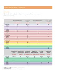
The world’s highest quality antibody provider has now extended its expertise to cytokine and growth factor production.<br />
<strong>Cell</strong> Signaling Technology (CST) is now offering a<br />
selection of cytokines and growth factors. These<br />
reagents are produced and bioassayed in-house, and<br />
are held to the same unparalleled quality standards as<br />
the CST antibodies you know and trust.<br />
:: Produced and bioassayed in-house with the<br />
highest purity and bioactivity.<br />
:: Comparison of multiple lots, stringent product<br />
specifications, and rigorous quality control<br />
ensures maximum lot-to-lot consistency.<br />
:: Endotoxin levels are routinely tested and are<br />
less than 0.01 ng/µg cytokine.<br />
:: Many products are produced in mammalian<br />
cells to maximize natural conformation and<br />
glycosylation.<br />
:: Multi-milligram quantities available.<br />
Visit our website for the most up-to-date<br />
product listing.<br />
Lot-to-Lot Consistency<br />
A<br />
B<br />
OD450-OD650<br />
0.6<br />
0.3<br />
0<br />
kDa<br />
200<br />
116<br />
97<br />
66<br />
55<br />
37<br />
31<br />
22<br />
14<br />
6<br />
4<br />
Lot 1 Lot 2 Lot 3<br />
0.6<br />
0.6<br />
0.01 0.1 1 10 100<br />
hIL-6 (ng/ml)<br />
+<br />
hIL-6<br />
OD450-OD650<br />
0.3<br />
0<br />
kDa<br />
200<br />
116<br />
97<br />
66<br />
55<br />
37<br />
31<br />
22<br />
14<br />
6<br />
4<br />
0.01 0.1 1 10 100<br />
hIL-6 (ng/ml)<br />
+<br />
0.01 0.1 1 10 100<br />
hIL-6 (ng/ml)<br />
Comparison of purity and bioactivity of three independent lots of Human Interleukin-6 (hIL-6) #8904:<br />
The proliferation of TF-1 cells was assessed after 48 hr treatment with increasing concentrations of hIL-6. <strong>Cell</strong>s were incubated with a<br />
tetrazolium salt and the OD 450 - OD 650 was determined (A). The purity of recombinant hIL-6 was determined by SDS-PAGE using 6 µg<br />
reduced (+) and non-reduced (-) recombinant hIL-6 and staining overnight with Coomassie Blue (B).<br />
hIL-6<br />
OD450-OD650<br />
0.3<br />
0<br />
kDa<br />
200<br />
116<br />
97<br />
66<br />
55<br />
37<br />
31<br />
22<br />
14<br />
6<br />
4<br />
+<br />
hIL-6<br />
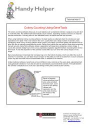
Human Tumor Necrosis Factor-α (hTNF-α) #8902 validation includes:<br />
A Purity<br />
B Bioactivity<br />
C Downstream Signaling,<br />
Western Blot<br />
D<br />
kDa<br />
116<br />
97<br />
66<br />
55<br />
37<br />
31<br />
22<br />
14<br />
6<br />
3<br />
+ –<br />
hTNF-α<br />
OD 595 nm<br />
0.4<br />
0.2<br />
0<br />
0 .01 .1 1 10 100<br />
hTNF-α (ng/ml)<br />
kDa<br />
100<br />
80<br />
60<br />
50<br />
100<br />
80<br />
60<br />
50<br />
–<br />
0.01<br />
0.1<br />
1<br />
10<br />
Phospho-NF-κB<br />
p65 (Ser536)<br />
(93H1) Rabbit mAb<br />
#3033<br />
NF-κB p65<br />
Antibody<br />
#3034<br />
hTNF-α (ng/ml)<br />
Human Tumor Necrosis Factor-α (hTNF-α) #8902: (A) 6 µg reduced (+) and non-reduced (-) hTNF-α; hTNF-α does not contain tags or additional<br />
amino acids. (B) The viability of L-929 cells treated with increasing amounts of hTNF-α in the presence of 2 ng/ml actinomycin D was determined. (C)<br />
Phosphorylation of NF-κB p65 was measured in response to treatment with hTNF-α. (D) HT-1080 cells were treated with increasing concentrations<br />
of hTNF-α to assess the effect on nuclear translocation of NF-κB, detected using NF-κB p65 (D14E12) XP ® Rabbit mAb #8242. All data points were<br />
performed in triplicate to ensure validity and reproducibility of the assay. The signal was analyzed using an <strong>Cell</strong>omics ArrayScan ® V TI . Inset: Confocal<br />
IF analysis of HT-1080 cells, untreated (left) or treated with hTNF-α (20 ng/ml, 20 min) (right), using NF-κB p65 (D14E12) XP ® Rabbit mAb #8242<br />
(green). Red = Actin filaments (DY-554 phalloidin); Blue = DRAQ5 ® #4084 (DNA dye).<br />
Normalized<br />
Nuclear Fluorescence Intensity<br />
170<br />
160<br />
150<br />
140<br />
130<br />
120<br />
110<br />
100<br />
Downstream Signaling,<br />
Immunofluorescent Analysis<br />
untreated<br />
hTNF-α-treated<br />
0.001 0.01 0.1<br />
hTNF-α (ng/ml)<br />
1 10<br />
USA Headquarters<br />
<strong>Cell</strong> Signaling Technology<br />
Technical Support:<br />
(toll-free) 1-877-678-8324<br />
Tel: 978-867-2300<br />
Fax: 978-867-2400<br />
E-mail: info@cellsignal.com<br />
International Subsidiaries<br />
<strong>Cell</strong> Signaling Technology China<br />
Technical Support:<br />
(toll-free) 4006-473287<br />
Tel: (86) 21-5835-6288<br />
Fax: (86) 21-5835-6116<br />
E-mail: info@cst-c.com.cn<br />
<strong>Cell</strong> Signaling Technology Europe<br />
Tel: +31 (0)71 568 1060<br />
Fax: +31 (0)71 568 1065<br />
E-mail: info@cellsignal.eu<br />
www.cellsignal.com<br />
<strong>Cell</strong> Signaling Technology Japan<br />
Tel: 03 (5652) 0213<br />
Fax: 03 (3249) 1170<br />
E-mail: info@cstj.co.jp<br />
www.cstj.co.jp<br />
Printed in the USA on 55% recycled paper (30% post-consumer waste fiber) using soy inks and processed chlorine free.<br />
© 2008-01/2012 <strong>Cell</strong> Signaling Technology, Inc.