Page: 51-53; FULL TEXT - Journal of IMAB
Page: 51-53; FULL TEXT - Journal of IMAB
Page: 51-53; FULL TEXT - Journal of IMAB

You also want an ePaper? Increase the reach of your titles
YUMPU automatically turns print PDFs into web optimized ePapers that Google loves.
From the dermatological status:<br />
The pathologic lesions affect posterior – lateral areas<br />
<strong>of</strong> both thighs. Morphologically they contain <strong>of</strong> grouped<br />
papules and nodules, reddish brown coloured and covered<br />
with gentle scales, predominantly at the peripheral zones.<br />
No enlarged lymphnodules are palpated.<br />
From the laboratory analysis: Hb 100, Ht 45, Leuc –<br />
6,8, Ly – 36, total protein 93, creatinin 120, urea 380<br />
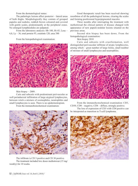
From the histopathological examination:<br />
Good therapeutic result has been received showing<br />
resorbtion <strong>of</strong> all the pathological lesions, clearing the scales<br />
and forming postlesional hyperpigmented macules.<br />
Three months after interrupting the treatment with<br />
methotrexad the clinical pattern <strong>of</strong> disease changed with<br />
appearance <strong>of</strong> new papulo-nodular lesions situated on the<br />
previous areas.<br />
Second skin biopsy has been down. From the<br />
histopathological examination:<br />
Skin biopsy 2010<br />
Cutis and subcutis with crustformation, well<br />
distinguished perivascular infiltrate <strong>of</strong> atypic lymphocytes,<br />
among which – great number <strong>of</strong> large forms, small number<br />
<strong>of</strong> mixture <strong>of</strong> small lymphocytes and neutrophiles.<br />
Skin biopsy – 2009.<br />
Cutis and subcutis with predominant perivascular as<br />
well periadnexial infiltration <strong>of</strong> large atypical lymphocytes,<br />
between witch a mixture <strong>of</strong> eosinophiles, neutrophiles and<br />
small lymphocytes is seen. There is no epidermotropism.<br />
From the immunohistochemical examination:<br />
From the immunohystochemical examination: CD3,<br />
CD30, CD8 – negative, CD4 – diffuse, strongly positive.<br />
The loss <strong>of</strong> expression <strong>of</strong> CD3 with CD4 positive can<br />
be interpreted as transition to T-cell lymphoma.<br />
The infiltrate is CD 3 positive and CD 30 positive.<br />
The treatment included low doses methotrexat (15 mg/<br />
weekly) for 10 weeks.<br />
52 / J<strong>of</strong><strong>IMAB</strong>; Issue: vol. 16, book 3, 2010 /