Page: 51-53; FULL TEXT - Journal of IMAB
Page: 51-53; FULL TEXT - Journal of IMAB
Page: 51-53; FULL TEXT - Journal of IMAB

You also want an ePaper? Increase the reach of your titles
YUMPU automatically turns print PDFs into web optimized ePapers that Google loves.
<strong>Journal</strong> <strong>of</strong> <strong>IMAB</strong> - Annual Proceeding (Scientific Papers) vol. 16, book 3, 2010<br />
LOCALIZED FORM OF LYMPHOMATOID<br />
PAPULOSIS, EVALUATING IN T-CELL LYMPHOMA<br />
– Case Report<br />
S. Shtilionova 1 , P. Drumeva 1 , M. Balabanova² 2 , I. Krasnaliev 3<br />
1) Department <strong>of</strong> dermatology and venereology, Medical University - Varna<br />
2) Department <strong>of</strong> dermatology and venereology, Medical University - S<strong>of</strong>ia<br />
3) Department <strong>of</strong> pathology, Medical University - Varna<br />
SUMMARY<br />
Lymphomatoid papulosis is presented by spontaneously<br />
regressing cutaneous infiltrates, that<br />
microscopically resemble a lymphomas and may evolve into<br />
Hodgkin or non- Hodgkin malignant lymphoma.<br />
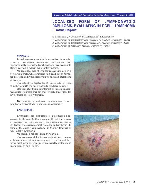
We present a case <strong>of</strong> Lymphomatoid papulosis in a<br />
64 years old male, who complains from reddish non painful<br />
papules, localized symmetrically on the back and lateral zone<br />
<strong>of</strong> the legs.<br />
The patient was treated for 10 weeks with low dose<br />
<strong>of</strong> methotrexat (15 mg per week) with good clinical result.<br />
One year after treatment interruption the same patient<br />
had a similar clinical changes and hystochemical signs for<br />
development <strong>of</strong> T-cell lymphoma.<br />
Key words: Lymphomatoid papulosis, T-cell<br />
lymphoma, hystopathology, immunohistochemistry<br />
CASE REPORT<br />
Lymphomatoid papulosis is a dermatological<br />
disorder firstly described by Dupont in 1965.It is presented<br />
by suddenly or spontaneously progressing cutaneous<br />
infiltrates , wich microscopically resemble a lymphoma . In<br />
some <strong>of</strong> the cases it can evoluate in Morbus Hodgkin or<br />
non-Hodgkin lymphoma.<br />
We present a patient – male 64 years old.<br />
The beginning <strong>of</strong> the disease starts about 1 year ago<br />
with appearance <strong>of</strong> non-painful, non – pruritic redish –<br />
brown small nodules, covering symmetrically posterior and<br />
lateral areas <strong>of</strong> both thighs.<br />
/ J<strong>of</strong><strong>IMAB</strong>; Issue: vol. 16, book 3, 2010 / <strong>51</strong>
From the dermatological status:<br />
The pathologic lesions affect posterior – lateral areas<br />
<strong>of</strong> both thighs. Morphologically they contain <strong>of</strong> grouped<br />
papules and nodules, reddish brown coloured and covered<br />
with gentle scales, predominantly at the peripheral zones.<br />
No enlarged lymphnodules are palpated.<br />
From the laboratory analysis: Hb 100, Ht 45, Leuc –<br />
6,8, Ly – 36, total protein 93, creatinin 120, urea 380<br />
From the histopathological examination:<br />
Good therapeutic result has been received showing<br />
resorbtion <strong>of</strong> all the pathological lesions, clearing the scales<br />
and forming postlesional hyperpigmented macules.<br />
Three months after interrupting the treatment with<br />
methotrexad the clinical pattern <strong>of</strong> disease changed with<br />
appearance <strong>of</strong> new papulo-nodular lesions situated on the<br />
previous areas.<br />
Second skin biopsy has been down. From the<br />
histopathological examination:<br />
Skin biopsy 2010<br />
Cutis and subcutis with crustformation, well<br />
distinguished perivascular infiltrate <strong>of</strong> atypic lymphocytes,<br />
among which – great number <strong>of</strong> large forms, small number<br />
<strong>of</strong> mixture <strong>of</strong> small lymphocytes and neutrophiles.<br />
Skin biopsy – 2009.<br />
Cutis and subcutis with predominant perivascular as<br />
well periadnexial infiltration <strong>of</strong> large atypical lymphocytes,<br />
between witch a mixture <strong>of</strong> eosinophiles, neutrophiles and<br />
small lymphocytes is seen. There is no epidermotropism.<br />
From the immunohistochemical examination:<br />
From the immunohystochemical examination: CD3,<br />
CD30, CD8 – negative, CD4 – diffuse, strongly positive.<br />
The loss <strong>of</strong> expression <strong>of</strong> CD3 with CD4 positive can<br />
be interpreted as transition to T-cell lymphoma.<br />
The infiltrate is CD 3 positive and CD 30 positive.<br />
The treatment included low doses methotrexat (15 mg/<br />
weekly) for 10 weeks.<br />
52 / J<strong>of</strong><strong>IMAB</strong>; Issue: vol. 16, book 3, 2010 /
DISCUSSION:<br />
The ethiology <strong>of</strong> Lymfomatoid papulosis is unclear<br />
and contradictory. Some authors have started that in 10-20%<br />
<strong>of</strong> the cases , the patients could be evolve in Lymphoma or<br />
primary giant – cell anaplastic T-cell Lymphoma . In rare cases<br />
the disease could evolve in Mycosis fungoides (1, 2).<br />
There are no objective criteria for the time, when the<br />
Lymphomatoid papulosis could evoluate in the mentioned<br />
above diseases, i. e to precede, attend, or to be there<br />
continuation. Even in the cases <strong>of</strong> regression <strong>of</strong><br />
Lymphomatoid papulosis, the moleculary- biologic studies<br />
show criteria for transformation to T-cell Lymphoma (2, 3, 4).<br />
Clinical features <strong>of</strong> the Lymphomatoid papulosis ara<br />
presented by multiple asymmetric papules and nodules with<br />
slight peripheral desquamation, solitary haemorrhagia ,<br />
crusts . In a few number <strong>of</strong> cases solitary tumour- like<br />
plaques are seen, but in other cases – a few number <strong>of</strong><br />
reddish-brown papules, strongly separated from the<br />
surrounding tissue. They are situated on the upper and lower<br />
extremites , buttocks .All the lesions have recurrent<br />
evaluation in weeks or months , but with tendency to<br />
relapse . Lymphadenopathy is extraordinary seen.<br />
Histopathological features <strong>of</strong> the disease are: primary<br />
non-epidermotropic mixedcell infiltrates, combined with<br />
eosinophiles , small lymphocytes , neutrophiles and<br />
histyocytes . In some <strong>of</strong> the cases the infiltrate is narrowbind<br />
shaped or nodular.<br />
The large lymphocytes resemble the cells <strong>of</strong> the<br />
Anaplastic giant –cell Lymphoma –they are CD positive<br />
.They can be positive for CD2,3,4,5 as well as for<br />
proliferative marker K67(6) .<br />
The laboratory analysis shows in rare cases<br />
lymphocytosis and hypregammaglobulinemia.<br />
The course <strong>of</strong> the disease is chronic, as the lack <strong>of</strong><br />
relapse more than 5 years means restoration to health. the<br />
clinical future that propose malignancy are ulceration,<br />
predominantly <strong>of</strong> the tumor like lesions and papulonecrotic<br />
changes, covering skin dermatoms(5).<br />
We comment a patient with localized form <strong>of</strong><br />
Lymphomatoid papulosis, in which the loss <strong>of</strong> expression<br />
<strong>of</strong> CD3, when having CD4 positive can be interpreted as<br />
transition to T – cell Lymphoma. Having in mind the<br />
possibility <strong>of</strong> transition <strong>of</strong> Lymphomatoid papulosis to<br />
malignant variant <strong>of</strong> lymphoma from great significance for<br />
the dermatologist is the clinical study <strong>of</strong> the patients as well<br />
as obligatory histopathological control <strong>of</strong> the eventual<br />
changes.<br />
CONCLUSIONS:<br />
1. Although the low percentage <strong>of</strong> the malignancy<br />
and relatively good prognosis for the course <strong>of</strong><br />
Lymphomatoide papulosis, the patients suffering it, must be<br />
clinically and histopathologically controlled.<br />
2. The localized form <strong>of</strong> Lymphomatoid papilosis is<br />
combatively rare, but brings the potential to evaluate in<br />
malignant form, as well as the standard disseminated variant.<br />
3. The patients with Lyphomatoid papulosis, having<br />
tumor - like plaques with ulceration, as well as<br />
papulonecrotik lesions must be considered as criteria for<br />
eventual malignant transformation in variant Mycisis<br />
fungoides, Morbus Hodgkin or malignant non – Hodgkin<br />
Lymphoma.<br />
REFERENCES:<br />
1. Burg G, Kerl H, Schmoeckel C.<br />
Differentiation between malignant B-cell<br />
lymphomas and pseudolymphomas <strong>of</strong> the<br />
skin. J Dermatol Oncol 1984; 10: 271-275.<br />
2. Burg G, Braun-Falco O. Cutaneous<br />
pseudolymphomas. In: Cutaneous<br />
lymphomas. Berlin: Springer-Verlag, 1983,<br />
415-464.<br />
3. Duncan SC, Evans HL, Winkelmann<br />
RK. Large cell lymphocytoma. Arch<br />
Dermatol 1980; 116: 1142-1146.<br />
4. Dorfman RF, Warnke R.<br />
Lymphadenopathy simulating the malignant<br />
lymphomas. Hum Pathol 1974.<br />
5. Englich JC, Smith NP, Wilson Jones<br />
E, Winkelmann RK. Large cell<br />
lymphocytoma [abstract]. J Cutan Pathol<br />
1986; 13: 441.<br />
6. Kerl H, Ackerman AB. Inflammatory<br />
diseases that simulate lymphomas:<br />
cutaneous pseudolymphomas. In:<br />
Fitzpatrick TB, Eisen AZ, Wolff K,<br />
Freedberg IM, Austen KF (editors).<br />
Dermatology in general medicine, 4th ed.<br />
New York: McGrew-Hill, 1993, 1315-<br />
1327.<br />
7. Landa N, Zelickson BD, Peters MS,<br />
et al. Cutaneous lymphoma versus<br />
pseudolymphoma: gene rearrangement<br />
studies <strong>of</strong> 21 cases with clinicopathologic<br />
correlation [Abstr.]. J Invest Dermatol<br />
Address for correspondence:<br />
Dr. Siana Shtilionova,<br />
Department <strong>of</strong> dermatology and venereology, Medical University - Varna<br />
55, Marin Drinov str., 9000 Varna, Bulgaria<br />
E-mail: shtilionova@abv.bg<br />
/ J<strong>of</strong><strong>IMAB</strong>; Issue: vol. 16, book 3, 2010 / <strong>53</strong>