ANA HEP-2 IFA Kit - Diagnostic Automation : Cortez Diagnostics
ANA HEP-2 IFA Kit - Diagnostic Automation : Cortez Diagnostics
ANA HEP-2 IFA Kit - Diagnostic Automation : Cortez Diagnostics

You also want an ePaper? Increase the reach of your titles
YUMPU automatically turns print PDFs into web optimized ePapers that Google loves.
DIAGNOSTIC AUTOMATION, INC.<br />
23961 Craftsman Road, Suite E/F, Calabasas, CA 91302<br />
Tel: (818) 591-3030 Fax: (818) 591-8383<br />
onestep@rapidtest.com<br />
technicalsupport@rapidtest.com<br />
www.rapidtest.com<br />
See external label 2°C-8°C Σ=36-60 tests cat#230596-SP<br />
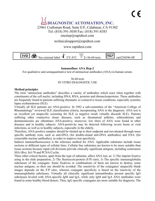
Inmunofluor <strong>ANA</strong> Hep-2<br />
For qualitative and semiquantitative test of antinuclear antibodies (<strong>ANA</strong>) in human serum.<br />
36-60 tests<br />
IN VITRO DIAGNOSTIC USE<br />
Method principles<br />
The term “antinuclear antibodies” describes a variety of antibodies which react when together with<br />
constituents of the cell nuclei, including DNA, RNA, proteins and ribonucleoproteins. These antibodies<br />
are frequently found in patients suffering rheumatic or connective tissue conditions, especially systemic<br />
lupus erythematosus (SLE).<br />
Virtually all SLE patients are <strong>ANA</strong>-positive. In 1982 a sub-committee of the “American College of<br />
Rheumatology” reviewed SLE classification criteria, incorporating <strong>ANA</strong> to the diagnosis. <strong>ANA</strong> test is<br />
an excellent yet unspecific screening for SLE (a negative result virtually discards SLE). Patients<br />
suffering other connective tissue diseases, such as rheumatoid arthritis, schlerodermia and<br />
dermatomiositis are oftentimes <strong>ANA</strong>-positive; moreover, low titers of <strong>ANA</strong> were found in other<br />
diseases and in healthy subjects. <strong>ANA</strong>-positivity may be detected following severe burns or viral<br />
infections, as well as in healthy subjects, especially in the elderly.<br />
Therefore, <strong>ANA</strong>-positive samples should be titrated up to their endpoint and reevaluated through more<br />
specific antibody tests, such as anti-DNA (for double-strand anti-DNA antibodies) and ENA (for<br />
extractable nuclear antibodies), in order to improve test specificity.<br />
Indirect immunofluorescence is the reference method for <strong>ANA</strong>. Applicable substrates include tissue<br />
sections or different types of cellular lines. Cellular line substrates are known to be more suitable than<br />
tissue sections because rapid cell divisions provide clinically significant antigens, including centromere<br />
SSA (Ro), Scl-70 and PCNA/Cyclin.<br />
Three other critical factors, apart from the type of substrate, affect <strong>ANA</strong> test, as: 1) The imprint fixative<br />
using in the slide preparation, 2) The fluorescein:protein (F/P) ratio, 3) The specific immunoglobulin<br />
subclasses of the conjugate. Some fixatives or combinations of them are known to destroy some<br />
nuclear antigens, so their use should be avoided. The sensitivity of the conjugates' unspecific blank<br />
images depends on the F/P ratio, whereas conjugate specificity is based on the reactivity of the<br />
immunoglobulin subclasses. Virtually all clinically significant autoantibodies present specific IgG<br />
subclasses leveled with <strong>ANA</strong>-specific IgM and IgA, while only IgM and IgA <strong>ANA</strong> antibodies were<br />
found in some healthy blood donors. Thus, IgG specific conjugates are more suitable for diagnosis. The
substrate used in the kit Inmunofluor <strong>ANA</strong> <strong>HEP</strong>-2 is a cellular line, and the conjugate is highly purified<br />
anti-human IgG, with a carefully selected F/P ratio.<br />
The reagents used in this kit are adjusted to detect clinically significant antibodies, including SSA and<br />
Scl-70, which cannot be detected by other commercial <strong>ANA</strong> tests. Furthermore, the specific IgG<br />
conjugate discards the physiologic positive results usually resulting from the low titers of specific IgM.<br />
Summary and explanation of the assay<br />
Indirect immunofluorescence technique involves sample incubation in the antigenic substrate and<br />
further rinsing of the non-reacting antibodies. The substrate is afterwards incubated along with the<br />
fluorescein-labeled specific antigammaglobulin and the unbound reagent is then rinsed. Reading is<br />
performed with fluorescence microscope. Antibody-positive samples present an apple-green<br />
fluorescence in the cell or nucleous areas which are bound to the antibodies.<br />
Test procedure<br />
1) Buffer preparation<br />
Reconstitute phosphate buffer saline (PBS) up to 1 liter with destilled water. A solution 0.01 M with a<br />
pH of 7.2 0.2 is obtained. If necessary adjust with (NaOH 1N or HCL 1N). Store at 2-8°C, in a clean<br />
covered container. In case of change of pH, cloudiness, or precipitation, discard.<br />
2) Sample dilution<br />
A) Initial screening: dilute the patients' samples with PBS 1/40 (E.G.: Add 20ml serum to 0.78 ml of<br />
PBS.<br />
NOTE: An initial screening dilution 1/20 is suggested for children under 10 years of age.<br />
B) Titration: dilute the positive samples with PBS to obtain graded dilution from the initial one (E.G.<br />
1/80, 1/160,... 1/2560).<br />
3) Samples inoculation<br />
Bring the slides to room temperature without unpacking. Once unpacked, place them in a suitable wet<br />
chamber and add one drop of positive and negative control to sites 1 and 2 respectively. Add one drop<br />
(50-75ml) of unknown serum 1-40 at the other site (one site per patient.) Incubate the slide for 30<br />
minutes in a wet chamber (plastic or glass flat-bottom container covered with a wet filter paper to keep<br />
the right humidity conditions.)<br />
NOTE: Incubation of samples and conjugates may be reduce to 10 minutes each without any sensitivity<br />
loss (especially during initial screening.).<br />
IMPORTANT: Avoid drying of the reaction areas over the following steps.<br />
4) Rinsing<br />
Following incubation with serum dilution, rinse with PBS, using plastic dropper or pipette. Bend the<br />
slide down and dry some PBS on it, minimizing sample contact between reactive areas. Do not drip<br />
PBS straight on the reaction sites, so as not to harm the substrate.<br />
Place the slides in a coplin jar and rinse twice or three times with buffer (5' each time), gently stirring<br />
during rinses.<br />
5) Application of the conjugate<br />
Dilute the labeled antigammaglobulin supplied with the kit (suggested dilution: ____ ) and add 10 ml.<br />
of Evans blue per ml. of gammaglobulin dilution.<br />
Stop rinsing the slides one at a time, stir them on blotting paper to drain off the excess PBS, drying<br />
around the reaction areas (if necessary.) Place all the slides back into the wet chamber.<br />
Immediately apply a drop of conjugate (50-75 ml.) per area, making sure that each zone is thoroughly<br />
covered.<br />
Incubate in wet chamber at room temperature for 30'. Protect from excessive light.
6) Rinsing<br />
Repeat step 4.<br />
7) Mounting<br />
Stop rinsing the slides one at a time, stir them on blotting paper to drain off the excess PBS, and add 3-<br />
4 drops of mounting medium along the slides.<br />
Gently place the cover on the slide, checking that no bubbles are formed. Drain the excess mounting<br />
medium off the preparation edges with blotting paper. Clean the rear side of the slide.<br />
8) Reading<br />
Read the slides in a fluorescence microscope as soon as possible. They should preferably be examined<br />
on the same day in which the above procedure is performed. Otherwise, store them in refrigerator at 2-<br />
8°C away from light, and read them on the following day. The mounting medium should not dry off<br />
between the slide and its cover. If it does, add some additional mounting medium.<br />
Contents of the kit<br />
For 36 tests<br />
1- 3 slides with 12 HEp-2 cell reaction areas.<br />
2- Antihuman antigamma globulin IgG labeled with fluorescein isothiocyanate (0.15 ml).<br />
3- Positive control human serum (1.0 ml). Ready to use.<br />
4- Negative control human serum (1.0 ml). Ready to use.<br />
5- Phosphate saline buffer (PBS): 2 foil bags. Each container is to be diluted in distilled water to<br />
obtain one liter.<br />
6- Mounting medium (5.0 ml).<br />
7- Evans blue (5.0 ml). Ready to use.<br />
8- Slide covers.<br />
9- User's guide.<br />
For 60 tests<br />
1- 5 slides with 12 HEp-2 cell reaction areas.<br />
2- Antihuman antigamma globulin IgG labeled with fluorescein isothiocyanate (0.20 ml).<br />
3- Positive control human serum (1.0 ml). Ready to use.<br />
4- Negative control human serum (1.0 ml). Ready to use.<br />
5- Phosphate saline buffer (PBS): 3 foil bags. Each container is to be diluted in distilled water to<br />
obtain one liter.<br />
6- Mounting medium (5.0 ml). Ready to use.<br />
7- Evans blue (5.0 ml).<br />
8- Slide covers.<br />
9- User's guide.<br />
Unstability of reconstituted sbstances<br />
Reconstituted phosphate buffer saline keeps stable for up to 15 days at 2-8ºC.<br />
For longer storage periods, add sodium azide 0.1% to prevent contamination.<br />
Required material (not supplied)<br />
1- Pasteur pipettes.<br />
2- Dropper.<br />
3- Immersion oil (optional).<br />
4- Accurate pipettes.<br />
5- A bowl for rinsing the slides.<br />
6- Fluorescence microscope with FITC filter system (maximum stimulation wavelength of 490<br />
mm, mean emission wavelength, 530 mm, excursions to 600-1000).
Warnings and precautions<br />
1- Sodium azide 0.1% is used as a preservative. When disposing of reagents, use large amounts of<br />
water to prevent waste azide from accumulating in the pipeline. Sodium azide can be poisonous if<br />
ingested.<br />
2- Control human serum components used in this kit have been found negative for anti-HIV<br />
antibodies and hepatitis B surface antigen (HBsAg), but because no diagnosis test can warrant absolute<br />
certainty as to the absence of these or other infectious agents, human controls and reagents should be<br />
treated as potentially infectious.<br />
3- Substitution of components with other not supplied in the system may originate inconsistent<br />
results.<br />
4- Keep the reaction areas wet throughout the whole technique (avoid any artificial drying<br />
techniques, such as air, stove, or heat drying). After rinsing, remove excess humidity with blotting<br />
paper; dry only the side outer edges of the slide. Immediately after drying, abundantly spread a<br />
properly diluted solution of antigammaglobulin and/or mounting medium, depending on the concerned<br />
stage.<br />
Limitations of the procedure<br />
1- High titers of <strong>ANA</strong> suggest connective tissue disease but has no diagnostic status. <strong>ANA</strong> results<br />
should be complemented with other serologic results and with the patient's medical record.<br />
2- <strong>ANA</strong> patterns may be altered if the sample is titrated up to its endpoint. This happens because low<br />
antibody titers reach the system sensitivity when a more diluted sample is tested.<br />
3- The sensitivity of this method depends upon a variety of external factors, including the type of<br />
microscope used, lamp intensity and period, the system of lenses, the system of filters, and the<br />
observer.<br />
Storage<br />
Store in refrigerator at 2-8°C.<br />
The kit components are stable up to the expiry date printed in the external and internal labels.<br />
The reagents may be hampered if exposed to light or frozen.<br />
Specimen collection<br />
Treat all blood, plasma and serum samples as though they were capable of transmitting infectious<br />
diseases. Antiseptically extract 5-8 ml. of venous blood. Keep the blood at room temperature until<br />
separated from serum, to prevent any hemolisis which might hamper test results. Patients should go on<br />
an 8-hour fast prior to sample collection.<br />
This test can only be performed on serum. Do not process hemolized, contaminated or lipemic samples.<br />
Sera may be kept in refrigerator (2-8°C) for up to 72 hours. For longer storage periods, freeze at -20°C.<br />
Avoid repeated freezing-unfreezing cycles.<br />
Interpretation of results<br />
Negative reaction<br />
A sample is considered negative if its specific nuclear image is equal or lower than the negative<br />
control. Samples may exhibit different degrees of unspecific fluorescence due to the presence of<br />
heterophil antibodies or low levels of cytoplasm autoantibodies, such as is the case with the contractile<br />
proteins.<br />
Positive reaction<br />
A sample is considered positive if its specific nuclear image is higher than the negative control.
Pattern interpretation<br />
A variety of patents may be found in nuclear and cytoplasmatic images depending on the types and<br />
relative amounts of sample autoantibodies, as:<br />
- Homogenous pattern: A solid nuclear taint image, with or without apparent masking of the nucleolus<br />
is observed.<br />
Nuclear antigens: ds DNA, ss DNA, histones.<br />
Related diseases: High titers are suggestive of SLE; low titers are suggestive of SLE or other<br />
connective tissue diseases.<br />
- Peripheral pattern: A solid taint image is seen around the nuclear membrane, becoming faint towards<br />
the nuclear center.<br />
Nuclear antigens: ds DNA, ss DNA, DNP, histones.<br />
Related diseases: High titers are suggestive of SLE; low titers are suggestive of SLE or other<br />
connective tissue diseases.<br />
- Speckled pattern: A fine or granulated taint is found at the nuclei, usually lacking fluorescence at the<br />
nucleoli.<br />
Nuclear antigens: Sm, RNP, Scl-70, SSA, SSB and other antigen-antibody systems, as yet<br />
uncharacterized.<br />
Related diseases: High titers are suggestive of SLE; low titers are suggestive of SLE (Sm antibody),<br />
mixed connective tissue disease (RNP antibody), schlerodermia (Scl-70 antibody) or Sjögren's<br />
syndrome sicca complex (antibody SSB), low titers are suggestive of other connective tissue diseases<br />
- Nucleolar pattern: A taint image presenting large and thick granulations, usually containing less than<br />
6 granulations per cell, with or without occasional fine granulations, is found in the nuclei.<br />
Nuclear antigens: RNA 4-6S and other unknown nuclear antibodies.<br />
Related diseases: High titers are predominant in schlerodermia and Sjögren's syndrome.<br />
- Centromeric pattern: A slightly granular taint image is observed. Granulations are highly discrete,<br />
usually appearing as a multiple of 46.<br />
Nuclear antigens: Chromosomal centromere (kinetochore).<br />
Related diseases: Highly suggestive of CREST, a variant of Progressive Sclerosis Syndrome (PSS).<br />
CREST is a kind of PSS with prominent calcinosis, Raynaud's phenomenon, esophagic dismotility and<br />
skin-restricted injury (usually in the fingers and the face), and telangiectasia.<br />
- Mitochondrial pattern: A discretely granulated taint image is found at the cytoplasm and relatively<br />
absent from the nuclear area.<br />
Antigens: Various types of mitochondrial antigens.<br />
Related disease: High titers are suggestive of primary biliary cirrhosis.<br />
Patterns should be used with caution in the test of specific antibodies, except in the case of nucleolar<br />
and centromeric images, which present distinctly defined antigens and a characteristic pattern. Because<br />
many autoantibodies or combinations of them may induce homogenous or speckled images, specific<br />
antibody tests, (such as a-DNA or ENA) are recommended in such cases.<br />
Characteristics of the test<br />
A number of patients presenting connective tissue disease were compared against 200 healthy blood<br />
donors. Following are the obtained results:<br />
Group of patients<br />
Positives with Inmunofluor<br />
No.<br />
<strong>ANA</strong> HEp-2<br />
SLE 105 105<br />
Drug-induced lupus 24 24<br />
Rheumatoid arthritis 40 28<br />
Sclerodermia 24 18<br />
Dermatomiositis 14 10<br />
Sjögren’s syndrome 14 10<br />
Healthy subjects 200 5
Bibliography<br />
1) Tan E.- Autoantibodies to nuclear antigens (<strong>ANA</strong>): Their inmunobiology and medicine.<br />
Advances in immunology 33:167-239,1982.<br />
2) Tan E, et al.: The 1982 Revised criteria for the classification of systemic lupus erythematosus,<br />
arthritis and rheumatism 25.-1271-1277,1982.<br />
3) Casalo SP, Friou GJ, Meyers LL: Significance of antibodies to DNA in systemic lupus<br />
erythematosus. Arthritis and Rheumatism 7:379-390, 1964.<br />
4) Gonzalea E, Rothfield N.- Immunoglobulin class and pattern of nuclear fluorescence in<br />
systemic lupus erythematosus. The New England J of Medicine 274:1333-1338,1966.<br />
5) Wilk A: Antinuclear factors in sera from healthy blood donors. Acta Path Microbiol Scand.<br />
84:215-220, 1976.<br />
6) Biosafety in Microbiological and Biomedical Laboratories. Center for Disease Control.<br />
National Institute of health, 1984 (HHS Pub. # (CDC) 84-8395).<br />
DIAGNOSTIC AUTOMATION, INC.<br />
23961 Craftsman Road, Suite E/F, Calabasas, CA 91302<br />
Tel: (818) 591-3030 Fax: (818) 591-8383<br />
ISO 13485-2003