Sacral rib : A rare congenital anomaly CASE REPORT - ResearchGate
Sacral rib : A rare congenital anomaly CASE REPORT - ResearchGate
Sacral rib : A rare congenital anomaly CASE REPORT - ResearchGate

Create successful ePaper yourself
Turn your PDF publications into a flip-book with our unique Google optimized e-Paper software.
Acta Orthop. Belg., 2008, 74, 429-431<br />
<strong>CASE</strong> <strong>REPORT</strong><br />
<strong>Sacral</strong> <strong>rib</strong> : A <strong>rare</strong> <strong>congenital</strong> <strong>anomaly</strong><br />
Muddassir RASHID, Mohd KHALID, Neelam MALIK<br />
From J.N. Medical College, Aligarh, India<br />
A 45-year-old patient reported for a follow-up visit<br />
after a motor vehicle accident with a history of vague<br />
right flank pain. He underwent a pelvic radiograph<br />
which revealed a long bony protuberance arising<br />
from the right sacral region ; the appearance was<br />
consistent with a sacral <strong>rib</strong>. However due to the limited<br />
nature of his complaints the patient denied any<br />
surgical treatment.<br />
Keywords : sacral <strong>rib</strong> ; <strong>congenital</strong>.<br />
INTRODUCTION<br />
<strong>Sacral</strong> <strong>rib</strong> is a <strong>rare</strong> <strong>congenital</strong> <strong>anomaly</strong> consisting<br />
of a bony formation that arises in soft tissue around<br />
the sacrum ; radiologically it resembles a <strong>rib</strong> or a<br />
phalanx (2). It is often an incidental finding and, in<br />
absence of symptoms, no specific treatment is<br />
required. It is important to identify this asymtopmatic<br />
variant on radiography to avoid unneccesary<br />
additional investigations.<br />
<strong>CASE</strong> <strong>REPORT</strong><br />
A 45-year-old patient reported for a follow-up<br />
visit after a motor vehicle accident with a history of<br />
vague right flank pain. His clinical examination was<br />
unremarkable except for deep tenderness in the<br />
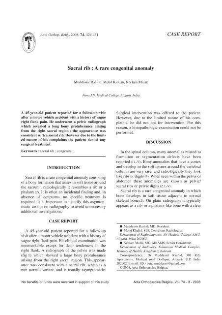
right flank. A radiograph of the pelvis was made<br />
(fig 1) which showed a large bony protuberance<br />
arising from the right sacral region. This appearance<br />
was consistent with a sacral <strong>rib</strong>, which is a<br />
<strong>rare</strong> normal variant, and is usually asymptomatic.<br />
Surgical intervention was offered to the patient.<br />
However, due to the limited nature of his complaints,<br />
he did not opt for intervention. For this<br />
reason, a histopathologic examination could not be<br />
performed.<br />
DISCUSSION<br />
In the spinal column, many anomalies related to<br />
formation or segmentation defects have been<br />
reported (11,13). Bony anomalies that have a cortex<br />
and develop in the soft tissues around the vertebral<br />
column are very <strong>rare</strong>, and radiologically they look<br />
like <strong>rib</strong>s or digits (9). When seen within the pelvis or<br />
abdomen these anomalies are known as pelvic/<br />
sacral <strong>rib</strong>s or pelvic digits (2,3,14).<br />
<strong>Sacral</strong> <strong>rib</strong> is a <strong>rare</strong> <strong>congenital</strong> <strong>anomaly</strong> in which<br />
bone develops in soft tissue adjacent to normal<br />
skeletal bone (2). On plain radiograph it typically<br />
appears as a <strong>rib</strong>- or a phalanx-like bone with a clear<br />
■ Muddassir Rashid, MD, Resident.<br />
■ Mohd Khalid, MD, Consultant Radiologist.<br />
Department of Radiodiagnosis, JN Medical College, AMU,<br />
Aligarh, India 202002.<br />
■ Neelam Malik, MD, MNAMS, Senior Consultant.<br />
Department of Radiology, Salmaniya Medical Complex,<br />
Ministry of Health, Kingdom of Bahrain.<br />
Correspondence : Dr Muddassir Rashid, 301 Rifa<br />
Apartments, Medical road Dodhpur, Aligarh, U.P, India<br />
202002. E-mail : ID - beighmuddassir@gmail.com<br />
© 2008, Acta Orthopædica Belgica.<br />
No benefits or funds were received in support of this study Acta Orthopædica Belgica, Vol. 74 - 3 - 2008
430 M. RASHID, M. KHALID, N. MALIK<br />
Fig. 1. — Anteroposterior radiograph of the pelvis showing a<br />
finger-like bony protuberance arising from the lower part of the<br />
sacrum on the right side.<br />
cortex and medulla connected to the pelvis, often<br />
with a characteristic pseudoarticulation at the<br />
base (9).<br />
The development of costal elements in humans<br />
takes place from a common primordial mass, with<br />
<strong>rib</strong>s progressing to full development in the thoracic<br />
region, whereas in the cervical, lumbar and sacral<br />
regions they fuse with the lateral masses or transverse<br />
processes during the fourth to twelfth week of<br />
development. Any defect in fusion may cause the<br />
<strong>rib</strong> to keep growing and form a supernumerary<br />
<strong>rib</strong> (8,14). McGlone et al postulated that in the pelvis<br />
the costal processes become incorporated into the<br />
sides of the sacrum and coccyx (9) and more laterally<br />
and anteriorly, the costal process mesenchyma<br />
normally degenerates and is lost – a process known<br />
as apoptosis (9). Failure of apoptosis may allow differentiation<br />
of costal process mesenchyma to form<br />
<strong>rib</strong> tissue (9). Clinically, cervical supernumerary<br />
<strong>rib</strong>s may cause neurovascular symptoms or thoracic<br />
outlet compression syndrome whereas pelvic <strong>rib</strong>s<br />
are usually asymptomatic and, in most cases, are<br />
found incidentally during examinations for other<br />
problems (8,12). In those <strong>rare</strong> cases in which sacral<br />
<strong>rib</strong>s cause symptoms, there is typically discomfort<br />
and diminished mobility during movement of the<br />
ipsilateral hip, and they can possibly cause compromise<br />
of the birth canal in females (4). Standard radiographs<br />
and computed tomography provide the best<br />
diagnostic information.<br />
The differential diagnosis includes myositis ossificans,<br />
avulsion injuries, heterotopic bone formation,<br />
Fong’s disease, and osteochondroma. Pelvic<br />
<strong>rib</strong> can usually be radiologically differentiated from<br />
posttraumatic myositis ossificans and from heterotopic<br />
bone formation by its well corticated appearance<br />
and the absence of a history of trauma (3,5). CT<br />
of pelvic <strong>rib</strong> confirms the presence of cortical<br />
bone (7) and is useful in equivocal cases (10).<br />
Myositis ossificans is characterized by a radiolucent<br />
core with a calcified periphery, which is clearly<br />
separated from adjacent bones (7). Avulsion<br />
injuries of the pelvis commonly occur during athletic<br />
activity, with peak incidence in adolescents and<br />
teenagers (1). Pain, diminished motion, and soft tissue<br />
haematoma correlate with a bony fragment (1).<br />
In some cases, new bone formation after surgery or<br />
ossification of the sacrotuberous ligament can<br />
resemble a pelvic <strong>rib</strong> (2). Fong’s disease is characterized<br />
by bilateral iliac horns arising posteriorly<br />
and centrally from the ilia (7). Osteochondroma is<br />
continuous with underlying bone (2) and a cartilaginous<br />
cap may be present (10). Pelvic <strong>rib</strong> is usually<br />
an asymptomatic, benign condition and is discovered<br />
incidentally (9). In the absence of symptoms,<br />
surgical excision is not required (5). Pelvic <strong>rib</strong>s are<br />
suggested as a <strong>rare</strong> cause of foetopelvic disproportion,<br />
and a case of scoliosis and hypoplasia of the<br />
ipsilateral gluteal musculature arising from a pelvic<br />
<strong>rib</strong> was previously desc<strong>rib</strong>ed (6,8).<br />
In conclusion, it is important to recognize and<br />
distinguish the pelvic <strong>rib</strong> from posttraumatic ossification<br />
and avulsion injuries and thus avoid unnecessary<br />
additional investigations. The radiographic<br />
entity of pelvic/sacral <strong>rib</strong> should be known by every<br />
radiologist as an incidental finding for which no<br />
further action is required.<br />
REFERENCES<br />
1. Fernbach SK, Wilkinson RH. Avulsion injuries of the<br />
pelvis and proximal femur. AJR Am J Roentgenol 1981 ;<br />
137 : 581-584.<br />
Acta Orthopædica Belgica, Vol. 74 - 3 - 2008
SACRAL RIB 431<br />
2. Goyen M, Barkhausen J, Markschies NA, Debatin JF.<br />
The pelvic digit – A <strong>rare</strong> developmental <strong>anomaly</strong>. A case<br />
report with CT correlation and review of the literature. Acta<br />
Radiol 2000 ; 41 : 317-319.<br />
3. Granieri GF, Bacarini L. The pelvic digit : five new<br />
examples of an unusual <strong>anomaly</strong>. Skeletal Radiol 1996 ;<br />
25 : 723-726.<br />
4. Greenspan A, Norman A. The pelvic digit. Bull Hosp Jt<br />
Dis 1984 ; 44 : 72-75.<br />
5. Hamilton S. Pelvic digit. Br J Radiol 1985 ; 58 : 1010-<br />
1011.<br />
6. Heligman D, Sullivan RC, Millar EA. <strong>Sacral</strong> <strong>rib</strong>s. A case<br />
report. Orthopedics 1987 ; 10 : 1439-1442.<br />
7. Hoeffel C, Hoeffel JC, Got I. Bilateral pelvic digits. A<br />
case report and review of the literature. Rofo 1993 ; 158 :<br />
275-276.<br />
8. Kaushal SP. <strong>Sacral</strong> <strong>rib</strong>s. Int Surg 1997 ; 62 : 37-38.<br />
9. McGlone BS, Hamilton S, FitzGerald MJ. Pelvic digit :<br />
an uncommon developmental <strong>anomaly</strong>. Eur Radiol 2000 ;<br />
10 : 89-91.<br />
10. Nguyen VD, Matthes JD, Wunderlich CC. The pelvic<br />
digit : CT correlation and review of the literature. Comput<br />
Med Imaging Graph 1990 ; 14 : 127-131<br />
11. O’Rahilly R, Muller F. Human Embryology and<br />
Teratology. 2nd ed, Wiley-Liss, New York, 1996, pp 329-<br />
360.<br />
12. Pais MJ, Levine A, Pais SO. Coccygeal <strong>rib</strong>s : development<br />
and appearance in two cases. Am J Roentgenol 1978 ;<br />
131 : 164-166.<br />
13. Tümer Y. Konjenital skolyoz. In : Ege R (ed). Vertebra<br />
Omurga. Türk Hava Kurumu Bas›mevi, Ankara ; 1992,<br />
pp 527-33.<br />
14. Van Derslice R, Gembala R, Zekavat PP. Case report :<br />
pelvic <strong>rib</strong>/digit. Spine 1992 ; 17 : 1264-6.<br />
Acta Orthopædica Belgica, Vol. 74 - 3 - 2008