Modified Essex-Lopresti / Westheus reduction for d... - ResearchGate
Modified Essex-Lopresti / Westheus reduction for d... - ResearchGate
Modified Essex-Lopresti / Westheus reduction for d... - ResearchGate
- No tags were found...

You also want an ePaper? Increase the reach of your titles
YUMPU automatically turns print PDFs into web optimized ePapers that Google loves.
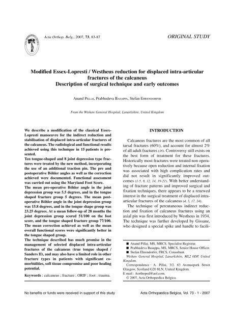
Acta Orthop. Belg., 2007, 73, 83-87ORIGINAL STUDY<strong>Modified</strong> <strong>Essex</strong>-<strong>Lopresti</strong> / <strong>Westheus</strong> <strong>reduction</strong> <strong>for</strong> displaced intra-articularfractures of the calcaneusDescription of surgical technique and early outcomesAnand PILLAI, Prabhudeva BASAPPA, Stefan EHRENDORFERFrom the Wishaw General Hospital, Lanarkshire, United KingdomWe describe a modification of the classical <strong>Essex</strong>-<strong>Lopresti</strong> manoeuvre <strong>for</strong> the indirect <strong>reduction</strong> andstabilisation of displaced intra-articular fractures ofthe calcaneus. The radiological and functional resultsachieved using this technique in 15 patients is presented.Ten tongue-shaped and 8 joint depression type fractureswere treated by the new method, incorporatingthe use of an additional traction pin. The pre andpostoperative Böhler angles as well as the correctionachieved were documented. Functional assessmentwas carried out using the Maryland Foot Score.The mean pre-operative Böhler angle in the jointdepression group was 5.5 degrees, and in the tongueshaped fracture group 5 degrees. The mean postoperativeBöhler angle in the joint depression groupwas 15.8 degrees, and in the tongue shape group was23.25 degrees. At a mean follow-up of 28 months thejoint depression group scored 51/100 on the footscore, and the tongue shaped fracture group 77/100.The mean correction achieved as well as the meanoverall functional scores were significantly better inthe tongue shaped group.The technique described has much promise in themanagement of selected displaced intra-articularfractures of the calcaneus (true tongue shaped /Sanders II), and may also have a limited role in otherfracture types in patients with significant comorbidities,soft tissue compromise and poor healingpotential.Keywords : calcaneus ; fracture ; ORIF ; foot ; trauma.INTRODUCTIONCalcaneus fractures are the most common of alltarsal fractures (60%), and account <strong>for</strong> almost 2%of all adult fractures (16). Controversy still exists onthe best <strong>for</strong>m of treatment <strong>for</strong> these fractures.Historically most fractures were treated non operativelybecause open <strong>reduction</strong> and internal fixationwas associated with high complication rates anddid not result in significantly improved outcomes(1-5, 8, 12, 14, 19-21). With better understandingof fracture patterns and improved surgical andfixation techniques, there appears to be a renewedinterest in the surgical treatment of displaced intraarticularfractures of the calcaneus (4, 5, 17, 24).The technique of percutaneous indirect <strong>reduction</strong>and fixation of calcaneus fractures using anaxial pin was first introduced by <strong>Westheus</strong> in 1934.The technique was further developed by Gissane,who designed a special spike and handle to facili-■ Anand Pillai, MS, MRCS, Specialist Registrar.■ Prabhudeva Basappa, MS, MRCS, Senior House Officer.■ Stefan Ehrendorfer, FRCS, Consultant.Wishaw General Hospital, Lanarkshire, ML2 0DP, UnitedKingdom.Correspondence : A. Pillai, 3/2. 63 Avenuepark StreetGlasgow, Scotland G20 8LN, United Kingdom.E mail : Aorthopod@aol.com.© 2007, Acta Orthopædica Belgica.No benefits or funds were received in support of this study Acta Orthopædica Belgica, Vol. 73 - 1 - 2007
84 A. PILLAI, P. BASAPPA, S. EHRENDORFERtate its use. The credit <strong>for</strong> describing the entire surgicaltechnique in sequence and <strong>for</strong> the introductionof a shoe plaster to incorporate the pin and tohold the <strong>reduction</strong> goes to <strong>Essex</strong>-<strong>Lopresti</strong> (6). Thetechnique has been further modified to include afull below-knee cast, percutaneous cancellousscrews, and arthroscopically assisted methods (7, 9,13, 18, 22, 23). The effectiveness of this technique inthe restoration of Böhler’s angle and calcanealheight in tongue shaped / Sanders type II fracturesis well documented (22, 23).We describe a modification of the classical<strong>Essex</strong>–<strong>Lopresti</strong> manoeuvre <strong>for</strong> the indirect <strong>reduction</strong>and stabilisation of displaced intra-articularfractures of the calcaneus and discuss the radiologicaland functional results achieved in a consecutiveseries of 15 patients.MATERIALS AND METHODSFifteen patients with displaced intra-articular fracturesof the calcaneus treated with the new techniquedescribed were prospectively followed-up. Open injuriesand patients with associated fractures were not includedin the study. Initial radiographic assessment of allpatients included standardised anterior-posterior, lateral,and axial views of the calcaneus. Further evaluation wasdone in all cases by computerised tomography. Allradiographic measurements were made on digitisedfilms on Picture Archival Communications System(PACS (Siemens), Magic Web).Fractures were classified according to the <strong>Essex</strong>-<strong>Lopresti</strong> system into tongue shaped (TS) and jointdepression types (JD). All surgical procedures were carriedout by the senior author (SE). Böhler’s angle wasmeasured preoperatively and post operatively, and thecorrection achieved was documented. All measurementswere independently verified by all the authors. Radiographicfollow-up was per<strong>for</strong>med at 6 weeks, 12 weeks,6 months and 12 months. Functional assessment wascarried out using the Maryland Foot Score.Surgical TechniqueWith the patient in the supine position, the affectedlimb was prepped and draped from the knee down. A 1-cm stab incision was made on the medial aspect of thecalcaneus, just anterior to the insertion of the Achillestendon. Blunt deep dissection was carried out carefullyFig. 1. — Intraoperative axial fluoroscopy viewavoiding tendons and neuro-vascular structures. A 4-mm Steinman pin was then introduced by hand from themedial wound and brought out laterally through anotherstab incision proximal and just anterior to the tendoAchillis insertion. The pin was attached and secured to aBöhler stirrup (fig 1). The leg was then flexed at theknee, and externally rotated at the hip into a figure offour- position, bringing the foot parallel to the surgicaltable. Longitudinal traction was then applied through theBohler’s stirrup in the line of the tibia, achieving an indirect<strong>reduction</strong> of the fracture. An assistant was requiredat this stage to apply counter-traction and to support thelimb. The <strong>reduction</strong> could then be checked by a lateralview of the foot taken through the table by an imageintensifier. If a radiolucent table is not available, then thefoot can be hung off the side of the table with tractionstill applied by the stirrup in the figure of four- position.The <strong>reduction</strong> achieved was then stabilised by a second4-mm Steinmann pin inserted using a power driverunder fluoroscopic control from the postero/lateralaspect of the calcaneus (fig 2). For this a third stab incisionwas made below the insertion of the Achilles tendon,and the pin introduced into the major posteriorfragment (posterior tuberosity fragment in tongueshaped fractures, and posterior body fragment in jointdepression fractures), parallel with the lateral border ofthe calcaneus and into the cuboid across the calcaneocuboidjoint. Further fine adjustments of the <strong>reduction</strong>were made if necessary by manipulating the fracturefragment directly with this pin, be<strong>for</strong>e it was passed intothe cuboid. The heel was then compressed between theActa Orthopædica Belgica, Vol. 73 - 1 - 2007
MODIFIED ESSEX-LOPRESTI/WESTHEUS REDUCTION 85Fig. 2. — Intraoperative lateral fluoroscopy viewsurgeon’s palms to reduce any lateral wall displacement.The first Steinman pin along with the Bohler’s stirrupwas removed, and the second Steinman pin cut shortoutside the skin. Sterile dressings were applied and thepin was incorporated in a below-knee lightweight cast.AP, lateral and axial views were then taken to confirmand document the final position achieved. A non-inflatedpneumatic tourniquet was routinely placed around thethigh, to be used if the procedure had to be converted toan open <strong>reduction</strong>.The pin and cast were maintained <strong>for</strong> 6 weeks and thepatient was kept non-weight bearing. The pins were routinelyremoved in the clinic with sedation. Following pinextraction, patients were referred to the physiotherapydepartment <strong>for</strong> foot and ankle mobilisation. Patientswere maintained non-weight bearing <strong>for</strong> a total of tenweeks post-operatively. This was followed by gradualweight bearing as tolerated.RESULTSThe study included 18 fractures in 15 patients (3bilateral injuries). The mean age was 39.1 years(range : 19 to 63). There was a clear male predominance(M:F = 4:1). The most common modeof injury was a fall from a height (80%).Radiologically, 10 fractures were of the tonguetype and 8 of the joint depression type. All patientsunderwent the procedure uneventfully. Only onepatient developed superficial wound infection atthe pin sites, which required oral antibiotics. Therewere no deep infections. Nine patients (60%) weresmokers (10 cigarettes or more per day), and allwere advised to quit or to cut down, with varyinglevels of compliance. Two patients were insulindependent diabetics. All patients retained their pins<strong>for</strong> 6 weeks, and none required any secondarysurgical procedure.The mean pre-operative Böhler angle in the jointdepression group was 5.5 degrees (range : 0 to 20),and in the tongue shaped fracture group 5 degrees(range : 0 to 15). There was no statistically significantdifference between the preoperative Bohler’sangle measurement between the two groups (p =0.8719 Mann-Whitney/Wilcoxon two sample test).The mean postoperative Böhler angle in the jointdepression group was 15.8 degrees (range : 10 to30). The mean postoperative Böhler angle in thetongue shape group was 23.25 degrees (range : 10to 32). The mean correction achieved in the jointdepression group was 10.5 degrees and in thetongue shaped group 19 degrees. The degree ofcorrection achieved in the tongue shaped group wassignificantly better than in the joint depressiongroup (p = 0.0025). There was no late collapsenoted in any patient.The mean duration of follow-up was 28 months(range : 24 to 48). All patients were available <strong>for</strong>final review. The mean Maryland Foot Score was67.3/100, with 67% of patients achieving fair toexcellent results. The mean functional score was77 (42-87/100) <strong>for</strong> the tongue shaped group and51 (42-73/100) <strong>for</strong> the joint depression group. Theoverall functional outcome in was significantlybetter in the tongue shaped group in comparison tothe joint depression group (p = 0.0027). The finalfunctional outcome correlated well with the postoperativeBöhler angle achieved.None of the patients in this series were onbenefits or were involved in workers compensationclaims.DISCUSSIONThe purpose of this paper is to describe a modified<strong>Essex</strong>-<strong>Lopresti</strong> / <strong>Westheus</strong> <strong>reduction</strong> technique<strong>for</strong> displaced intra-articular fractures of the calcaneus.The original technique requires the patient tobe prone, and <strong>reduction</strong> is achieved by tractionActa Orthopædica Belgica, Vol. 73 - 1 - 2007
86 A. PILLAI, P. BASAPPA, S. EHRENDORFERapplied to the talus by lifting the leg off the operatingtable with the knee flexed, and manoeuvringthe fragment with a Gissane os calcis spike (6). The<strong>reduction</strong> is held by incorporating the spike in aplaster cast. Modifications of the original techniquehave been reported with the patient supine and alsoin the lateral positions (13).There are several advantages to the technique wepropose :1) Supine position of the patient. Patients with calcanealfractures may indeed have other associatedinjuries, the most common being fracturesof the spine and the pelvis. In such patientsachieving a prone or even lateral position maybe difficult.2) The figure of four- position makes imaging witha modern C-arm easy. If a radiolucent table isnot available, then the foot can be simplybrought over the edge of the table, or even madeto rest on the C arm itself.3) Traction is applied by means of a pin passedproximal to the os calcis, and no leverage isapplied to the intraosseous pin. This may preventloosening and pin site infection.4) The technique allows the axial pin to be introducedby power under image guidance.5) Advancing the pin into the cuboid, providesadditional stability to the fixation.6) The technique is suitable <strong>for</strong> use with cannulatedcancellous screws.Bohler’s tuber joint angle is commonly assessedwhen evaluating calcaneal fractures. A severe heelfracture will result in significant decrease or loss ofthe angle. A number of studies clearly indicate thatBöhler’s angle is a good predictor of long termfunctional outcome in calcaneal fractures (4, 5, 15).Patients with angles less than 15 degrees did significantlyworse than those with greater angles.This was the reason <strong>for</strong> choosing Böhler’s angle asthe measure of surgical <strong>reduction</strong> in our patients. Inour group of patients, a mean correction of10.5 degrees was achieved in the JD group and19 degrees in the TS group. A mean post operativeBöhler angle of greater than 15 degrees wasachieved in both groups of patients. The plain radiographfracture classification by <strong>Essex</strong>-<strong>Lopresti</strong>based on the configuration of the secondary fractureline through the posterior facet, as used in ourstudy was also found to be highly prognostic (p =0.035) (4, 5).Compared to open procedures, percutaneous<strong>reduction</strong> and fixation offers lower complicationrates, shorter operating times and more rapid healingdue to the undisturbed soft tissue envelope (1, 8,11). For carefully selected patients, this techniqueprovides good results comparable with open <strong>reduction</strong>and internal fixation. Ten out of 15 patients inour series were either smokers or diabetics, whowould be considered high risk <strong>for</strong> open <strong>reduction</strong>and internal fixation (4, 5). The overall infection ratein our series is only 6%, and we did not encounterany additional complications in this vulnerablegroup.Eighty percent (12 patients) of our patients weremale and 40% (6 patients) were involved in heavymanual work. Both factors are considered to lead topoor outcomes after open fixation.The scope of percutaneous techniques <strong>for</strong> thetreatment of intra-articular fractures of the calcaneusis limited. The goals of this approach are toimprove the alignment of the calcaneus and <strong>reduction</strong>of the posterior facet. In tongue-shaped fractures,the posterior articular facet is contiguouswith the posterior tuberosity. Direct manipulationof the facet is possible by traction and a pin insertedinto the posterior tuberosity. This allows thefracture to be reduced accurately percutaneously(10). In joint depression type, the posterior articularfacet is depressed, rotated and often impactedinto the posterior body fragment. Since directmanipulation of the facet is not possible, by a pininserted into the posterior body fragment, itbecomes difficult to achieve an accurate <strong>reduction</strong>by this technique (10). Our results thus support earlierobservations that the percutaneous technique isbest suited <strong>for</strong> tongue shaped fractures (22, 23). Themean correction achieved as well as the mean overallfunctional scores were significantly better inthis group. However, our results clearly show that acorrection of the Böhler angle was achieved in bothtongue shaped and joint depression types of fracturesby the technique described. The addition of asecond traction pin allows greater <strong>for</strong>ces to beActa Orthopædica Belgica, Vol. 73 - 1 - 2007
MODIFIED ESSEX-LOPRESTI/WESTHEUS REDUCTION 87applied across the subtalar joint to disimpact thefracture fragments. Also passing the pin past thecalcaneo-cuboid joint af<strong>for</strong>ds greater stability tothe fixation. Hence we must disagree with previousreports that the method is suitable only <strong>for</strong> tongueshaped calcaneal fractures.Deciding how to manage a displaced intra-articularfracture of the calcaneus is challenging.Assessment of the fracture, patient status, and functionalneeds are paramount in dictating treatment.Although our report is only from a single unit, andinvolves a relatively small number of patients, wefeel that the technique described has much promisein the management of selected displaced intraarticularfractures (true tongue shaped / Sanders II)and may also have a limited role in other fracturetypes in patients with significant co-morbidities,soft tissue compromise and poor healing. Howeverthe technique allows only limited exposure andcontrol of fracture fragments, and if anatomical<strong>reduction</strong> is sought then open <strong>reduction</strong> should bepreferred.REFERENCES1. Abidi NA, Dhawan S, Gruen GS et al. Wound healingrisk factors after open <strong>reduction</strong> and internal fixation ofcalcaneal fractures. Foot Ankle Int 1998 ; 19 : 856-861.2. Bridgeman SA, Dunn KM, McBride DJ, Richards PJ.Interventions <strong>for</strong> treating calcaneal fractures. CochraneDatabase Syst Rev 2002 ; 2 : CD001161.3. Buckley RE, Meek RN. Comparison of open vs. closed<strong>reduction</strong> of intraarticular calcaneal fractures : a matchedcohort in workmen. J Orthop Trauma 1992 ; 6 : 216-222.4. Buckley R, Tough S, McCormack R et al. Operativecompared with non operative treatment of displaced intraarticularcalcaneal fractures. A prospective, randomised,controlled multicenter trial. J Bone Joint Surg 2002 ; 84-A : 1733-1744.5. Buckley R, Tough S. Displaced intra-articular calcanealfractures. J Am Acad Othop Surg 2004 ; 12 : 172-178.6. <strong>Essex</strong>-<strong>Lopresti</strong> P. The mechanism, <strong>reduction</strong> technique,and results in fractures of the os calcis. Br J Surg 1952 ;39 : 395-419.7. Fernandez DL, Koella C. Combined percutaneous and“mimimal” internal fixation displaced articular fractures ofthe calcaneus. Clin Orthop 1993 ; 290 : 108-116.8. Folk JW, Starr AJ, Early JS. Early wound complicationsof operative treatment of calcaneus fractures : analysis of190 fractures. J Orthop Trauma 1999 ; 13 : 369-372.9. Gavlik JM, Rammelt S, Zwipp H. Percutaneous, arthroscopically-assistedosteosynthesis of calcaneus fractures.Arch Orthop Trauma Surg 2002 ; 122 : 424-428.10. Hammesfahr R, Fleming L. Calcaneal fractures : A goodprognosis. Foot Ankle 1981 ; 2 : 161-171.11. Howard JL, Buckley R, McCormack R et al. Complicationsfollowing management of displaced intraarticularcalcaneal fractures. A prospective randomised trial comparingopen <strong>reduction</strong> internal fixation with non operativemanagement. J Orthop Trauma 2003 ; 17 : 241-249.12. Jarvholm U, Komer L, Thoren O, Wildund LM.Fractures of the calcaneus. A comparison of open andclosed treatment. Acta Ortop Scand 1984 ; 55 : 652-656.13. King RE. Axial pin fixation of fractures of the os calcis(Method of <strong>Essex</strong>-<strong>Lopresti</strong>). Orthop Clin N Am 1973 ; 4 :185-188.14. Kundel K, Funk E, Brutscher M, Bickel R. Calcanealfractures : operative versus non-operative treatment.J Trauma 1996 ; 41 : 839-845.15. Loucks C, Buckley R. Böhler’s angle : correlation withoutcome in displaced intraarticular calcaneal fractures.J Orthop Trauma 1999 ; 13 : 554-558.16. Nicklebur S, Dixon TB, Probe R. Calcaneus fractures.EMedicine, July 21, 2004. http://www.emedicine.com/orthoped/topic33.htm.17. Palmer I. The mechanism and treatment of fractures of thecalcaneus. Open <strong>reduction</strong> with the use of cancellousgrafts. J Bone Joint Surg 1948 ; 30-A : 2-8.18. Rammelt S, Amlang M, Barthel S, Zwipp H. Minimallyinvasivetreatment of calcaneal fractures. Injury 2004 ; 35Suppl 2 : SB55-63.19. Randle JA, Kreder HJ, Stephan D et al. Shouldcalcaneal fractures be treated surgically ? A meta analysis.Clin Orthop 2002 ; 377 : 217-227.20. Thermann H, Krettek C, Hufner T et al. Management ofcalcaneal fractures in adults. Conservative versus operativetreatment. Clin Orthop 1998 ; 353 : 107-124.21. Thordarson DB, Krieger LE. Operative vs. non operativetreatment of intraarticular fractures of the calcaneus : aprospective randomised trial. Foot Ankle Int 1996 ; 17 : 2-9.22. Tornetta P 3 rd . <strong>Essex</strong>-<strong>Lopresti</strong> <strong>reduction</strong> <strong>for</strong> calcanealfractures revisited. J Orthop Trauma 1998 ; 12 : 469-473.23. Tornetta P 3 rd . Percutaneous treatment of calcaneal fractures.Clin Orthop 2000 ; 375 : 91-96.24. Zwipp H, Tscherne H, Thermann H, Weber T.Osteosynthesis of displaced intraarticular fractures of thecalcaneus. Results in 123 cases. Clin Orthop 1993 ; 290 :76-86.Acta Orthopædica Belgica, Vol. 73 - 1 - 2007