ORIGINAL STUDY Acromioclavicular joint reconstruction using the ...
ORIGINAL STUDY Acromioclavicular joint reconstruction using the ...
ORIGINAL STUDY Acromioclavicular joint reconstruction using the ...

You also want an ePaper? Increase the reach of your titles
YUMPU automatically turns print PDFs into web optimized ePapers that Google loves.
Acta Orthop. Belg., 2008, 74, 167-172<strong>ORIGINAL</strong> <strong>STUDY</strong><strong>Acromioclavicular</strong> <strong>joint</strong> <strong>reconstruction</strong> <strong>using</strong> <strong>the</strong> Nottingham Surgilig :A preliminary reportRajarshi BHATTACHARYA, Lorna GOODCHILD, Amar RANGANFrom James Cook University Hospital, Middlesbrough, United KingdomEleven patients with a chronic acromio-clavicular<strong>joint</strong> disruption underwent acromio-clavicular <strong>joint</strong>stabilisation <strong>using</strong> <strong>the</strong> Nottingham Surgilig and werefollowed up clinically and radiologically for an averageof 24 months post operation.The mean post-operative Constant score was 83.1,<strong>the</strong> Imatani score was 81.2 and <strong>the</strong> Walsh score was14.1. Eighty-two per cent of patients were satisfiedwith <strong>the</strong> operation. There was one case of rupturethrough <strong>the</strong> central portion of <strong>the</strong> Surgilig, and followinglaboratory analysis, <strong>the</strong> ligament has beenmodified since. In 4 patients <strong>the</strong>re was evidence ofloosening of <strong>the</strong> screw but only one complained ofthis being a problem.This short-term outcome analysis of <strong>the</strong> NottinghamSurgilig is <strong>the</strong> first such report outside <strong>the</strong>Nottingham unit where <strong>the</strong> pros<strong>the</strong>sis was originallydeveloped. Our results are encouraging and justifyfur<strong>the</strong>r use and evaluation of this relatively new technique.Keywords : acromioclavicular <strong>joint</strong> disruption ;chronic ; <strong>reconstruction</strong> ; Surgilig.INTRODUCTIONAcromio-clavicular (A-C) <strong>joint</strong> disruption is arelatively common injury (10). There is a significantmale preponderance of this injury and <strong>the</strong> mostcommon mechanism is a direct force occurringfrom a fall on <strong>the</strong> point of <strong>the</strong> shoulder (18). A-C<strong>joint</strong> injuries have been classified by Tossy et al (21)and Allman (1) as incomplete (Grades I and II) andcomplete (Grade III). Rockwood et al (18) expanded<strong>the</strong> original classification to include six types ofinjuries. The ideal treatment of <strong>the</strong> Type 3 injuryremains a matter of debate.Although various methods of stabilising <strong>the</strong> disruptionof <strong>the</strong> A-C <strong>joint</strong> have been described <strong>using</strong><strong>the</strong> coracoacromial ligament (4,6,11,13,20,24,25), <strong>the</strong>need to preserve this ligament where possible hasalso been acknowledged in <strong>the</strong> literature (12,14).Over <strong>the</strong> years, <strong>the</strong> methods that have been used on<strong>the</strong>ir own or in conjunction with <strong>the</strong> coraco-acromialligament for <strong>the</strong> <strong>reconstruction</strong> of <strong>the</strong> coracoclavicularligament include PDS (16), merselene(17), Dacron (5,22) or Dacrylene (15), carbonfibre (2) and coraco-clavicular screw (24).More recently, a braided syn<strong>the</strong>tic ligament, <strong>the</strong>Nottingham Surgilig, was developed for this purpose,<strong>using</strong> polyester fibres, similar to that used for■ Rajarshi Bhattacharya, MBBS, MRCS (Edin), MRCS(Glas), MSc (Orth), Specialist Registrar in Orthopaedics.■ Lorna Goodchild, BSc (Psysio) MCSP, MACP, SeniorPhysio<strong>the</strong>rapist.■ Amar Rangan, FRCS (Glas) MCH (Orth) FRCS Ed(Orth), Consultant Orthopaedic Surgeon.Department of Trauma and Orthopaedic Surgery, JamesCook University Hospital, Middlesbrough, United Kingdom.Correspondence : R. Bhattacharya, 11 Carham Close,Gosforth, Newcastle Upon Tyne, NE3 5DX, United Kingdom.E-mail : drbhattacharya@yahoo.com© 2008, Acta Orthopædica Belgica.No benefits or funds were received in support of this study Acta Orthopædica Belgica, Vol. 74 - 2 - 2008
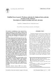
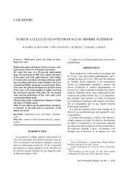
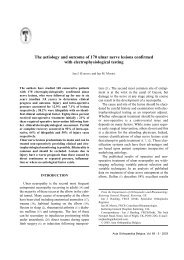
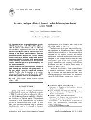
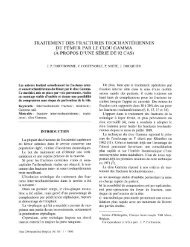
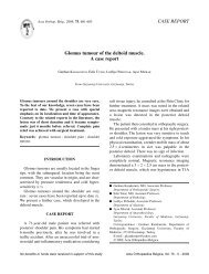
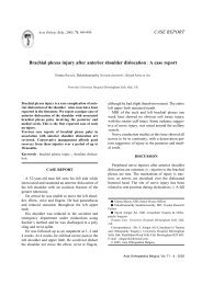
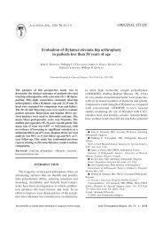
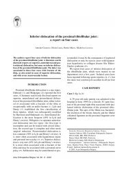
168 R. BHATTACHARYA, L. GOODCHILD, A. RANGANFig. 2. — Instruments for <strong>the</strong> Surgilig from above down :1. loop tensioner, 2. tubular introducer, 3. curved rasp, 4. cannulatedpositioner.Fig. 1. — The Nottingham Surgilig with 2 loops and fixationscrew.anterior cruciate ligament <strong>reconstruction</strong> in <strong>the</strong>knee. We report our experience with <strong>the</strong> use of thisligament for A-C <strong>joint</strong> <strong>reconstruction</strong> especially incases of longstanding A-C <strong>joint</strong> instability.MATERIALS AND METHODSThe Nottingham Surgilig is a braided polyester ligamentwith a loop at each end (fig 1). One of <strong>the</strong> loops isstiff and called <strong>the</strong> hard loop, which is used for screwfixation. The o<strong>the</strong>r loop, called <strong>the</strong> soft loop is used tothread <strong>the</strong> ligament through itself. The ligament ispassed around <strong>the</strong> coracoid process threaded throughitself, passed around <strong>the</strong> back of <strong>the</strong> clavicle and <strong>the</strong>nanchored to <strong>the</strong> clavicle <strong>using</strong> a screw. The pros<strong>the</strong>ticligament is available in sizes from 5 to 20 cm in incrementsof 1 cm. The standard instruments available forpros<strong>the</strong>sis insertion include a curved rasp, a cannulatedpositioner, a tubular introducer and a loop tensioner(fig 2) along with a Surgilig length gauge with a metalleader (fig 3).Eleven patients (10 male and 1 female) underwent A-C <strong>joint</strong> stabilisation <strong>using</strong> <strong>the</strong> Nottingham Surgilig. Theaverage gap period between <strong>the</strong> injury and <strong>the</strong> stabilisationprocedure was 21 months (range : 5 to 41). Only inone case was <strong>the</strong> operation done relatively early, at5 months following injury, due to impending breakdownof <strong>the</strong> overlying skin as a result of pressure from <strong>the</strong> lateralend of <strong>the</strong> clavicle. All <strong>the</strong> remaining patients hadlong standing A-C <strong>joint</strong> instability, of at least a year ormore, with associated symptoms mainly of pain andFig. 3. — Tubular introducer with Surgilig length gaugefunctional disability, following an initial disruption ofRockwood Type 3 or above and had a course of conservativetreatment.All <strong>the</strong> patients received a similar postoperative follow-upregime involving immobilisation in a poly slingfor 2 weeks followed by supervised physio<strong>the</strong>rapy tofull mobilisation as tolerated.Operative techniqueA vertical skin incision is made from above <strong>the</strong> clavicleto <strong>the</strong> coracoid. An incision is <strong>the</strong>n made along <strong>the</strong>lateral 2 cm of <strong>the</strong> clavicle, dividing <strong>the</strong> periosteum asfar as <strong>the</strong> displaced lateral end of <strong>the</strong> clavicle. The lateral1 cm of <strong>the</strong> clavicle is excised.The base of <strong>the</strong> coracoid is identified with a bluntinstrument and <strong>the</strong>n <strong>the</strong> Surgilig Tubular Introducer(fig 2) is passed around <strong>the</strong> base of <strong>the</strong> coracoid with itstip passed adjacent to <strong>the</strong> bone from <strong>the</strong> medial sidedownwards staying close to <strong>the</strong> bone. The SurgiligLength Gauge, which has a loop at each end and a metalleader in front of one of <strong>the</strong> loops (fig 2), is <strong>the</strong>n fed intoActa Orthopædica Belgica, Vol. 74 - 2 - 2008
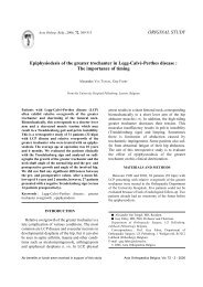
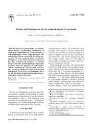
ACROMIOCLAVICULAR JOINT RECONSTRUCTION 169Fig. 4. — Surgilig with loop tensionerFig. 5. — Final position of Surgilig in an anatomical model<strong>the</strong> Tubular Introducer (fig 3) <strong>using</strong> <strong>the</strong> metal leader as<strong>the</strong> advancing end.Holding <strong>the</strong> Surgilig Length Gauge that has exitedfrom <strong>the</strong> Tubular Introducer, <strong>the</strong> Introducer is <strong>the</strong>nremoved, leaving <strong>the</strong> measuring tape of <strong>the</strong> Gaugearound <strong>the</strong> coracoid. The measuring tape is <strong>the</strong>n loopedaround <strong>the</strong> coracoid in <strong>the</strong> same way as <strong>the</strong> ligamentwould be and passed up and behind <strong>the</strong> lateral end of <strong>the</strong>clavicle and <strong>the</strong> clavicle is reduced to its normal alignment,flush with <strong>the</strong> level of <strong>the</strong> acromion and <strong>the</strong> appropriatelength of <strong>the</strong> pros<strong>the</strong>sis can be chosen from <strong>the</strong>measuring tape.Two fixation positions are acceptable, ei<strong>the</strong>r aimingfor <strong>the</strong> hard loop of <strong>the</strong> Surgilig to lie at <strong>the</strong> posterior orsuperior edge of <strong>the</strong> clavicle. The aim is to achieve aslight degree of over correction of <strong>the</strong> clavicular reduction.The appropriate length of Surgilig now replaces <strong>the</strong>Surgilig Length Gauge by daisy chaining <strong>the</strong> Surgiligonto <strong>the</strong> Length Gauge. The Surgilig is <strong>the</strong>n passedaround <strong>the</strong> base of <strong>the</strong> coracoid and <strong>the</strong> hard loop isthreaded through <strong>the</strong> soft loop, so that <strong>the</strong> soft loop sitson <strong>the</strong> clavicular (superior) side of <strong>the</strong> coracoid. TheSurgilig should sit flat on <strong>the</strong> coracoid and not be twisted.Any bony spikes are gently removed from <strong>the</strong> claviclewith <strong>the</strong> Curved Rasp (fig 2). The Surgilig is snuggedup to <strong>the</strong> coracoid <strong>using</strong> <strong>the</strong> Surgilig Loop Tensioner(fig 4).The Surgilig is <strong>the</strong>n passed behind <strong>the</strong> clavicle. TheTubular Introducer is now inserted through <strong>the</strong> loop ofSurgilig and used to apply traction on <strong>the</strong> Surgilig toallow accurate reduction of <strong>the</strong> clavicle.A drill hole is now made in <strong>the</strong> clavicle at <strong>the</strong> positionof <strong>the</strong> hard loop of <strong>the</strong> Surgilig and <strong>the</strong> Surgilig is fixedwith a bicortical screw inserted through <strong>the</strong> loop andinto <strong>the</strong> clavicle. It is important that <strong>the</strong> Surgilig is nottwisted and <strong>the</strong>re is no slack in <strong>the</strong> ligament (fig 5).Postoperatively, <strong>the</strong> arm is supported in a sling for2 weeks and <strong>the</strong>n mobilised with supervised physio<strong>the</strong>rapy.Follow-upThis was a prospective cohort study involving11 patients all of whom were followed-up to an averageperiod of 24 months (range : 6 to 76) post operation.Except for one patient who had a revision procedure at6 months, all <strong>the</strong> o<strong>the</strong>r patients had a minimum followupof 12 months. The follow-up protocol consisted ofclinical and radiological examinations (fig 6), with plainradiographs of <strong>the</strong> A-C <strong>joint</strong>, and collection of data for<strong>the</strong> Constant-Murley, <strong>the</strong> Imatani (7) and <strong>the</strong> Walsh (23)scores. The average age of <strong>the</strong> patient cohort was35.1 years (range : 21 to 56). There were an equalnumber of operations on <strong>the</strong> right and left sides and ondominant and non-dominant sides.RESULTSAt follow-up, clinical examination showed satisfactoryoutcome in most of <strong>the</strong> patients. Nine(82%) were satisfied with <strong>the</strong> overall procedure,with good functional outcome. This was corroboratedby <strong>the</strong> good scores obtained in <strong>the</strong> outcomemeasurement questionnaires.The detailed scores are presented in table I.Clinically, <strong>the</strong>re was prominence of <strong>the</strong> fixationscrew in 4 cases and radiographs showed lucencyaround <strong>the</strong> screws, but only one of <strong>the</strong> cases complainedof any pain or discomfort. However, hedeclined surgery for screw removal. Unfortunately,his individual low scores affected <strong>the</strong> mean scoresof <strong>the</strong> study despite all <strong>the</strong> o<strong>the</strong>r patients scoringActa Orthopædica Belgica, Vol. 74 - 2 - 2008
170 R. BHATTACHARYA, L. GOODCHILD, A. RANGANTable I. — Postoperative scoresMean Standard RangedeviationPost-op Constant score 83.1 12 61-100Post-op Imatani score 81.2 19.4 51-98Post-op Walsh scores 14.1 4.3 8-20Constant score : 0 to 100. Normal is 100.Imatani score : 0 to 100. Normal is 100.Walsh score : 0 to 20. Normal is 20.Fig. 6. — Pre-operative and post-operative x-raysreasonably well in all three scoring systems(table II). There was a rupture of <strong>the</strong> central portionof <strong>the</strong> ligament in one patient after 6 months andthis had to be revised with a clavicular hook plate,hence this patient’s follow-up was considered onlytill 6 months. All <strong>the</strong> patients in <strong>the</strong> above studyhad been treated with <strong>the</strong> unmodified ligament butonly one had <strong>the</strong> ligament rupture. Followingdetailed analysis of this retrieved ligament, <strong>the</strong>pros<strong>the</strong>sis has since been reinforced and streng<strong>the</strong>nedin <strong>the</strong> central portion.DISCUSSIONSurgery for A-C <strong>joint</strong> separation has remained adebated issue over <strong>the</strong> years. When surgical treatmentis indicated, <strong>the</strong>re are a variety of operationsto choose from that have varying degrees of successreported in <strong>the</strong> literature (3).The Weaver-Dunn procedure, where <strong>the</strong> coracoacromialligament is detached from its acromialinsertion and reattached within <strong>the</strong> intramedullarycavity of <strong>the</strong> clavicle has gained considerable popularity.O<strong>the</strong>r methods of A-C <strong>joint</strong> fixation <strong>using</strong><strong>the</strong> coraco-acromial ligament have also been proposed(4,6,11,13,20,25). More recently, <strong>the</strong> importanceof preserving <strong>the</strong> subacromial arch andspecifically <strong>the</strong> coraco-acromial ligament has beenstressed (12,14). Biomechanical studies have shownthat release of <strong>the</strong> coraco-acromial ligament canlead to increased glenohumeral <strong>joint</strong> translationand laxity (14) while phylomorphic analyses havestressed <strong>the</strong> role of <strong>the</strong> coraco-acromial ligamentto provide increased mechanical stability of <strong>the</strong>shoulder (12). The coraco-acromial ligament alsoacts as a buffer between <strong>the</strong> acromion and <strong>the</strong>rotator cuff and this buffering action is lost incoraco-acromial ligament transection (19). Anadvantage of <strong>the</strong> Surgilig is <strong>the</strong> sparing of <strong>the</strong>coraco-acromial arch. Besides, occasionally, patientspresent with failed Weaver-Dunn procedures or arepeat A-C <strong>joint</strong> disruption after o<strong>the</strong>r types ofprevious stabilisation with <strong>the</strong> coraco-acromialligament. The Nottingham Surgilig would be auseful alternative to consider in <strong>the</strong>se situations.Our series is based mainly on <strong>the</strong> results of late<strong>reconstruction</strong>s of chronic Rockwood Type 3 A-C<strong>joint</strong> disruption. Although <strong>the</strong> literature describesvarious forms of surgery in A-C <strong>joint</strong> disruption,very few papers deal with <strong>the</strong> results of surgery insymptomatic chronic injuries having late <strong>reconstruction</strong>(3,4,6,15,25) and even fewer look exclusivelyat grade 3 injuries (25). Most of <strong>the</strong> papers reporta mixed population of injuries. While some of <strong>the</strong>sehave a very few chronic cases (24), some fail toreport <strong>the</strong> results of <strong>the</strong> chronic group separately(9). Besides <strong>the</strong> lack of homogeneity in <strong>the</strong> studyActa Orthopædica Belgica, Vol. 74 - 2 - 2008
ACROMIOCLAVICULAR JOINT RECONSTRUCTION 171Table II. — Detailed results of <strong>the</strong> patientsPatient Age Injury to operation Follow-up Constant Imatani Walshtime (months) (months)1 37 23 76 88 86 162 56 25 34 94 97 153 22 5 33 78 51 94 25 8 29 90 85 205 30 13 22 78 76 126 24 41 14 95 93 147 43 25 14 61 52 88 46 24 14 79 97 179 51 18 12 92 98 1910 21 33 6 71 67 1011 31 12 14 88 91 15groups, <strong>the</strong> outcome measures used in <strong>the</strong> differentpapers also vary widely. Most of <strong>the</strong> authors haveassessed patients <strong>using</strong> self-prepared evaluationsystems. Very few papers have used A-C <strong>joint</strong>specific scores and even in those that have, <strong>the</strong>re isno uniformity of <strong>the</strong> scoring system used. As aresult of this variation in study group and outcomemeasures, a direct comparison of <strong>the</strong> results withour series becomes very difficult.The overall success rate in A-C <strong>joint</strong> surgery,both acute and chronic, is around 90% as reportedby various authors (4,6,13,15,24,25), while in <strong>the</strong> caseof late <strong>reconstruction</strong> <strong>the</strong> success rate has beenreported at around 78% (4,6,25). In our unit, <strong>the</strong> useof <strong>the</strong> Surgilig ligament in case of chronic AC <strong>joint</strong>disruptions has yielded comparable overall resultsto <strong>the</strong> o<strong>the</strong>r techniques of late <strong>reconstruction</strong>sreported in <strong>the</strong> literature. Additionally, <strong>the</strong> Surgiligcan be loaded immediately and mobilised earlyunlike <strong>the</strong> Weaver-Dunn procedure. The initialresting period is purely for scar healing. Unlikeprevious published studies where patients haduntoward reactions to <strong>the</strong> syn<strong>the</strong>tic materials usedfor coracoclavicular ligamentoplasty (5,15), none ofour patients reported any tolerance problems to <strong>the</strong>material used in Surgilig.CONCLUSIONThe Nottingham Surgilig is a relatively newtechnique for A-C <strong>joint</strong> <strong>reconstruction</strong>. A recentseries from <strong>the</strong> Nottingham Unit, where <strong>the</strong> ligamentwas initially developed, has shown promisingresults with this technique (8). Our series is however,<strong>the</strong> first report from an independent centre outsideNottingham, and despite <strong>the</strong> relatively smallnumber of cases, based on our experience of thisartificial ligament, <strong>the</strong>se short term results wouldjustify fur<strong>the</strong>r use and evaluation of this technique.REFERENCES1. Allman FL Jr. Fractures and ligamentous injuries of <strong>the</strong>clavicle and its articulation. J Bone Joint Surg 1967 ;49-A : 774-784.2. Burri C, Neugebauer R. Carbon fibre replacement of<strong>the</strong> ligaments of <strong>the</strong> shoulder girdle and <strong>the</strong> treatment oflateral instability of <strong>the</strong> ankle <strong>joint</strong>. Clin Orthop 1985 ;196 : 112-117.3. Dewar FP, Barrington TW. The treatment of chronicacromioclavicular dislocation. J Bone Joint Surg 1965 ;47-B : 32-35.4. Dumontier C, Sautet A, Man M, Apoil A.<strong>Acromioclavicular</strong> dislocations : treatment by coracoacromialligamentoplasty. J Shoulder Elbow Surg 1995 ; 4 :130-134.5. Goldberg JA, Viglione W, Cumming WJ et al. Review ofcoracoclavicular ligament <strong>reconstruction</strong> <strong>using</strong> Dacrongraft material. Austr N Z J Surg 1987 ; 57 : 441-445.6. Guy DK, Wirth MA, Griffin JL, Rockwood CA Jr.Reconstruction of chronic and complete dislocations of <strong>the</strong>acromioclavicular <strong>joint</strong>. Clin Orthop 1998 ; 347 : 138-149.7. Imatani RJ, Hanlon JJ, Cady GW. Acute, completeacromioclavicular separation. J Bone Joint Surg 1975 ;57-A : 328-331.Acta Orthopædica Belgica, Vol. 74 - 2 - 2008
172 R. BHATTACHARYA, L. GOODCHILD, A. RANGAN8. Jeon IH, Dewnany G, Hartley R et al. Chronic acromioclavicularseparation : The medium term results of coracoclavicularligament <strong>reconstruction</strong> <strong>using</strong> braided polyesterpros<strong>the</strong>tic ligament. Injury 2007 ; 38 : 1247-1253.9. Kawabe N, Watanabe R, Sato M. Treatment of completeacromioclavicular separation by coracoacromial ligamenttransfer. Clin Orthop 1984 ; 185 : 222-227.10. Koval KJ, Zuckerman JD. Handbook of Fractures.2 nd edition. Lippincott Williams and Wilkins, London,2002, p 68.11. Kumar S, Sethi A, Jain AK. Surgical treatment ofcomplete acromioclavicular dislocation <strong>using</strong> <strong>the</strong> coracoacromialligament and coracoclavicular fixation : report ofa technique in 14 patients. J Orthop Trauma 1995 ; 9 : 507-510.12. Kummer FJ, Blank K, Zuckerman JD. Coracoacromialligament function : a phylomorphic analysis. Bull HospitalJoint Disease 1996 ; 55 : 72-74.13. Kutschera HP, Kotz RI. Bone-ligament transfer ofcoracoacromial ligament for acromioclavicular dislocation.A new fixation method used in 6 cases. Acta OrthopScand 1997 ; 68 : 246-248.14. Lee TQ, Black AD, Tibone JE, McMahon PJ. Release ofcoracoacromial ligament can lead to glenohumeral laxity :a biomechanical study. J Shoulder Elbow Surg 2001 ; 10 :68-72.15. Mathieu L, Rongieras F, Fascia P et al. [<strong>Acromioclavicular</strong>dislocations treated by syn<strong>the</strong>tic coraco-clavicularligamentoplasty.] (French) Rev Chir Orthop 2007 ; 93 :116-125.16. Motamadi AR, Blevins FT, Willis MC et al.Biomechanics of <strong>the</strong> coracoclavicular ligament complexand augmentations used in its repair and <strong>reconstruction</strong>.Am J Sports Med 2000 ; 28 : 380-384.17. Pearsall AW 4 th , Hollis JM, Russell GV Jr, Stokes DA.Biomechanical comparison of <strong>reconstruction</strong> techniquesfor disruption of <strong>the</strong> acromioclavicular and coracoclavicularligaments. J South Orthop Assoc 2002 ; 11 : 11-17.18. Rockwood CA Jr, Williams GR, Young DC. Injuries to<strong>the</strong> acromioclavicular <strong>joint</strong>. In : Rockwood and Green(eds). Fractures in Adults. 4 th edition. Lippincott-Raven,London, 1996.19. Salter EG Jr, Nasca RJ, Shelley BS. Anatomical observationson <strong>the</strong> acromioclavicular <strong>joint</strong> and supporting ligaments.Am J Sports Med 1987 ; 15 : 199-206.20. Tienan TG, Oyen JF, Eggen PJ. A modified technique of<strong>reconstruction</strong> for complete acromioclavicular dislocation :a prospective study. Am J Sports Med 2003 ; 31 : 655-659.21. Tossy JD, Mead MC, Simmond HM. <strong>Acromioclavicular</strong>separations : Useful and practical classification for treatment.Clin Orthop 1963, 28 : 111-119.22. Verhaven E, DeBoeck H, Haentjens P et al. Surgicaltreatment of acute type-V acromioclavicular injuries inathletes. Arch Orthop Trauma Surg 1993 ; 112 : 189-192.23. Walsh WM, Peterson DA, Shelton G, Neumann RD.Shoulder strength following acromioclavicular injury.Am J Sports Med 1985 ; 13 : 153-158.24. Weaver JK, Dunn HK. Treatment of acromioclavicularinjuries, especially complete acromioclavicular separation.J Bone Joint Surg 1972 ; 54-A : 1187-1194.25. Weinstein DM, McCann PD, McIlveen SJ et al. Surgicaltreatment of complete acromioclavicular dislocations.Am J Sports Med 1995 ; 23 : 324-331.Acta Orthopædica Belgica, Vol. 74 - 2 - 2008