Free Beta HCG - Diagnostic Automation : Cortez Diagnostics
Free Beta HCG - Diagnostic Automation : Cortez Diagnostics
Free Beta HCG - Diagnostic Automation : Cortez Diagnostics

Create successful ePaper yourself
Turn your PDF publications into a flip-book with our unique Google optimized e-Paper software.
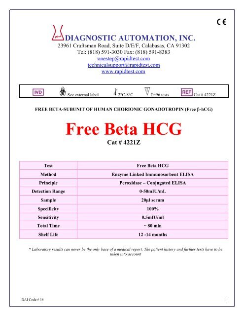
DIAGNOSTIC AUTOMATION, INC.<br />
23961 Craftsman Road, Suite D/E/F, Calabasas, CA 91302<br />
Tel: (818) 591-3030 Fax: (818) 591-8383<br />
onestep@rapidtest.com<br />
technicalsupport@rapidtest.com<br />
www.rapidtest.com<br />
See external label 2°C-8°C Σ=96 tests Cat # 4221Z<br />
FREE BETA-SUBUNIT OF HUMAN CHORIONIC GONADOTROPIN (<strong>Free</strong> β-hCG)<br />
<strong>Free</strong> <strong>Beta</strong> <strong>HCG</strong><br />
Cat # 4221Z<br />
Test<br />
<strong>Free</strong> <strong>Beta</strong> <strong>HCG</strong><br />
Method<br />
Enzyme Linked Immunosorbent ELISA<br />
Principle<br />
Peroxidase – Conjugated ELISA<br />
Detection Range<br />
0-50mIU/mL<br />
Sample<br />
20µl serum<br />
Specificity 100%<br />
Sensitivity<br />
0.5mIU/ml<br />
Total Time<br />
~ 80 min<br />
Shelf Life<br />
12 -14 months<br />
* Laboratory results can never be the only base of a medical report. The patient history and further tests have to be<br />
taken into account<br />
DAI Code # 16 1
Intended use<br />
The free beta hCG-subunit quantitative assay is designed for in vitro quantitative measurement of human chorionic<br />
gonadotropin free beta-subunit in patient’s serum. (For professional use only).<br />
Introduction<br />
Human Chorionic Gonadotropin (hCG) is a glycoprotein hormone normally produced by placenta during<br />
pregnancy. The hormone is present in blood and urine around seven to thirteen days following implantation of the<br />
fertilized ovum. Structurally intact hCG molecules consist of two non-covalently linked polypeptide subunits, the<br />
alpha and beta chain subunits. Measurement of intact hCG and of the alpha subunit of hCG appears to give similar<br />
results in blood and urine but not the levels of beta subunit. In the normal second-trimester maternal sera, the level<br />
of intact hCG range from 20,000 mIU/ml to 50,000 mIU/ml. In contrast, the levels of either free - or free -hCG<br />
are on average one half of 1% of hCG levels. hCG and the free subunits appear not to be useful as serological<br />
markers for nontrophoblastic tumors; however, the absolute increase of -hCG level in choriocarcinoma patients<br />
clearly differentiates it from normal pregnancy.<br />
Recent studies showed a significant increase in the level of free -hCG subunit in trisomy 21 cases as compared<br />
with controls. Hence, it has been suggested that free -hCG subunit assay in a combination of maternal serum AFP<br />
could be effective in a screening protocol for trisomy 21.<br />
Principle of the test<br />
The free β-hCG Quantitative Test Kit is based on a solid phase enzyme-linked immunosorbent assay. The assay<br />
system utilizes one anti-β-hCG antibody for solid phase (microtiter wells) immobilization and another mouse<br />
monoclonal anti-β-hCG antibody in the antibody-enzyme (horseradish peroxidase) conjugate solution. The test<br />
specimen (serum) is added to the β-hCG antibody coated microtiterwells and incubated with the Zero Buffer. If β-<br />
hCG is present in the specimen, it will combine with the antibody on the well. The well is then washed to remove<br />
any residual test specimen, and β-hCG antibody labeled with horseradish peroxidase (conjugate) is added. The<br />
conjugate will bind immunologically to the β-hCG on the well, resulting in the β-hCG molecules being sandwiched<br />
between the solid phase and enzyme- linked antibodies. After an incubation at room temperature, the wells are<br />
washed with water to remove unbound labeled antibodies. A solution of TMB is added and incubated for 20<br />
minutes, resulting in the development of a blue color. The color development is stopped with the addition of 2N<br />
HCl, and the color is changed to yellow and measured spectrophotometrically at 450 nm. The concentration of β-<br />
hCG is directly proportional to the color intensity of the test sample.<br />
Materials and components<br />
Materials provided with the test kits:<br />
1. Antibody-coated microtiter wells.<br />
2. Reference standards,0,2.5,5,10,25,50 mIU/ml, in the sample diluent against WHO IRP 75/551. (1 mIU/ml<br />
= 1 ng/ml for β-hCG).Lyophilized standards, reconstitute with 0.5 ml distilled water before use.<br />
3. Zero Buffer (Sample diluent), 20 ml<br />
4. Enzyme Conjugate Reagent, 18 ml<br />
5. TMB Substrate, 12 ml.<br />
6. Wash Buffer Concentrate(50X), 15ml.<br />
7. Stop Solution , 12 ml.<br />
Materials required but not provided:<br />
1. Precision pipettes: 0.04~ 0.2ml, 0.2~1.0ml<br />
2. Disposable pipette tips<br />
3. Distilled water<br />
4. Vortex mixer or equivalent<br />
5. Absorbent paper or paper towel<br />
DAI Code # 16 2
6. Graph paper<br />
7. Microtiter well reader<br />
Specimen collection and preparation<br />
Serum should be prepared from a whole blood specimen obtained by acceptable medical techniques. This kit is for<br />
use with serum samples without additives only.<br />
Storage of test kits and instrumentation<br />
1. Unopened test kits should be stored at 2-8 o C upon receipt and the microtiter plate should be kept in a sealed bag<br />
with desiccants to minimize exposure to damp air. The test kit may be used throughout the expiration date of<br />
the kit (One year from the date of manufacture). Refer to the package label for the expiration date.<br />
2. Opened test kits will remain stable until the expiring date shown, provided it is stored as prescribed above.<br />
3. A microtiter plate reader with a bandwidth of 10nm or less and an optical density range of 0-2 OD or greater at<br />
450nm wavelength is acceptable for use in absorbance measurement.<br />
Reagent preparation<br />
1. All reagent should be brought to room temperature (18-22 o C) before use.<br />
2. Reconstitute each lyophilized standard with 0.5 ml distilled water. Allow the reconstituted material to stand for<br />
at least 20 minutes. Reconstituted standards should be stored sealed at 2-8 o C, and it will be stable for at least<br />
two weeks at that conditions.<br />
3. Dilute 1 volume of Wash Buffer (50x) with 49 volumes of distilled water. For example, Dilute 15 ml of Wash<br />
Buffer Concentrate(50x) into distilled water to prepare 750 ml of washing buffer(1x). Mix well before use.<br />
Assay procedures<br />
1. Secure the desired number of coated wells in the holder.<br />
2. Dispense 50µl of standard, specimens, and controls into appropriate wells.<br />
3. Dispense 100µl of Zero Buffer into each well.<br />
4. Thoroughly mix for 10 seconds. It is very important to have completed mixing in this setup.<br />
5. Incubate at 37 o C for 30 minutes.<br />
6. Remove the incubation mixture by flicking plate content into a sink.<br />
7. Rinse and empty the microtiter plate 4 times with washing buffer(1X) and 1 time with distilled water.<br />
8. Strike the wells sharply onto absorbent paper or paper towels to remov all residual water droplets.<br />
9. Dispense 150µl of Enzyme Conjugate Reagent into each well. Gently mix for 5 seconds.<br />
10. Incubate at 37°C for 30 minutes. Remove the incubation mixture by flicking plate contents into sink.<br />
11. Rinse and empty the microtiter plate 4 times with washing buffer(1X and 1 time with distilled water.<br />
12. Strike the wells sharply onto absorbent paper or paper towels to remove all residual water droplets.<br />
13. Dispense 100µl TMB solution into each well. Gently mix for 5 seconds.<br />
14. Incubate at room temperature in the dark for 20 minutes.<br />
15. Stop the reaction by adding 100µl of Stop Solution to each well.<br />
16. Gently mix for 5~30 seconds. It is very important to make sure that the blue color changes to yellow<br />
color completely.<br />
17. Read optical density at 450nm with a microtiter plate reader within 30 minutes.<br />
Important Note:<br />
The wash procedure is critical. Insufficient washing will result in poor precision and falsely elevated absorbance<br />
reading.<br />
DAI Code # 16 3
Limitations and Precaution:<br />
1. The free beta hCG-subunit quantitative assay is designed for in vitro use only. The components in this kit are<br />
intended for use as an integral unit. The components of different lots should not be mixed.<br />
2. The absorbance of this quantitative assay is to 50 mIU/ml of β-hCG. It is recommended that samples falling<br />
above the range should be diluted with sample diluent (Zero Buffer) to absorbance that within the standard<br />
curve range.<br />
Calculation of results<br />
Calculate the mean absorbance value (A 450 ) for each set of reference standards, specimens, controls and patient<br />
samples. Constructed a standard curve by plotting the mean absorbance obtained from each reference standard<br />
against its concentration in ng/ml on log-log graph paper, with absorbance values on the vertical or Y axis and<br />
concentrations on the horizontal or X axis. Use the mean absorbance values for each specimen to determine the<br />
corresponding concentration of β-hCG in mIU/ml from the standard curve.<br />
Example of standard curve<br />
Results of typical standard run with optical density reading at 450nm shown in the Y axis against hCG<br />
concentrations shown in the X axis. This standard curve is for the purpose of illustration only, and should not be<br />
used to calculate unknowns. Each user should obtain his or her own data and standard curve.<br />
β-hCG (mIU/ml)<br />
Absorbance (450nm)<br />
0 0.021<br />
2.5 0.122<br />
5.0 0.247<br />
10.0 0.573<br />
25.0 1.662<br />
50.0 3.231<br />
Expected Values and Indications for Quantitative <strong>Free</strong> β-hCG Assay:<br />
1. In early pregnancy, free β-hCG concentration was found 10~80 mIU/ml. The free β-hCG /intact hCG ratio was<br />
found 3.08~`3.28 percents. After 6 to 7 weeks the free β-hCG and the ratio value declined. During the second<br />
and third trimester, a constant ratio was observed about 1 percent.<br />
2. Serum samples from 40 normal subjects were assayed, in this population, 99% of the values were less than 0.4 mIU/ml.<br />
Serum hCG and free subunit levels in sera from patients with gestational choriocarcinoma were reported as following:<br />
(Ozuturk et al. Endocrinology, 1987)<br />
Patient No <strong>HCG</strong> (mIU/ml) α-hCG (mIU/ml) β-hCG(mIU/ml)<br />
1 210,000 112 8,000<br />
2 22,195 20 1,300<br />
3 6,840 1 232<br />
4 36,000 44 3,900<br />
5 4,200 2 350<br />
DAI Code # 16 4
Performance Characteristics<br />
1. Precision<br />
1]. Intra-Assay:<br />
Replicates Mean S.D. % CV<br />
Level I 16 4.22 0.09 2.23<br />
Level II 16 8.54 0.21 2.49<br />
Level III 16 20.16 1.23 6.09<br />
2]. Inter-Assay:<br />
Replicates Mean S.D. % CV<br />
Level I 16 4.36 0.29 6.70<br />
Level II 16 9.22 0.65 7.10<br />
Level III 16 20.05 1.42 7.10<br />
2. Linearity<br />
Two patient sera were serially diluted with Sample Diluent in a linearity study. The average recovery was 103.4 %.<br />
Sample A<br />
Dilution Expected Reading % Rec.<br />
Undiluted 26.1 26.1<br />
x2 13.1 12.9 98.7<br />
x4 6.5 5.9 90.8<br />
x8 3.3 3.1 93.6<br />
x16 1.6 1.8 113.0<br />
Avg.: 99.0<br />
Sample B<br />
Dilution Expected Reading % Rec.<br />
Undiluted 36.6 36.6<br />
x2 18.3 21.3 116.3<br />
x4 9.2 9.3 101.8<br />
x8 4.6 4.4 96.0<br />
x16 2.3 2.4 103.4<br />
Avg.: 104.4<br />
3. Recovery<br />
Various patient serum samples of known -hCG levels were mixed and assayed in duplicate. The average recovery<br />
was 99.95 %.<br />
Expected<br />
Observed<br />
% Recovery<br />
Concentration<br />
Concentration<br />
37.47 35.59 95.0<br />
21.40 19.99 93.4<br />
11.36 11.68 102.8<br />
6.07 5.90 102.9<br />
3.04 2.98 102.0<br />
2.02 1.95 103.6<br />
DAI Code # 16 5
Average Recovery: 94.9 %<br />
4. Sensitivity<br />
The minimum detectable concentration of this assay is estimated to be 0.5 mIU/mL.<br />
5. Cross-reactivity<br />
The following human materials were tested for cross reactivity of the assay:<br />
Antigens Conc. Equivalent <strong>HCG</strong> % Cross-Reactivity<br />
TSH 1,000 IU/ml 0.0 mIU/ml 0.00<br />
FSH 5,000 mIU/ml 0.0 mIU/ml 0.00<br />
Prolactin 1,000 ng/ml 0.68 mIU/ml 0.07<br />
LH 1,000 mIU/ml 4.88 mIU/ml 0.49<br />
α-hCG 5,000 ng/ml 1.25 mIU/ml 0.03<br />
Intact hCG 1,000 mIU/ml 7.24 mIU/ml 0.72<br />
6. Hook Effect<br />
No hook effect was observed in this assay.<br />
References<br />
1. Brizot ML, Jauniaux E, Mckie AT, Farzaneh F, and Nicolaides KH. Hum Reprod 1995; 10: 2506-9<br />
2. Forest JC, Masse J, Rousseau F, Moutquin JM, Brideau NA, and Belanger M. Clin Biochem 1995; 28: 443-9<br />
3. Breimer L. Ann Clin Biochem 1995: 32: 233<br />
4. Loncar K, Barnabei VM, and Larsen JW Jr. Obstet Gynecol Surv 1995; 50: 316-20<br />
5. Densem J, and Wald NJ. Prenat Diagn 1995; 15: 94-5<br />
6. Ozturk M, Berkowitz R, Goldstein D, Bellet D, Wands JR. Am J Obstet Gynecol 1988; 158:193-8<br />
7. Wald NJ, Cuckle HS, Densem JW, et al. Br. Med J 1988; 297:883-7<br />
8. Hay DL. Br. J Obstet Gynaecol 1988; 95:1268-75<br />
9. Macri JN, et al. Am J Obstet Gynecol 1990; 163:1248-53<br />
10. Ozturk M et al. Endocrinology 1987; 120:499-508<br />
11. Cole LA, et al. Endocrinology 1983; 113:1176<br />
12. Gaspard UJ et al. Clin Endocrinol (OXF) 1980;13:319<br />
Date Adopted Reference No.<br />
2008-05-01 DA-<strong>Free</strong> B-hCG-2009<br />
DIAGNOSTIC AUTOMATION, INC.<br />
23961 Craftsman Road, Suite D/E/F, Calabasas, CA 91302<br />
Tel: (818) 591-3030 Fax: (818) 591-8383<br />
ISO 13485-2003<br />
Revision Date: 10/10/2009<br />
DAI Code # 16 6