DIAGNOSTIC AUTOMATION, INC. - Diagnostic Automation : Cortez ...
DIAGNOSTIC AUTOMATION, INC. - Diagnostic Automation : Cortez ...
DIAGNOSTIC AUTOMATION, INC. - Diagnostic Automation : Cortez ...

Create successful ePaper yourself
Turn your PDF publications into a flip-book with our unique Google optimized e-Paper software.
<strong>DIAGNOSTIC</strong> <strong>AUTOMATION</strong>, <strong>INC</strong>.<br />
23961 Craftsman Road, Suite E/F, Calabasas, CA 91302<br />
Tel: (818) 591-3030 Fax: (818) 591-8383<br />
onestep@rapidtest.com<br />
technicalsupport@rapidtest.com<br />
www.rapidtest.com<br />
See external label 2°C-8°C Σ=96 tests #8206-3<br />
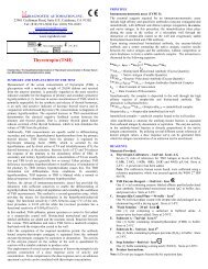
Toxocara Microwell Serum ELISA<br />
Catalog No. 8206-3<br />
Intended Use<br />
For the qualitative screening of serum IgG antibodies to Toxocara using an Enzyme-Linked Immunosorbent Assay (ELISA)<br />
technique.<br />
Summary<br />
Toxocariasis is the infection caused by roundworm of the genus Toxocara (usually T. canis, rarely T. cati) and is acquired by<br />
ingesting soil contaminated with embryonated eggs from an animal’s feces. These eggs become embryonated after 2 to 5 weeks<br />
after being passed by the animal. Thus, human infection does not occur by contact with fresh feces. In addition, infected humans<br />
cannot pass the infection to other humans. 6<br />
The disease manifests itself as visceral larval migrans (VLM) and ocular larval migrans (OLM). Signs and symptoms of VLM<br />
may vary from an asymptomatic state with mild eosinophilia to a severe and potentially fatal disorder. Patients with OLM also<br />
vary widely in presentation, from acute lesions in the eye to asymptomatic infections. Toxocara larva migrans is believed to be<br />
the second most common helminth infection in developed countries. 1,2<br />
There is no definitive method to diagnose Toxocara infections, thus true sensitivity and specificity of serologic tests cannot be<br />
accurately determined. The diagnosis is further complicated by the fact that the antibody response varies depending on worm<br />
burden and location. However, numerous studies have shown that immunoassays using a purified excretory antigen from the<br />
larval stage, as in this ELISA, have shown dramatically improved sensitivities and specificities when compared to assays using<br />
crude antigens. 1-6<br />
Principle of Procedure<br />
The micro test wells are coated with an excretory/secretory antigen from the Toxocara larvae. During the first incubation with<br />
the diluted patients’ sera, any antibodies which are reactive with the antigen will bind to the coated wells. After washing to<br />
remove the rest of the sample, the Enzyme Conjugate is added. If antibodies have been bound to the wells, the Enzyme<br />
Conjugate will then bind to these antibodies. After another series of washes, a chromogen (tetramethlybenzidine or TMB) is<br />
added. If the Enzyme Conjugate is present, the peroxidase will catalyze a reaction that consumes the peroxide and turns the<br />
chromogen from clear to blue. Addition of the Stop Solution ends the reaction and turns the blue color to a bright yellow color.<br />
The reaction may then be read visually or with an ELISA reader.<br />
Reagents<br />
• Test strips: microwells containing Toxocara antigens – 96 or 48 test wells in a test strip holder.<br />
• Enzyme conjugate: One (1) bottle containing 11or 6 ml of Protein A conjugated to peroxidase.<br />
• Positive control serum: One (1) vial containing 1 ml of diluted positive rabbit serum.<br />
• Negative control serum: One (1) vial containing 1 ml of diluted negative human serum.
• Chromogen: One (1) bottle containing 11 ml of the chromogen tetramethylbenzidine (TMB).<br />
• Wash concentrate solution (20X): One (1) bottle containing 25 ml of concentrated buffer and surfactant.<br />
• Dilution buffer: Two (2) or one (1) bottle(s) containing 30 ml of buffered protein solution.<br />
• Stop solution: One (1) bottle containing 11 ml of 1 M phosphoric acid.<br />
Precautions<br />
Do not use solutions if they precipitate or become cloudy. Wash concentrate may show crystallization upon storage at 2-8º C.<br />
Crystallization will disappear after dilution to working strength.<br />
Do not use serum that may have supported microbial growth, or is cloudy due to high lipid content. Samples high in lipids<br />
should be clarified before use.<br />
Treat all sera as if capable of being infectious. Negative control has been tested and found negative for Hepatitis B surface<br />
antigen and for the antibody to HIV by required test methods. This product should be used under appropriate safety conditions<br />
that would be used for any potentially infectious agent.<br />
Do not add azides to the samples or any of the reagents.<br />
Storage Conditions<br />
Reagents, strips and bottled components:<br />
Store between 2 - 8º C.<br />
Squeeze bottle containing diluted wash buffer may be stored at room temperature.<br />
Preparation<br />
Wash Buffer - Remove cap and add contents of bottle to 475 ml of reagent grade water. Place diluted wash buffer into a squeeze<br />
bottle with a narrow tip opening.<br />
Note: Washings consist of filling to the top of each well, shaking out the contents and refilling.<br />
Avoid generating bubbles in the wells during the washing steps.<br />
Collection and Preparation of Serum<br />
Coagulate blood and remove serum. Freeze sample at -20º C or lower if not used immediately.<br />
Do not heat inactivate serum and avoid repeated freezing and thawing of samples.<br />
Test samples: Make a 1:64 dilution of patients sera using the dilution buffer (e.g. 5 µl sera and 315 µl dilution buffer).<br />
Materials Provided<br />
Toxocara Serology Microwell ELISA Kit<br />
Procedure<br />
Materials Required But Not Provided<br />
Pipettes<br />
Squeeze bottle for washing strips (narrow tip is recommended)<br />
Reagent grade water and graduated cylinder<br />
Tubes for sample dilution<br />
Absorbent paper
Suggested Materials<br />
ELISA plate reader with a 450 nm and a 650 to 620 nm filter (optional if results are read visually)<br />
Performance of Test<br />
1. Break off number of wells needed (two for controls plus number of samples) and place in strip holder.<br />
2. Add 100 µl (or two drops) of the negative control to well #1, 100 µl of the positive control to well #2 and 100 µl of the<br />
diluted (1:64) test samples to the remaining wells.<br />
Note: Negative and positive controls are supplied prediluted. Do not dilute further.<br />
3. Incubate at room temperature (15 to 25º C) for 10 minutes.<br />
4. Shake out contents and wash 3 times with the diluted wash buffer.<br />
5. Add 2 drops of Enzyme Conjugate to each well.<br />
6. Incubate at room temperature for 5 minutes.<br />
7. Shake out contents and wash 3 times with wash buffer. Slap plates against paper toweling to remove excess moisture.<br />
8. Add 2 drops of the Chromogen to every well.<br />
9. Incubate at room temperature for 5 minutes.<br />
10. Add 2 drops of the Stop Solution and mix by tapping strip holder.<br />
Reading of Results<br />
Visually: Look at each well against a white background (e.g. paper towel) and record as clear or +,++ or +++ reaction.<br />
ELISA Reader: Zero reader on air. Set for bichromatic readings at 450/650-620 nm.<br />
Test Limitations<br />
Serologic results are an aid in diagnosis but cannot be used as the sole method of diagnosis.<br />
Quality Control<br />
The use of controls allows validation of kit stability. The kit should not be used if any of the controls are out of range.<br />
Expected values for the controls are:<br />
Negative - 0.0 to 0.3 OD units<br />
Positive - 0.5 OD units and above<br />
Troubleshooting<br />
Negative control has excessive color after development.<br />
Reason: inadequate washings.<br />
Correction: wash more vigorously. Remove excessive liquid from the wells by tapping against an absorbent towel.<br />
Do not allow test wells to dry out.<br />
Zero ELISA reader on air. Read all wells at 450/650-620 nm.<br />
Interpretation of Results - ELISA Reader
Positive - Absorbance reading equal to or greater than 0.3 OD units.<br />
Negative - Absorbance reading less than 0.3 OD units.<br />
A negative OD reading indicates that the patient has no detectable level of antibodies. This may be due to lack of infection or<br />
poor immune response by the patient.<br />
Interpretation of Results -Visual<br />
Compare results to the controls. A sample should be interpreted as positive if the degree of color development is significant<br />
and obvious.<br />
Expected Results<br />
The number of individuals showing positive results can vary significantly between populations and geographic regions. If<br />
possible, each laboratory should establish an expected range for its patient population.<br />
Performance Data<br />
Study #1 – Canadian Reference Center<br />
Compared <strong>DIAGNOSTIC</strong> <strong>AUTOMATION</strong>, <strong>INC</strong>. ELISA to another commercial ELISA. Found<br />
concordance of 84% (n=82).<br />
Study #2 – Mayo Clinic<br />
Specificity of 87.5% (21/24)<br />
Sensitivity of 93.3% (14/15)<br />
References<br />
1. Glickman, L., P. Schantz, and R. Grieve. “Toxocariasis”. Immunodiagnosis of Parasitic Diseases. Vol.1, Helminthic<br />
Diseases. Ed. Walls and Schantz. Academic Press, 1986. pp. 201-231.<br />
2. Schantz, P. “Toxocara Larva Migrans Now.” Am J Trop Med Hyg. Vol. 41 (Sup 3), 1989, pp. 21-34.<br />
3. Jacquier, P. et. al. “Immunodiagnosis of Toxocarosis in Humans: Evaluation of a New Enzyme-Linked Immunosorbent<br />
Assay Kit.” J Clin Micro. Vol. 29 #9, Sept. 1991, pp. 1831-1835.<br />
4. Carlier, Y. et. al. “The Use of an Excretory-Secretory Antigen for an ELISA Specific Serodiagnosis of Visceral Larva<br />
Migrans.” Biomedicine. Vol. 36, 1982, pp. 39-42.<br />
5. Brunello, F., P. Falagiani, and C. Genchi. “Enzyme Immunoassay (ELISA) for the Detection of Specific IgG Antibodies<br />
to Toxocara canis ES Antigens.” Boll. Ist. sieroter. Milan. Vol. 65 #1, 1986, pp. 54-60.<br />
6. Josephson, S.L., “Toxocariasis”, Laboratory Diagnosis of Infectious Diseases – Principles and Practices, Vol. 1, 1988, pp.<br />
993-997.<br />
<strong>DIAGNOSTIC</strong> <strong>AUTOMATION</strong>, <strong>INC</strong>.<br />
23961 Craftsman Road, Suite E/F, Calabasas, CA 91302<br />
Tel: (818) 591-3030 Fax: (818) 591-8383<br />
ISO 13485-2003<br />
Revision Date: 4-10-06