(PROTEIN) WATER MOLECULE AMINO GROUP
(PROTEIN) WATER MOLECULE AMINO GROUP
(PROTEIN) WATER MOLECULE AMINO GROUP

Create successful ePaper yourself
Turn your PDF publications into a flip-book with our unique Google optimized e-Paper software.
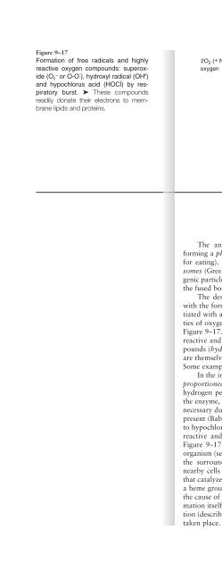
Figure 9–17<br />
Formation of free radicals and highly<br />
reactive oxygen compounds: superoxide<br />
(O 2 – or O-O ˜ ), hydroxyl radical (OH •˜ )<br />
and hypochlorus acid (HOCl ˜ ) by respiratory<br />
burst. ➤ These compounds<br />
readily donate their electrons to membrane<br />
lipids and proteins.<br />
2O2 (+ NADPH)<br />
oxygen<br />
NADP oxidase 2O2 - . (+ NADP + + H +)<br />
superoxide<br />
Fe<br />
H2O (+ 2 O )<br />
2<br />
hydrogen<br />
peroxide<br />
+2 (or Cu +1 )<br />
OH .<br />
hydroxyl<br />
OCl<br />
radical<br />
-<br />
* *<br />
myeloperoxidase<br />
*spontaneous,<br />
no enzyme used<br />
Ocular Immunochemistry • 265<br />
superoxide<br />
dismutase<br />
Cl -<br />
HOCl<br />
hypochlorous<br />
acid (biological<br />
"chlorox")<br />
1 O2 (+ H 2 O + Cl - )<br />
singlet<br />
oxygen<br />
The antigens become surrounded by the leucocyte membranes<br />
forming a phagosome (Greek: fagosoma- phagosoma—literally: a body<br />
for eating). The phagosome itself is internalized and fused with lysosomes<br />
(Greek lusosoma: literally: a body for breaking) where the antigenic<br />
particles or even whole bacteria or viruses are destroyed. Note that<br />
the fused bodies are this point are called phagolysosomes.<br />
The destruction of antigenic organisms begins in the phagosome<br />
with the formation of highly active forms of oxygen. This process is initiated<br />
with a respiratory burst in which leucocytes take up large quantities<br />
of oxygen (O 2) to form superoxide radicals (O 2 –• ). This is shown in<br />
Figure 9–17. Superoxide radicals have unpaired electrons that are highly<br />
reactive and unstable. The radicals are rapidly converted to other compounds<br />
(hydrogen peroxide, hydroxyl radicals, and singlet oxygen) that<br />
are themselves highly reactive with tissue proteins and membrane lipids.<br />
Some examples are shown in Figure 9–18.<br />
In the initial reaction of Figure 9–17, superoxide is dismuted or disproportioned<br />
(one molecule is reduced while the other is oxidized) to<br />
hydrogen peroxide by the enzyme superoxide dismutase. The need for<br />
the enzyme, in spite of the highly reactive species, has been shown to be<br />
necessary due to the relatively low concentrations of superoxide that are<br />
present (Babior, 2000). Some of the hydrogen peroxide is also converted<br />
to hypochlorous acid (commercially known as “chlorox”), a very highly<br />
reactive and destructive chemical for nearly any living organism (see<br />
Figure 9–17). All these oxidative species attack the membranes of the<br />
organism (see Figure 9–18) within the phagosome and then spill out into<br />
the surrounding cellular environment to cause collateral damage to<br />
nearby cells or even the phagocytes themselves eventually. The enzyme<br />
that catalyzes the formation of hypochlorous acid, myeloperoxidase, has<br />
a heme group at its active site that has a green color. The green color is<br />
the cause of the green hue that is associated with pus formation. Pus formation<br />
itself is the buildup of cellular debris from the oxidative destruction<br />
(described here) and enzymatic onslaught (described below) that has<br />
taken place.