LCP Clavicle Hook Plate. The fixation system with ... - Osteosyntese
LCP Clavicle Hook Plate. The fixation system with ... - Osteosyntese
LCP Clavicle Hook Plate. The fixation system with ... - Osteosyntese

You also want an ePaper? Increase the reach of your titles
YUMPU automatically turns print PDFs into web optimized ePapers that Google loves.
Technique Guide<br />
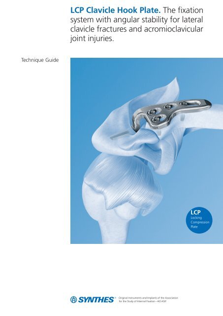
<strong>LCP</strong> <strong>Clavicle</strong> <strong>Hook</strong> <strong>Plate</strong>. <strong>The</strong> <strong>fixation</strong><br />
<strong>system</strong> <strong>with</strong> angular stability for lateral<br />
clavicle fractures and acromioclavicular<br />
joint injuries.
Table of Contents<br />
Introduction<br />
Features and Benefits 2<br />
AO ASIF Principles 4<br />
Indications 5<br />
Surgical Technique<br />
Lateral <strong>Clavicle</strong> Fractures 7<br />
Dislocation of the Acromioclavicular Joint 14<br />
Implant Removal 21<br />
Ordering Information<br />
<strong>Plate</strong>s 22<br />
Screws 23<br />
Instruments 24<br />
Stardrive<br />
Hex drive<br />
Image intensifier control<br />
Warning<br />
This description is not sufficient for immediate application of<br />
the instrumentation. Instruction by a surgeon experienced in<br />
handling this instrumentation is highly recommended.<br />
Synthes 1
Features and Benefits<br />
One solution for two indications<br />
<strong>The</strong> <strong>LCP</strong> <strong>Clavicle</strong> <strong>Hook</strong> <strong>Plate</strong> provides a<br />
single solution for <strong>fixation</strong> of both lateral<br />
clavicle fractures and acromioclavicular<br />
joint injuries.<br />
Lateral clavicle fracture<br />
Acromioclavicular joint dislocation<br />
Anatomically pre-contoured<br />
<strong>The</strong> plate design facilitates optimal<br />
implant placement and surgery in order<br />
to provide an improved outcome.<br />
– Soft radius, smooth hook design<br />
and<br />
posterior hook offset<br />
– Rounded shaft profile<br />
Minimize the risk of conflicts between the<br />
plate and surrounding soft tissue, the<br />
acromioclavicular joint and the rotator<br />
cuff.<br />
Undercuts in shaft<br />
Reduce impairment of blood<br />
supply<br />
12º bend in shaft<br />
Eases implant placement<br />
2 Synthes <strong>LCP</strong> <strong>Clavicle</strong> <strong>Hook</strong> <strong>Plate</strong> Technique Guide
Intra-operative choice of hook size<br />
Optimized size matching<br />
6 trial implants help determine the proper<br />
hook size.<br />
– <strong>Hook</strong> depths 12, 15 and 18 mm<br />
– Left and right version<br />
<strong>The</strong> <strong>LCP</strong> <strong>Clavicle</strong> <strong>Hook</strong> <strong>Plate</strong> is available in<br />
different lengths and hook sizes <strong>with</strong> a<br />
left and right version for optimal sizing<br />
and screw positioning for each individual<br />
patient.<br />
4 different lengths<br />
4–7 holes<br />
<strong>LCP</strong> Locking Compression <strong>Plate</strong><br />
Angular stable <strong>fixation</strong> of fragments<br />
regardless of bone quality<br />
Minimised risk of primary and secondary<br />
loss of reduction, even under<br />
high dynamic loading<br />
Reduced impairment of periosteal<br />
blood supply due to the limited plate<br />
contact<br />
Good purchase also in osteoporotic<br />
bone and in multifragment factures<br />
3 different hook depths<br />
12, 15 and 18 mm<br />
<strong>LCP</strong> combi-hole<br />
Intraoperative choice between compression<br />
and angular stable locking<br />
With standard screws:<br />
interfragmental or dynamic-axial<br />
compression<br />
With locking screws:<br />
stable plate-screw connection <strong>with</strong>out<br />
loss of reduction, regardless of plate<br />
modelling<br />
Synthes 3
AO ASIF Principles<br />
In 1958, the AO ASIF (Association for the Study of Internal<br />
Fixation) formulated four basic principles 1 , which have become<br />
the guidelines for internal <strong>fixation</strong>.<br />
Anatomic reduction<br />
Fixation of lateral clavicle fractures and dislocations <strong>with</strong> the<br />
anatomically pre-contoured <strong>LCP</strong> <strong>Clavicle</strong> <strong>Hook</strong> <strong>Plate</strong> allows<br />
for anatomic reduction.<br />
Stable <strong>fixation</strong><br />
<strong>The</strong> anatomically pre-contoured shaft <strong>with</strong> a 12° bend eases<br />
implant placement. <strong>The</strong> <strong>LCP</strong> <strong>Clavicle</strong> <strong>Hook</strong> <strong>Plate</strong> is available<br />
in 4 different lengths and 3 different hook sizes for optimal<br />
sizing and screw positioning for each individual patient.<br />
Preservation of blood supply<br />
<strong>The</strong> well-proven <strong>LCP</strong> concept and the undercuts on the shaft<br />
of the <strong>LCP</strong> <strong>Clavicle</strong> <strong>Hook</strong> <strong>Plate</strong> allow preservation of the<br />
blood supply through minimal bone-to-plate contact. <strong>The</strong><br />
rounded shaft profile, soft radius, smooth hook design and<br />
posterior hook offset minimize the risk of conflicts between<br />
the plate and surrounding soft tissue, the acromioclavicular<br />
joint and the rotator cuff.<br />
Early, active mobilization<br />
<strong>The</strong> <strong>LCP</strong> <strong>Clavicle</strong> <strong>Hook</strong> <strong>Plate</strong>, combined <strong>with</strong> AO technique,<br />
provides stable fracture <strong>fixation</strong> <strong>with</strong> minimal trauma to vascular<br />
supplies. This helps create an improved environment for<br />
bone healing, accelerating the patient’s return to previous<br />
mobility and function.<br />
1 M.E. Müller, M. Allgöwer, R. Schneider, and R. Willenegger (1991)<br />
AO Manual of Internal Fixation, 3rd Edition. Berlin: Springer.<br />
4 Synthes <strong>LCP</strong> <strong>Clavicle</strong> <strong>Hook</strong> <strong>Plate</strong> Technique Guide
Indications<br />
Indications<br />
Lateral clavicle fractures: Neer type II or Jäger and Breitner<br />
type II<br />
Acromioclavicular joint dislocation type: Tossy III or<br />
Rockwood III to V<br />
Not indicated<br />
Stable lateral clavicle fractures<br />
Tossy Type I and II<br />
Rockwood Type I and II<br />
Acute infection<br />
Caution: It is recommended that the <strong>LCP</strong> <strong>Clavicle</strong> <strong>Hook</strong> <strong>Plate</strong><br />
is removed after healing to prevent potential irritation of the<br />
acromion or impinging on the rotator cuff.<br />
Synthes 5
6 Synthes <strong>LCP</strong> <strong>Clavicle</strong> <strong>Hook</strong> <strong>Plate</strong> Technique Guide
Lateral <strong>Clavicle</strong> Fractures<br />
Experience in the use of <strong>LCP</strong> plates or instruction from an experienced<br />
surgeon is recommended (see Synthes application<br />
notes for <strong>LCP</strong> plates, Art. No. 036.000.019)<br />
1<br />
Position the patient<br />
Place the patient in the beach-chair position. Tilt the head<br />
away from the operated side, <strong>with</strong> care for the position of<br />
the neck. A sandbag under the thoracic spine allows the<br />
scapula to fall backwards: this aids realignment and reduction<br />
of the fracture. Excessive extension of the neck should<br />
be avoided.<br />
2<br />
Access to fracture<br />
If image intensification is to be used, determine that access<br />
for the C-arm is sufficient for the anteroposterior and<br />
cephalic tilt views.<br />
Through either a superior (sabre cut) or transacromial incision,<br />
the delto-trapezial fascia is exposed. Care is taken not<br />
to injure the lateral supraclavicular nerves. <strong>The</strong> fracture is<br />
usually marked by bruising and a rent in the deltoid fascia<br />
and/or trapezius. <strong>The</strong> acromioclavicular joint may be identified<br />
<strong>with</strong> a needle.<br />
Synthes 7
3<br />
Reduce the fracture and provide temporary <strong>fixation</strong><br />
Temporary <strong>fixation</strong> of the fracture <strong>with</strong> Kirschner wires or the<br />
pointed reduction forceps may be undertaken.<br />
<strong>The</strong> posterior aspect of the acromioclavicular joint capsule<br />
is identified and a 5 mm detachment of the extracapsular fibres<br />
of the trapezius from the medial border of the acromion<br />
is performed, to allow passage of the hook of the plate under<br />
the acromion.<br />
8 Synthes <strong>LCP</strong> <strong>Clavicle</strong> <strong>Hook</strong> <strong>Plate</strong> Technique Guide
4<br />
Determine hook size and plate length<br />
Required instruments<br />
329.930 Trial Implant for <strong>LCP</strong> <strong>Clavicle</strong> <strong>Hook</strong><br />
<strong>Plate</strong>, <strong>Hook</strong> depth 12 mm, left<br />
329.931 Trial Implant for <strong>LCP</strong> <strong>Clavicle</strong> <strong>Hook</strong><br />
<strong>Plate</strong>, <strong>Hook</strong> depth 12 mm, right<br />
329.932 Trial Implant for <strong>LCP</strong> <strong>Clavicle</strong> <strong>Hook</strong><br />
<strong>Plate</strong>, <strong>Hook</strong> depth 15 mm, left<br />
329.933 Trial Implant for <strong>LCP</strong> <strong>Clavicle</strong> <strong>Hook</strong><br />
<strong>Plate</strong>, <strong>Hook</strong> depth 15 mm, right<br />
329.934 Trial Implant for <strong>LCP</strong> <strong>Clavicle</strong> <strong>Hook</strong><br />
<strong>Plate</strong>, <strong>Hook</strong> depth 18 mm, left<br />
329.935 Trial Implant for <strong>LCP</strong> <strong>Clavicle</strong> <strong>Hook</strong><br />
<strong>Plate</strong>, <strong>Hook</strong> depth 18 mm, right<br />
Trial implants are provided to aid in determining the appropriate<br />
hook size.<br />
Use the trial implant <strong>with</strong> a 12 mm hook, and pass the hook<br />
under the acromion. Place the shaft of the trial implant onto<br />
the superior aspect of the clavicle. If it is difficult to lower the<br />
shaft onto the reduced clavicle, then use an implant <strong>with</strong><br />
15 mm or 18 mm hook size. Once the plate shaft is placed<br />
on the clavicle, the end of the hook should be in contact<br />
<strong>with</strong> the underside of the acromion.<br />
Confirm that the correct anatomic alignment of the clavicle<br />
and acromion has been restored <strong>with</strong>out impinging on the<br />
rotator cuff. Use the C-arm to verify that full shoulder motion,<br />
particularly in abduction and external rotation, can be<br />
achieved <strong>with</strong>out impinging on the humeral head by the<br />
hook.<br />
<strong>The</strong> plate length must ensure appropriate <strong>fixation</strong> on the<br />
medial side of the fracture.<br />
Note: Do not bend or implant the trial implants.<br />
Synthes 9
5<br />
Option: Fixate the plate temporarily<br />
Once the hook size is determined, remove the trial implant<br />
and position the implant. After confirming the correct plate<br />
position under the image intensifier, the plate can be fixed<br />
temporarily using a Kirschner wire. Drill the wire through the<br />
drill sleeve in the distal hole to fix the distal part of the plate.<br />
10 Synthes <strong>LCP</strong> <strong>Clavicle</strong> <strong>Hook</strong> <strong>Plate</strong> Technique Guide
6<br />
Option: Adapt plate to the patient’s anatomy<br />
Possible instruments<br />
329.040/050 Bending Iron<br />
329.150 Bending Pliers, length 230 mm<br />
Important: Since the plate shaft is anatomically pre-contoured<br />
(12°), bending of the plate is not necessary, but can<br />
be done if required.<br />
Contour the plate using the appropriate bending instruments<br />
(as <strong>with</strong> standard plates). <strong>The</strong> combi-holes should not be deformed<br />
excessively during bending, as this may hinder the<br />
subsequent insertion of locking screws. If possible, bend the<br />
plate between the combi-holes.<br />
Notes<br />
Do not bend the shaft between the holes more than<br />
20 to 25°.<br />
Do not bend the hook more than 10 to 15°.<br />
Do not bend the plate and hook back and forth.<br />
Take care that the plate surface does not get scratched.<br />
Sharp edges can irritate soft tissue.<br />
If only non-locking cortex screws are used, the plate needs to<br />
be congruent <strong>with</strong> the surface of the bone, and bending or<br />
twisting may be required.<br />
It is recommended that the most proximal plate hole is<br />
aligned over the axis of the shaft of the clavicle and provisional<br />
cortical screw <strong>fixation</strong> using either a locking screw or<br />
cortical screw is performed first. By alignment of the medial<br />
and lateral fracture fragments <strong>with</strong> the plate using reduction<br />
forceps, the fracture is indirectly reduced and definitive <strong>fixation</strong><br />
can then be carried out.<br />
Synthes 11
7a<br />
Fixation <strong>with</strong> Locking Screws 3.5 mm<br />
Required instruments<br />
310.284 <strong>LCP</strong> Drill Bit 2.8 mm<br />
311.431 Handle <strong>with</strong> Quick Coupling<br />
314.030 Screwdriver Shaft, hexagonal<br />
or<br />
314.116 Screwdriver Shaft Stardrive T15<br />
319.010 Depth Gauge for Screws 2.7 to 4.0 mm<br />
323.027 <strong>LCP</strong> Drill Sleeve 3.5 for Drill Bits 2.8 mm<br />
323.360 Universal Drill Guide 3.5<br />
397.705 Handle for Torque Limiter 1.5 Nm<br />
511.770/773 Torque Limiter 1.5 Nm<br />
Once the hook size is determined, remove the trial implant<br />
and position the implant. After confirmation of the correct<br />
plate position under the image intensifier, start definitive<br />
<strong>fixation</strong> <strong>with</strong> screws.<br />
Carefully screw the <strong>LCP</strong> drill sleeve into the threaded central<br />
hole of the plate.<br />
With an <strong>LCP</strong> drill bit 2.8 mm, predrill the screw hole<br />
through both cortices. Read the required screw length directly<br />
from the drill bit. Use the depth gauge to check the<br />
length of screw.<br />
It is recommended that the final turns are done manually.<br />
Notes<br />
<strong>The</strong> screw length should be carefully observed in order to<br />
avoid neurovascular injuries.<br />
Do not mix titanium and steel implants (e.g. titanium plate<br />
<strong>with</strong> steel screws).<br />
To ensure a stable <strong>fixation</strong> of the implant, use at least two<br />
screws in the medial part of the plate. One or two screws<br />
can be used to fix the lateral fragments.<br />
Insert the locking screw <strong>with</strong> the screwdriver (hexagonal or<br />
Stardrive recess) mounted on the torque limiter 1.5 Nm.<br />
Insert the screw manually or by power until a click is heard. If<br />
a power tool is used, reduce speed when tightening the<br />
head of the locking screw into the plate.<br />
Repeat the procedure until all pre-determined shaft holes are<br />
used. Perform a final check to confirm all screws are locked.<br />
12 Synthes <strong>LCP</strong> <strong>Clavicle</strong> <strong>Hook</strong> <strong>Plate</strong> Technique Guide
7b<br />
Fixation <strong>with</strong> Cortex Screws 3.5 mm<br />
Required instruments<br />
310.250 Drill bit 2.5 mm<br />
311.431 Handle <strong>with</strong> Quick Coupling<br />
314.030 Screwdriver Shaft, hexagonal<br />
319.010 Depth Gauge for Screws 2.7 to 4.0 mm<br />
323.360 Universal Drill Guide 3.5<br />
397.705 Handle for Torque Limiter 1.5 Nm<br />
511.770/773 Torque Limiter 1.5 Nm<br />
Once the hook size is determined, remove the trial implant<br />
and position the implant. After confirmation of the correct<br />
plate position under the image intensifier, start definitive <strong>fixation</strong><br />
<strong>with</strong> screws.<br />
Use the drill guide and the drill bit 2.5 mm to pre-drill<br />
both cortices.<br />
Determine the required length of the cortex screw <strong>with</strong> the<br />
depth gauge.<br />
Insert the self-tapping cortex screw 3.5 mm by using the<br />
screwdriver shaft mounted on a power tool or on a handle<br />
<strong>with</strong> quick coupling.<br />
Notes<br />
<strong>The</strong> screw length should be carefully observed in order to<br />
avoid neurovascular injuries.<br />
Do not mix titanium and steel implants (e.g. titanium plate<br />
<strong>with</strong> steel screws).<br />
To ensure a stable <strong>fixation</strong> of the implant, use at least two<br />
screws in the medial part of the plate. One or two screws<br />
can be used to fix the lateral fragments.<br />
Repeat the procedure until all pre-determined shaft holes are<br />
used.<br />
Synthes 13
Dislocation of the Acromioclavicular<br />
Joint<br />
Experience in the use of <strong>LCP</strong> plates or instruction from an experienced<br />
surgeon is recommended (see Synthes application<br />
notes for <strong>LCP</strong> plates, Art.No. 036.000.019)<br />
1<br />
Position the patient<br />
Place the patient in the beach-chair position. Tilt the head<br />
away from the operated side, <strong>with</strong> care for the position of<br />
the neck. A sandbag under the thoracic spine allows the<br />
scapula to fall backwards: this aids realignment and reduction<br />
of the acromioclavicular joint. Excessive extension of the<br />
neck should be avoided.<br />
2<br />
Access<br />
If image intensification is to be used, determine that access<br />
for the C-arm is sufficient for the anteroposterior and<br />
cephalic tilt views.<br />
Through either a superior (sabre cut) or transacromial incision,<br />
the delto-trapezial fascia is exposed. Care is taken not<br />
to injure the lateral supraclavicular nerves. <strong>The</strong> acute dislocation<br />
is marked by a rupture through the superior acromioclavicular<br />
ligament, <strong>with</strong> prolapse of the intra-articular disc<br />
remnants which usually remain partially attached to the clavicle,<br />
and incomplete rupture of the acromial fibres of the<br />
trapezius. <strong>The</strong> coracoclavicular ligaments and the periosteum<br />
are also ruptured in Rockwood Type V injuries.<br />
14 Synthes <strong>LCP</strong> <strong>Clavicle</strong> <strong>Hook</strong> <strong>Plate</strong> Technique Guide
3<br />
Reduce the dislocation and fixate it temporarily<br />
<strong>The</strong> arm, and therefore the scapula, is elevated towards the<br />
clavicle and supported by the assistant or on a side table.<br />
<strong>The</strong> acromion is reduced to the clavicle in the horizontal and<br />
vertical planes. Temporary <strong>fixation</strong> of the acromioclavicular<br />
joint may be achieved by a transacromial Kirschner wire<br />
passed into the distal clavicle.<br />
<strong>The</strong> posterior aspect of the acromioclavicular joint capsule is<br />
identified and a 5 mm detachment of the extracapsular fibres<br />
of the trapezius from the medial border of the acromion<br />
is performed, to allow passage of the hook of the plate under<br />
the acromion.<br />
In the fresh dislocation the superior acromioclavicular and<br />
coracoclavicular ligaments may be repaired.<br />
In the chronic dislocation repair of the ligaments is generally<br />
impossible; reconstruction is necessary. A transfer of the<br />
coraco-acromial ligament to the distal clavicle, and augmentation<br />
of scapuloclavicular suspension by autogenous ligament<br />
grafts (plantaris, palmaris longus, or hamstring tendons)<br />
or artificial ligaments should be considered at this<br />
stage. Preparation of the trapezial envelope for later reefed<br />
repair is undertaken.<br />
Synthes 15
4<br />
Determine hook size and plate length<br />
Required instruments<br />
329.930 Trial Implant for <strong>LCP</strong> <strong>Clavicle</strong> <strong>Hook</strong><br />
<strong>Plate</strong>, <strong>Hook</strong> depth 12 mm, left<br />
329.931 Trial Implant for <strong>LCP</strong> <strong>Clavicle</strong> <strong>Hook</strong> <strong>Plate</strong>,<br />
<strong>Hook</strong> depth 12 mm, right<br />
329.932 Trial Implant for <strong>LCP</strong> <strong>Clavicle</strong> <strong>Hook</strong> <strong>Plate</strong>,<br />
<strong>Hook</strong> depth 15 mm, left<br />
329.933 Trial Implant for <strong>LCP</strong> <strong>Clavicle</strong> <strong>Hook</strong> <strong>Plate</strong>,<br />
<strong>Hook</strong> depth 15 mm, right<br />
329.934 Trial Implant for <strong>LCP</strong> <strong>Clavicle</strong> <strong>Hook</strong> <strong>Plate</strong>,<br />
<strong>Hook</strong> depth 18 mm, left<br />
329.935 Trial Implant for <strong>LCP</strong> <strong>Clavicle</strong> <strong>Hook</strong> <strong>Plate</strong>,<br />
<strong>Hook</strong> depth 18 mm, right<br />
Trial implants are provided to help determine the proper<br />
hook size.<br />
Use the trial implant <strong>with</strong> a 12 mm hook, and pass the hook<br />
under the acromion. Place the shaft of the trial implant onto<br />
the superior aspect of the clavicle. If it is difficult to lower the<br />
shaft onto the clavicle, then use an implant <strong>with</strong> 15 mm or<br />
18 mm hook size. Once the plate shaft is placed on the clavicle,<br />
the end of the hook should be in contact <strong>with</strong> the underside<br />
of the acromion.<br />
Confirm that the correct anatomic alignment of the clavicle<br />
and acromion has been restored <strong>with</strong>out impinging on the<br />
rotator cuff. Use the C-arm to verify that full shoulder motion,<br />
particularly in abduction and external rotation, can be<br />
achieved <strong>with</strong>out impinging on the humeral head by the<br />
hook.<br />
<strong>The</strong> plate length must ensure appropriate <strong>fixation</strong> on the<br />
medial side of the reduced joint.<br />
Note: Do not bend or implant the trial implants.<br />
16 Synthes <strong>LCP</strong> <strong>Clavicle</strong> <strong>Hook</strong> <strong>Plate</strong> Technique Guide
5<br />
Option: Fixate the plate temporarily<br />
Once the hook size is determined, remove the trial implant<br />
and position the implant. After confirmation of the correct<br />
plate position under the image intensifier, <strong>fixation</strong> can be<br />
achieved temporarily by using a Kirschner wire. Drill the wire<br />
through the drill sleeve in the distal hole to fix the distal part<br />
of the plate.<br />
Fixation can also be achieved using a cortical screw in the<br />
most medial plate hole.<br />
Synthes 17
6<br />
Option: Adapt plate to the patient’s anatomy<br />
Possible instruments<br />
329.040/050 Bending Iron<br />
329.150 Bending Pliers, length 230 mm<br />
Important: Since the plate shaft is anatomically pre-contoured<br />
(12°), bending of the plate is not necessary, but can<br />
be done if required.<br />
Contour the plate using the appropriate bending instruments<br />
(as <strong>with</strong> standard plates). <strong>The</strong> combi-holes should not be deformed<br />
excessively during bending, as this may hinder the<br />
subsequent insertion of locking screws. If possible, bend the<br />
plate between the combi-holes.<br />
Notes<br />
Do not bend the shaft between the holes more than<br />
20 to 25°.<br />
Do not bend the hook more than 10 to 15°.<br />
Do not bend the plate and hook back and forth.<br />
Take care that the plate surface does not get scratched.<br />
Sharp edges can irritate soft tissue.<br />
If only non-locking cortex screws are used, the plate needs to<br />
be congruent <strong>with</strong> the surface of the bone, and bending or<br />
twisting may be required.<br />
It is recommended that the most distal plate hole is aligned<br />
over the axis of the shaft of the clavicle and provisional cortical<br />
screw <strong>fixation</strong> using either a locking screw or cortical<br />
screw is performed first.<br />
18 Synthes <strong>LCP</strong> <strong>Clavicle</strong> <strong>Hook</strong> <strong>Plate</strong> Technique Guide
7a<br />
Fixation <strong>with</strong> Locking Screws 3.5 mm<br />
Required instruments<br />
310.284 <strong>LCP</strong> Drill Bit 2.8 mm<br />
311.431 Handle <strong>with</strong> Quick Coupling<br />
314.030 Screwdriver Shaft, hexagonal<br />
or<br />
314.116 Screwdriver Shaft Stardrive T15<br />
319.010 Depth Gauge for Screws 2.7 to 4.0 mm<br />
323.027 <strong>LCP</strong> Drill Sleeve 3.5 for Drill Bits 2.8 mm<br />
323.360 Universal Drill Guide 3.5<br />
397.705 Handle for Torque Limiter 1.5 Nm<br />
511.770/773 Torque Limiter 1.5 Nm<br />
Once the hook size is determined, remove the trial implant<br />
and position the implant. After confirmation of the correct<br />
plate position under the image intensifier, start definitive <strong>fixation</strong><br />
<strong>with</strong> screws.<br />
Carefully screw the <strong>LCP</strong> drill sleeve into the threaded central<br />
hole of the plate.<br />
Predrill the screw hole <strong>with</strong> an <strong>LCP</strong> drill bit 2.8 mm<br />
through both cortices. Read the required screw length directly<br />
from the drill bit. Use depth gauge to check the length<br />
of screw.<br />
It is recommended that the final turns are done manually.<br />
Notes<br />
<strong>The</strong> screw length should be carefully observed in order to<br />
avoid neurovascular injuries.<br />
Do not mix titanium and steel implants (e.g. titanium plate<br />
<strong>with</strong> steel screws).<br />
Insert the locking screw <strong>with</strong> the screwdriver (hexagonal or<br />
Stardrive recess) mounted on torque limited attachment 1.5<br />
Nm. Insert the screw manually or by power until a click is<br />
heard. If a power tool is used, reduce speed when tightening<br />
the head of the locking screw into the plate.<br />
To ensure a stable <strong>fixation</strong> of the implant, use at least two<br />
screws in the medial part of the plate. Perform a final check<br />
to confirm all screws are locked.<br />
Synthes 19
7b<br />
Fixation <strong>with</strong> Cortex Screws 3.5 mm<br />
Required instruments<br />
310.250 Drill bit 2.5 mm<br />
311.431 Handle <strong>with</strong> Quick Coupling<br />
314.030 Screwdriver Shaft, hexagonal<br />
319.010 Depth Gauge for Screws 2.7 to 4.0 mm<br />
323.360 Universal Drill Guide 3.5<br />
397.705 Handle for Torque Limiter 1.5 Nm<br />
511.770/773 Torque Limiter 1.5 Nm<br />
Once the hook size is determined, remove the trial implant<br />
and position the implant. After confirmation of the correct<br />
plate position under the image intensifier, start definitive<br />
<strong>fixation</strong> <strong>with</strong> screws.<br />
Use the drill guide and the drill bit 2.5 mm to pre-drill<br />
both cortices.<br />
Determine the required length of the cortex screw <strong>with</strong> the<br />
depth gauge.<br />
Notes<br />
<strong>The</strong> screw length should be carefully observed in order to<br />
avoid neurovascular injuries.<br />
Do not mix titanium and steel implants (e.g. titanium plate<br />
<strong>with</strong> steel screws).<br />
Insert the self-tapping cortex screw 3.5 mm by using the<br />
screwdriver shaft mounted on a power tool or on a handle<br />
<strong>with</strong> quick coupling.<br />
To ensure a stable <strong>fixation</strong> of the implant, use at least two<br />
screws in the medial part of the plate.<br />
20 Synthes <strong>LCP</strong> <strong>Clavicle</strong> <strong>Hook</strong> <strong>Plate</strong> Technique Guide
Implant Removal<br />
Required instruments<br />
309.521 Extraction Screw<br />
311.430 T-Handle<br />
314.030 Screwdriver Shaft, hexagonal<br />
or<br />
314.116 Screwdriver Shaft Stardrive T15<br />
Implant removal is usually done 3 months after implantation.<br />
Caution: It is recommended that the <strong>LCP</strong> <strong>Clavicle</strong> <strong>Hook</strong> <strong>Plate</strong><br />
is removed after healing to prevent potential irritation of the<br />
acromion or impinging on the rotator cuff.<br />
To remove the implants, unlock all locking screws before removing<br />
them completely. <strong>The</strong> plate may otherwise rotate<br />
while the last screw is being removed, which can damage<br />
the soft tissue.<br />
If the locking screws cannot be removed <strong>with</strong> the screw<br />
driver (e.g. the recess of the screw is damaged or the locking<br />
screw is jammed in the plate), use an extraction screw <strong>with</strong><br />
left-handed thread. Loosen the screw by turning the handle<br />
in a counter-clockwise direction.<br />
Important: It is very important to have correct instrumentation<br />
to ensure trouble-free implant removal. <strong>The</strong> correct<br />
screwdrivers (hex or Stardrive) and the extraction screws are<br />
especially important (e.g. see brochure „Screw extraction<br />
Set“, Art. No. 036.000.917).<br />
Synthes 21
<strong>Plate</strong>s<br />
<strong>LCP</strong> <strong>Clavicle</strong> <strong>Hook</strong> <strong>Plate</strong> 3.5<br />
Available in:<br />
– 4 different lengths<br />
– 3 hook sizes<br />
– left and right version<br />
– titanium and stainless steel<br />
– non-sterile and sterile packed<br />
Right Left Number <strong>Hook</strong> depth<br />
of holes (mm)<br />
X41.072 X41.073 4 12<br />
X41.074 X41.075 4 15<br />
X41.076 X41.077 4 18<br />
X41.082 X41.083 5 12<br />
X41.084 X41.085 5 15<br />
X41.086 X41.087 5 18<br />
X41.094 X41.095 6 15<br />
X41.096 X41.097 6 18<br />
X41.104 X41.105 7 15<br />
X41.106 X41.107 7 18<br />
X=2: stainless steel<br />
X=4: titanium<br />
All plates are available sterile packed. <strong>The</strong> article number of sterile packed implants<br />
is followed by (S)<br />
22 Synthes <strong>LCP</strong> <strong>Clavicle</strong> <strong>Hook</strong> <strong>Plate</strong> Technique Guide
Screws<br />
Locking Screws 3.5 mm, length 12-60 mm, selftapping<br />
– Stardrive (X12.102-124)<br />
– Hex drive (X13.012-060)<br />
Cortex Screws 3.5 mm, length 14-60 mm, selftapping<br />
– Hex drive (X04.814-860)<br />
X=2: stainless steel<br />
X=4: titanium<br />
All plates are available sterile packed. <strong>The</strong> article number of sterile packed implants<br />
is followed by (S)<br />
Synthes 23
Instruments<br />
Trial Implant for <strong>LCP</strong> <strong>Clavicle</strong> <strong>Hook</strong> <strong>Plate</strong><br />
Art. No. <strong>Hook</strong> depth (mm) Direction<br />
329.931 12 right<br />
329.933 15 right<br />
329.935 18 right<br />
329.930 12 left<br />
329.932 15 left<br />
329.934 18 left<br />
Available instrument sets<br />
Without screws<br />
– 182.460 <strong>LCP</strong> Small Fragment Instrument Set and Standard<br />
Instruments in Vario Case<br />
With hex screws<br />
– 182.466 <strong>LCP</strong> Small Fragment Instrument Set <strong>with</strong> Locking<br />
Screws 3.5 mm (Titanium Alloy) in Vario Case<br />
– 182.467 <strong>LCP</strong> Small Fragment Instrument Set <strong>with</strong> Locking<br />
Screws 3.5 mm (Stainless Steel) in Vario Case<br />
With Stardrive screws<br />
– 182.468 <strong>LCP</strong> Small Fragment Instrument Set <strong>with</strong> Locking<br />
Screws 3.5 mm Stardrive (Titanium Alloy) in Vario Case<br />
– 182.469 <strong>LCP</strong> Small Fragment Instrument Set <strong>with</strong> Locking<br />
Screws 3.5 mm Stardrive (Stainless Steel) in Vario Case<br />
Note: <strong>The</strong> <strong>LCP</strong> <strong>Clavicle</strong> <strong>Hook</strong> <strong>Plate</strong> is compatible <strong>with</strong> 3.5<br />
<strong>LCP</strong> instruments and standard small fragment instruments.<br />
<strong>The</strong>se additionally required instruments are not explicitly<br />
shown in this technique guide, but are essential for the application<br />
of the plate.<br />
24 Synthes <strong>LCP</strong> <strong>Clavicle</strong> <strong>Hook</strong> <strong>Plate</strong> Technique Guide
Presented by:<br />
0123<br />
036.000.473 SE_034295 AA 30050092 © Synthes 2006 <strong>LCP</strong> and Stardrive are trademarks of Synthes Subject to modifications