Cytoplast™ PTFE Membrane Product Brochure - Osteogenics
Cytoplast™ PTFE Membrane Product Brochure - Osteogenics
Cytoplast™ PTFE Membrane Product Brochure - Osteogenics

Create successful ePaper yourself
Turn your PDF publications into a flip-book with our unique Google optimized e-Paper software.
4 | Cytoplast Technique<br />
The Cytoplast Ridge Preservation Technique<br />
1.<br />
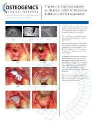
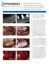
Figure 1. A minimally invasive, atraumatic extraction technique<br />
should be used. The use of periotomes or surgical sectioning is<br />
encouraged to minimize mechanical trauma to the thin cortical<br />
bone. All soft tissue remnants should be removed with a sharp<br />
curettage. Special care should be taken to remove residual soft<br />
tissues at the apical extent of the socket of endodontically treated<br />
teeth. Bleeding from the socket walls should be noted and, if<br />
necessary, decortication of the socket wall can be done with a #2<br />
round burr to increase early vascularization and access to osteoprogenitor<br />
cells.<br />
2.<br />
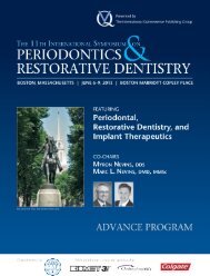
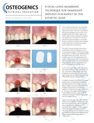
Figure 2. A subperiosteal pocket is created with a small periosteal<br />
elevator or curette, extending 3-5 mm beyond the socket margins<br />
(or defect margins) on the palatal and the facial aspect of the<br />
socket. In the esthetic zone, rather than incising and elevating<br />
the interdental papilla, it is left intact and undermined in a similar<br />
fashion. The d-<strong>PTFE</strong> membrane will be tucked into this subperiosteal<br />
pocket.<br />
3.<br />
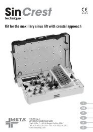
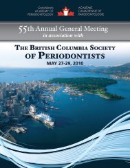
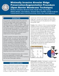
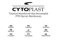
Figure 3 - 4. Particulate augmentation material is placed into the<br />
socket with a syringe or curette. Ensure that the material is evenly<br />
distributed throughout the socket, but not condensed or packed<br />
too tightly. This will only reduce the available space between particles,<br />
which is critical for vascular ingrowth and subsequent bone<br />
formation.<br />
4.