Cytoplast™ PTFE Membrane Product Brochure - Osteogenics
Cytoplast™ PTFE Membrane Product Brochure - Osteogenics
Cytoplast™ PTFE Membrane Product Brochure - Osteogenics

You also want an ePaper? Increase the reach of your titles
YUMPU automatically turns print PDFs into web optimized ePapers that Google loves.
High-Density <strong>PTFE</strong> <strong>Membrane</strong>s
2 | Why use d<strong>PTFE</strong><br />
Why Use Dense <strong>PTFE</strong> as a <strong>Membrane</strong><br />
<strong>PTFE</strong>: Polytetrafluoroethylene (<strong>PTFE</strong>) is comprised of a carbon chain with two fluorine atoms for every carbon atom. The complete<br />
fluorination of the carbon chain, along with the strength of the carbon-to-fluorine bonds, makes <strong>PTFE</strong> highly stable. This stability results<br />
in a synthetic polymer that is non-resorbable, biologically inert and chemically non-reactive, and therefore an ideal material for many<br />
medical device applications. In addition to its long history in the field of guided tissue regeneration (GTR), <strong>PTFE</strong> has been used for over 30<br />
years in cardiovascular applications such as suture, vascular grafts and heart valves.<br />
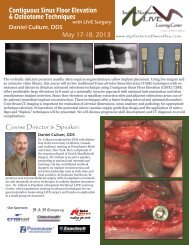
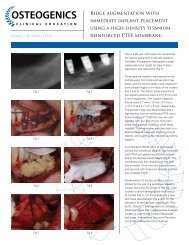
Expanded <strong>PTFE</strong>: <strong>PTFE</strong> as a biomaterial differs in porosity based on the amount of expansion applied during manufacturing.<br />
Heating <strong>PTFE</strong> and then applying force expands the material’s microstructure to make expanded <strong>PTFE</strong> (e<strong>PTFE</strong>). Under scanning electron<br />
microscopy, we see a network of dense nodes connected by fibrils. As the nodes and fibrils are expanded, the porosity of the material<br />
continues to increase.<br />
Expanded <strong>PTFE</strong> has a long history of success in GTR procedures, particularly in periodontics. However, the highly porous structure of<br />
e<strong>PTFE</strong> allows ingrowth of bacteria when the membrane is exposed in the mouth. Exposure results in high rates of infection and frequently<br />
requires early removal of the device. In addition, the highly porous structure allows soft tissue ingrowth, which complicates removal, often<br />
requiring sharp dissection and extensive surgery. Expanded <strong>PTFE</strong> must be completely buried and primary closure must be maintained<br />
to ensure predictability. While expanded <strong>PTFE</strong> is useful and quite predictable in deep, buried sites for guided tissue regeneration, there is<br />
currently no role for this material in extraction site grafting where exposure is likely.<br />
Dense <strong>PTFE</strong>: Dense <strong>PTFE</strong>, also known as high-density <strong>PTFE</strong> or d<strong>PTFE</strong>, is manufactured to eliminate expansion of the nodes and fibrils,<br />
resulting in a micro-porous material that is impervious to bacteria while still allowing diffusion of gases and small molecules. Dense<br />
<strong>PTFE</strong> was designed to withstand exposure in the oral environment, which represents an improvement to earlier versions of e<strong>PTFE</strong> in many<br />
applications, especially socket preservation where deliberate membrane exposure offers several advantages.<br />
Upon implantation, dense <strong>PTFE</strong> is immediately coated with plasma proteins, facilitating cellular adhesion to the smooth, biocompatible<br />
surface. This cellular adhesion is observed to form a hermetic seal, providing resistance to migration of bacteria and epithelial cells<br />
around and under the membrane when it is exposed in the mouth. Plasma protein adsorption also facilitates diffusion of soluble organic<br />
molecules across the membrane. Removal of dense <strong>PTFE</strong> is simplified due to the lack of tissue ingrowth into the surface structure.<br />
A textured dense <strong>PTFE</strong> is available. Texturing the membrane results in an increase in surface area and may increase the pull-out strength<br />
of the material through three dimensional attachment of soft tissue. The increased stability in the wound may result in less flap retraction<br />
and reduce risk of membrane movement and loosening. The primary advantage of dense <strong>PTFE</strong> is the ability to remain exposed in the<br />
mouth while protecting the underlying defect and bone graft. The membrane is soft, flexible and easy to handle. Primary closure is not<br />
required, and the membrane may be removed without additional surgery if exposed. If primary closure technique is used, the membrane<br />
may be easily removed through a small incision in a flapless technique.<br />
Dense <strong>PTFE</strong> is also available with titanium reinforcement, which increases the stiffness of the material for use in defects where spacemaking<br />
is required. The embedded titanium framework allows the membrane to be shaped to fit a variety of defects without rebounding<br />
and provides additional stability in large, non-space-making osseous defects.<br />
500X MAGNIFICATION<br />
200 µm<br />
20,000X MAGNIFICATION<br />
5 µm<br />
Expanded <strong>PTFE</strong> (e<strong>PTFE</strong>)<br />
High-Density <strong>PTFE</strong> (d<strong>PTFE</strong>)
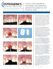
Evolution | 3<br />
The Evolution of <strong>PTFE</strong> <strong>Membrane</strong>s<br />
1980s<br />
Gore-Tex® creates the gold standard<br />
in barrier membranes.<br />
1994<br />
TefGen-FD®, a smooth bacteria-resistant dense <strong>PTFE</strong><br />
membrane, is introduced to withstand exposure. 1,2<br />
1997-Present<br />
Cytoplast dense <strong>PTFE</strong> membrane becomes an industry leader with advancements<br />
such as Regentex textured surface technology, multiple shapes and sizes, simple<br />
atraumatic removal, and optional titanium reinforcement. 3-7<br />
Unique Properties of Dense <strong>PTFE</strong><br />
Impervious to Bacteria: A microbial barrier (strike-through) test was completed by an independent third party lab in accordance<br />
with US FDA regulations. The purpose of the test was to verify that the dense <strong>PTFE</strong> membranes were impervious to bacteria in an<br />
accelerated environment. E. faecalis was chosen as the challenge organism for its common presence in the oral environment, its spherical<br />
morphology, rapid growth, and its small size of 0.5 to 1.0 µm.<br />
The challenge organism was placed on the dense <strong>PTFE</strong> membranes at a concentration of 2 x 10 7 (two million) colony forming units per membrane.<br />
Ten samples were placed on agar plates and incubated for 48 hours. Following incubation, membranes were removed and agar plates<br />
were further incubated for 48 hours, and then bacterial counts were completed on the area underneath the membranes. While all positive<br />
controls exhibited growth, all ten test articles exhibited zero growth on the agar plates underlying the dense <strong>PTFE</strong> membranes. *Reference<br />
data on file.<br />
Cell Attachment: Although <strong>PTFE</strong> is inherently a non-stick material, cells attach to the outside<br />
of the d<strong>PTFE</strong> membranes. Scanning electron micrographs of removed d<strong>PTFE</strong> membranes reveal<br />
attached fibroblasts to the surface of the d<strong>PTFE</strong> membranes. Additionally, membrane removal of<br />
exposed d<strong>PTFE</strong> membranes at 21-28 days often results in slight bleeding, which would indicate a biological<br />
attachment to the d<strong>PTFE</strong> membrane. Cellular attachment is important to create a seal around<br />
the edges of exposed d<strong>PTFE</strong> membranes or to support primary closure in larger grafting applications.<br />
1. Bartee BK, Carr JA. Evaluation of a high-density polytetrafluoroethylene (n-<strong>PTFE</strong>) membrane as a barrier material to facilitate guided bone regeneration in the rat mandible.<br />
J Oral Implantol 1995;21:88-95. 2. Bartee BK. The use of high-density polytetrafluoroethylene membrane to treat oral osseous defects: Clinical reports. Implant Dentistry<br />
4:21-32, 1995. 3. Bartee BK. Evaluation of a new polytetrafluoroethylene guided tissue regeneration membrane in healing extraction sites. Compend Contin Educ Dent<br />
1998;19:1256-1264. 4. Barber HD, Lignelli J, Smith BM, Bartee BK. Using a dense <strong>PTFE</strong> membrane without primary closure to achieve bone and tissue regeneration. J Oral Maxillofac<br />
Surg 2007;65:748-752. 5. Hoffman O, Bartee BK, Beaumont C, Kasaj A, Deli G, Zafiropoulos GG. Alveolar bone preservation in extraction sockets using non-resorbable<br />
d<strong>PTFE</strong> membranes: A retrospective non-randomized study. J Periodontol 2008; 79: 1355-1369. 6. Fotek PD, Neiva RF, Wang HL. Comparison of dermal matrix and polytetrafluoroethylene<br />
membrane for socket bone augmentation: A clinical and histologic study. J Periodontol 2009;80:776-785. 7. Barboza EP, Stutz B, Ferreira VF, Carvalho W. Guided<br />
bone regeneration using nonexpanded polytetrafluoroethylene membranes in preparation for dental implant placements – A report of 420 cases. Implant Dent 2010; 19:2-7.<br />
Gore-Tex® is a registered trademark of W. L. Gore and Associates, Inc. TefGen-FD® is a registered trademark of Keystone Dental, Inc.
4 | Cytoplast Technique<br />
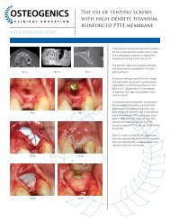
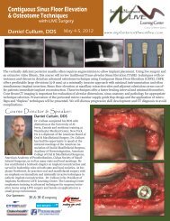
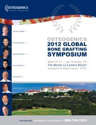
The Cytoplast Ridge Preservation Technique<br />
1.<br />
Figure 1. A minimally invasive, atraumatic extraction technique<br />
should be used. The use of periotomes or surgical sectioning is<br />
encouraged to minimize mechanical trauma to the thin cortical<br />
bone. All soft tissue remnants should be removed with a sharp<br />
curettage. Special care should be taken to remove residual soft<br />
tissues at the apical extent of the socket of endodontically treated<br />
teeth. Bleeding from the socket walls should be noted and, if<br />
necessary, decortication of the socket wall can be done with a #2<br />
round burr to increase early vascularization and access to osteoprogenitor<br />
cells.<br />
2.<br />
Figure 2. A subperiosteal pocket is created with a small periosteal<br />
elevator or curette, extending 3-5 mm beyond the socket margins<br />
(or defect margins) on the palatal and the facial aspect of the<br />
socket. In the esthetic zone, rather than incising and elevating<br />
the interdental papilla, it is left intact and undermined in a similar<br />
fashion. The d-<strong>PTFE</strong> membrane will be tucked into this subperiosteal<br />
pocket.<br />
3.<br />
Figure 3 - 4. Particulate augmentation material is placed into the<br />
socket with a syringe or curette. Ensure that the material is evenly<br />
distributed throughout the socket, but not condensed or packed<br />
too tightly. This will only reduce the available space between particles,<br />
which is critical for vascular ingrowth and subsequent bone<br />
formation.<br />
4.
Cytoplast Technique | 5<br />
The Cytoplast Ridge Preservation Technique<br />
5.<br />
Figure 5 - 6. The d-<strong>PTFE</strong> membrane is trimmed to extend 3-5 mm<br />
beyond the socket walls and then tucked subperiosteally under<br />
the palatal flap, the facial flap, and underneath the interdental<br />
papilla with a curette. The membrane should rest on bone 360°<br />
around the socket margins, if possible. Note that minimal flap reflection<br />
is necessary to stabilize the membrane. Prior to suturing,<br />
ensure that there are no folds or wrinkles in the membrane and<br />
that it lies passively over the socket. Remove any stray bone graft<br />
particles that may be present between the membrane and the flap.<br />
To prevent bacterial leakage under the membrane, take care to<br />
avoid puncturing the membrane, and do not overlap two adjacent<br />
membranes.<br />
Figure 7. The membrane is further stabilized with a criss-cross<br />
<strong>PTFE</strong> suture. It is not recommended to suture through the membrane.<br />
Alternatively, interrupted sutures may be placed. The <strong>PTFE</strong><br />
sutures, which cause minimal inflammatory response, are left in<br />
place for 10 to 14 days.<br />
6.<br />
7.<br />
Figure 8. The membrane is removed, non-surgically, in 21 - 28 days.<br />
With intact sockets, the membrane may be removed as early as 3<br />
weeks. Studies have shown that by 21-28 days there is a dense, vascular<br />
connective tissue matrix in the socket and early osteogenesis is<br />
observed in the apical 2/3 of the socket. Sockets with missing walls<br />
may benefit from a longer time frame. Topical anesthetic is applied,<br />
and then the membrane is grasped with a tissue forcep and simply<br />
removed with a gentle tug.<br />
8.<br />
Figure 9 - 10. Immediately following membrane removal, a dense<br />
highly vascular osteoid matrix is observed filling the socket. Adjacent<br />
gingival epithelium migrates across the osteoid matrix upon<br />
removal of the membrane. At 6 weeks, thick keratinized gingiva<br />
is beginning to form over the grafted socket. The natural soft<br />
tissue architecture is preserved, including the interdental papillae.<br />
New bone is beginning to form in the socket.<br />
9.<br />
10.
6 | Clinical Evidence & References<br />
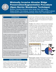
Clinical Evidence<br />
Predictability: In two separate studies treating a total of 696 extraction sites using Cytoplast d<strong>PTFE</strong> membranes in an<br />
exposed technique, there were no reported infections. 3,5<br />
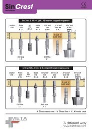
Efficacy: Bone loss 1-year post-extraction using<br />
The Cytoplast Technique for socket preservation. 4<br />
Soft tissue regeneration after extraction using The<br />
Cytoplast Technique for socket preservation. 6<br />
0.5 100 %<br />
0.4 80 %<br />
N=10 N=15<br />
0.3 60 %<br />
0.25 mm<br />
Vertical Bone Loss<br />
*Loss of vertical bone<br />
height measured at crest.<br />
0.3 mm<br />
Horizontal Bone Loss<br />
*Loss of horizontal bone width measured<br />
from stent to buccal plate.<br />
59.68 %<br />
0.2 40 %<br />
N=15<br />
0.1 20 %<br />
18.25 %<br />
0 0 %<br />
Cytoplast TXT-200<br />
No <strong>Membrane</strong><br />
**Measurements taken at time of extraction<br />
*Measurements taken at time of ex-<br />
and 90 days post extraction. traction and 90 days post<br />
extraction.<br />
Applicable References<br />
1. Chesnoiu-Matei I, Texeira HS, Tovar N, Marin C,<br />
Dragan IF, Coelho PG. Alveolar bone dimensional<br />
changes following tooth extraction in sites treated<br />
with or without <strong>PTFE</strong> membranes: A histological<br />
study in dogs. Presented at the 2012 research<br />
forum poster session. Annual meeting of the<br />
American Academy of Periodontology (AAP) in Los<br />
Angeles, CA, September 30 – October 2, 2012.<br />
2. Yamashita M, Horita S, Takei N, Sasada Y, Shibato<br />
W, Ishikawa Y, Takao K, Maki K, Funakoshi E.<br />
Minimally invasive alveolar ridge preservation/<br />
augmentation procedure (open barrier membrane<br />
technique). Presented at the 2010 Research Forum<br />
Poster Session. Annual Meeting of the American<br />
Academy of Periodontology (AAP) in Honolulu, HI,<br />
October 30 – November 2, 2010.<br />
3. Barboza EP, Stutz B, Ferreira VF, Carvalho W.<br />
Guided bone regeneration using nonexpanded<br />
polytetrafluoroethylene membranes in preparation<br />
for dental implant placements – A report of 420<br />
cases. Implant Dent 2010;19:2-7.<br />
4. Fotek PD, Neiva RF, Wang HL. Comparison of<br />
dermal matrix and polytetrafluoroethylene membrane<br />
for socket bone augmentation: A clinical and<br />
histologic study. J Periodontol 2009;80:776-785.<br />
5. Hoffman O, Bartee BK, Beaumont C, Kasaj A, Deli<br />
G, Zafiropoulos GG, Alveolar bone preservation<br />
in extraction sockets using non-resorbable d<strong>PTFE</strong><br />
membranes: A retrospective non-randomized<br />
study. J Periodontol 2008;79:1355-1369.<br />
6. Barboza EP, Francisco BS, Ferreira VF. Soft<br />
tissue enhancement using non-expanded <strong>PTFE</strong><br />
membranes without primary closure [abstract].<br />
Presented at the 2008 Research Forum Poster<br />
Session. Annual Meeting of the American Academy<br />
of Periodontology (AAP) in Seattle, WA, September<br />
6-9, 2008.<br />
7. Barber HD, Lignelli J, Smith BM, Bartee BK. Using<br />
a dense <strong>PTFE</strong> membrane without primary closure<br />
to achieve bone and tissue regeneration. J Oral<br />
Maxillofac Surg 2007;65:748-752.<br />
8. Walters SP, Greenwell H, Hill M, Drisko C, Pickman<br />
K, Scheetz JP. Comparison of porous and<br />
non-porous teflon membranes plus a xenograft in<br />
the treatment of vertical osseous defects: A clinical<br />
reentry study. J Periodontol 2003;74:1161-1168.<br />
9. Bartee BK. Extraction site reconstruction for alveolar<br />
ridge preservation Part 1: Rationale and material<br />
selection. J Oral Implantol 2001;27:187-193.<br />
10. Bartee BK. Extraction site reconstruction for<br />
alveolar ridge preservation Part 2: <strong>Membrane</strong>assisted<br />
surgical technique. J Oral Implantol<br />
2001;27:194-197.<br />
11. Lamb JW III, Greenwell H, Drisko C, Henderson<br />
RD, Scheetz JP, Rebitski G. A comparison of porous<br />
and non-porous teflon membranes plus demineralized<br />
freeze-dried bone allograft in the treatment of<br />
class II buccal/lingual furcation defects: A clinical<br />
reentry study. J Periodontol 2001;72:1580-1587.<br />
12. Bartee BK. Evaluation of a new polytetrafluoroethylene<br />
guided tissue regeneration membrane<br />
in healing extraction sites. Compend Contin Educ<br />
Dent 1998;19:1256-1264.<br />
13. Bartee BK. The use of high-density polytetrafluoroethylene<br />
membrane to treat osseous defects:<br />
Clinical reports. Implant Dent 1995;4:21-26.
<strong>Product</strong> | 7<br />
Cytoplast Dense <strong>PTFE</strong> <strong>Membrane</strong>s<br />
<strong>Membrane</strong>s shown actual size<br />
Cytoplast Titanium-Reinforced Dense <strong>PTFE</strong> <strong>Membrane</strong>s<br />
Anterior<br />
Narrow<br />
Anterior<br />
Singles<br />
Buccal<br />
Posterior<br />
Singles<br />
Posterior Large<br />
XL<br />
XLK<br />
Extraction Site Reconstruction<br />
Ridge Augmentation<br />
Cytoplast Dense <strong>PTFE</strong> <strong>Membrane</strong>s<br />
TXT-200<br />
Singles<br />
TXT-200
4620 71st Street | Building 78-79<br />
Lubbock, TX 79424<br />
www.osteogenics.com | +1 806.796.1923