chapter 10, early development and axis formation in amphibians part-2
chapter 10, early development and axis formation in amphibians part-2
chapter 10, early development and axis formation in amphibians part-2

Create successful ePaper yourself
Turn your PDF publications into a flip-book with our unique Google optimized e-Paper software.
CHAPTER <strong>10</strong>, EARLY<br />
DEVELOPMENT AND AXIS<br />
FORMATION IN AMPHIBIANS<br />
PART-2<br />
ZOO3603C
Progressive Determ<strong>in</strong>ation of the<br />
Amphibian Axes
The specification of axes <br />
Vegetal prote<strong>in</strong>s<br />
VegT destroyed--- entire embryo develops as an<br />
epidermis<br />
Vg1 lacked --- no endoderm nor dorsal mesoderm<br />
The axes are specified by events triggered at<br />
fertilization <strong>and</strong> realized dur<strong>in</strong>g gastrulation.
Concept of regulative <strong>development</strong><br />
Blastomere has a potency greater than its normal<br />
embryonic fate.<br />
Fate is determ<strong>in</strong>ed (<strong>in</strong>duced) by <strong>in</strong>teractions<br />
between neighbor<strong>in</strong>g cells.<br />
How is it <strong>in</strong>duced<br />
What factor cause the <strong>in</strong>duction
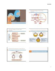
Spemann’s demonstration of nuclear<br />
equivalence <strong>in</strong> newt cleavage<br />
Ligature at 8-cell stage<br />
Enter<strong>in</strong>g a s<strong>in</strong>gle nucleus<br />
to the other side at 16-<br />
cell stage<br />
Nuclei at 16-cell stage are genetically identical<br />
<strong>and</strong> still totipotent <strong>in</strong> the newt. <br />
After 14 days, each side had<br />
a normal embryo.
Asymmetry <strong>in</strong> the amphibian egg<br />
Separation at<br />
left-right <strong>axis</strong><br />
Separation at<br />
ventral-dorsal <strong>axis</strong><br />
Conta<strong>in</strong><strong>in</strong>g<br />
epidermal, blood,<br />
mesenchymal <strong>and</strong><br />
gut cells. <br />
Gray crescent is<br />
essential for<br />
normal<br />
<strong>development</strong>.
Determ<strong>in</strong>ation of ectoderm dur<strong>in</strong>g newt<br />
gastrulation<br />
Normal <strong>development</strong><br />
Regulative <strong>development</strong><br />
What does cause the determ<strong>in</strong>ation <br />
Secondary neural plate<br />
Autonomous <strong>development</strong>
Hans Spemann <strong>and</strong> Hilde Mangold:<br />
Primary Embryonic Induction
Organization of a secondary <strong>axis</strong> by<br />
dorsal blastopore lip tissue<br />
Dorsal blastopore is the<br />
self-differentiat<strong>in</strong>g<br />
tissue.<br />
• Organizer<br />
• Primary embryonic <strong>in</strong>duction
Mechanisms of Axis Determ<strong>in</strong>ation <strong>in</strong><br />
Amphibians<br />
• How does the organizer form<br />
• The dorsal signal: -caten<strong>in</strong><br />
• The vegetal TGF--like signal<br />
• The mesodermal signal
Questions on the mechanisms<br />
How did the organizer get its properties<br />
What caused the dorsal blastopore lip to differ from<br />
any other region of the embryo<br />
What factors were be<strong>in</strong>g secreted from the<br />
organizer to cause the <strong>formation</strong> of the neural tube<br />
<strong>and</strong> to create the anterior-posterior, dorsal-ventral<br />
<strong>and</strong> left-right axes
Mesodermal <strong>in</strong>duction by vegetal endoderm<br />
Animal cap cells generate<br />
mesodermal tissue.<br />
Factors from vegetal cells
Mesodermal <strong>in</strong>duction by vegetal endoderm<br />
On dorsal side, a signal is<br />
released by Nieuwkoop center.
Signals for the axial determ<strong>in</strong>ation<br />
The dorsal signal: -caten<strong>in</strong><br />
The vegetal TGF--like signal<br />
The mesodermal signal, Nodal-relat<strong>in</strong>g prote<strong>in</strong>s
The vegetal cells are responsible for caus<strong>in</strong>g<br />
the <strong>in</strong>itiation of gastrulation<br />
Rescue by transplantation of dorsal vegetal blastomeres which<br />
can <strong>in</strong>duce another <strong>axis</strong> <strong>in</strong> the embryo.<br />
Vegetal cells <strong>in</strong>duce the<br />
organizer which <strong>in</strong>duces <strong>axis</strong>.
The regional specificity of mesoderm <strong>in</strong>duction<br />
D1 most<br />
<strong>in</strong>duced dorsal<br />
mesoderm.<br />
Nieuwkoop Center<br />
is <strong>in</strong> D1 cell<br />
What makes<br />
dorsal/ventral<br />
mesoderm
The role of Wnt pathway <strong>in</strong> ventral-dorsal<br />
<strong>axis</strong> specification<br />
2-Cell<br />
-caten<strong>in</strong><br />
Blastula<br />
-caten<strong>in</strong> localize <strong>in</strong> nuclei on<br />
dorsal, but not on ventral.<br />
Dorsal<br />
Ventral<br />
-caten<strong>in</strong> dorsal localization<br />
persists through gastrula stage.<br />
How this localization happen
Model of the mechanism of localization of<br />
-caten<strong>in</strong> <strong>in</strong> the dorsal portion<br />
Dsh, Dishevelled prote<strong>in</strong><br />
GBP, GSK3-b<strong>in</strong>d<strong>in</strong>g prote<strong>in</strong><br />
GSK3, glycogen synthase k<strong>in</strong>ase 3<br />
on microtubules<br />
cortical rotation<br />
Cortical rotation causes the sift<br />
of the Dsh complex.
Model of the mechanism of localization of<br />
-caten<strong>in</strong> <strong>in</strong> the dorsal portion<br />
1. Dsh <strong>in</strong>hibits GSK3<br />
2. GSK3 degrade -caten<strong>in</strong><br />
3. -caten<strong>in</strong> <strong>in</strong>itiate the organizer<br />
Dsh localization cause the ventraldorsal<br />
<strong>axis</strong>
Model of the mechanism of localization of<br />
-caten<strong>in</strong> <strong>in</strong> the dorsal portion<br />
Inactivation of GSK3 on both<br />
blastomeres of 2 cell.<br />
Formation of 2 nd <strong>axis</strong>.<br />
GSK3 is one of the key to<br />
determ<strong>in</strong>e the ventral-dorsal <strong>axis</strong><br />
through -caten<strong>in</strong>.
Induction of the organizer <strong>in</strong> the dorsal<br />
mesoderm<br />
• Tcf3, ubiquitous transcription factor<br />
• -caten<strong>in</strong>/Tcf3 complex can<br />
activate the transcription.<br />
• Goosecoid prote<strong>in</strong>, transcription<br />
factor which can activate genes <strong>in</strong><br />
organizer.<br />
Difference of -caten<strong>in</strong><br />
expression cause the ventraldorsal<br />
<strong>axis</strong>.
Mesoderm <strong>in</strong>duction <strong>and</strong> organizer <strong>formation</strong><br />
by -caten<strong>in</strong> <strong>and</strong> TGF- prote<strong>in</strong>s<br />
Xnr, nodal-related gene<br />
<strong>in</strong>duced by -caten<strong>in</strong>,<br />
Veg1 <strong>and</strong> Vg1. <br />
Goosecoid <strong>in</strong>duction
Functions of the Organizer<br />
• Induction of neural ectoderm <strong>and</strong> dorsal<br />
mesoderm: BMP <strong>in</strong>hibitors<br />
• Nogg<strong>in</strong><br />
• Chord<strong>in</strong><br />
• Follistat<strong>in</strong>
Four major ability of the organizer<br />
Self-differentiate dorsal mesoderm.<br />
Dorsalize the surround<strong>in</strong>g mesoderm <strong>in</strong>to paraxial<br />
mesoderm.<br />
Dorsalize the ectoderm, <strong>in</strong>clud<strong>in</strong>g the <strong>formation</strong> of<br />
neural tube.<br />
Initiate the movements of gastrulation.
Ability of goosecoid mRNA to <strong>in</strong>duce a<br />
new <strong>axis</strong><br />
Gastrula<br />
Control Injected<br />
goosecoid mRNA<br />
+goosecoid<br />
S<strong>in</strong>gle<br />
blastopore<br />
Double<br />
blastopore<br />
Control<br />
Two dorsal axes Two heads<br />
Goosecoid can <strong>in</strong>duce the <strong>axis</strong>.
Neural structures <strong>in</strong>duced <strong>in</strong> presumptive<br />
ectoderm<br />
Dorsal<br />
lip<br />
Ectoderm<br />
Filter membrane<br />
Incubation of ectoderm with dorsal lip<br />
without the direct contacts <strong>in</strong>duced<br />
neural structures. <br />
Secreted factors from dorsal<br />
lip <strong>in</strong>duced the neural<br />
differentiation. <br />
What is the secreted factors<br />
from dorsal lip
Rescue of dorsal structures by Nogg<strong>in</strong><br />
prote<strong>in</strong><br />
Control<br />
Exposure to UV causes no the<br />
cortical rotation.<br />
<br />
No organizer<br />
+ nogg<strong>in</strong> mRNA<br />
Nogg<strong>in</strong> mRNA <strong>in</strong>jection rescue the<br />
dorsal structure <strong>in</strong> dosagerelated<br />
fashion
Localization of nogg<strong>in</strong> mRNA <strong>in</strong> the<br />
organizer tissue<br />
In situ Hybridization<br />
Gastrulation<br />
Involution<br />
Blastopore lip<br />
Convergent<br />
extension<br />
Prechordal plate<br />
& pharyngeal<br />
endoderm<br />
Extend<br />
beneath the<br />
ectoderm
Localization of chord<strong>in</strong> mRNA<br />
In situ Hybridization<br />
Just prior to<br />
gastrulation<br />
Chord<strong>in</strong>, one of the organizer prote<strong>in</strong><br />
Beg<strong>in</strong>n<strong>in</strong>g gastrulation,<br />
Dorsal blastopore lip<br />
Later gastrulation,<br />
organizer tissues<br />
Chord<strong>in</strong> expression is activated by -caten<strong>in</strong>
Model for the action of the organizer<br />
Organizer molecules block<br />
the action of BMP4.<br />
Immuno-histo chemistry of Smad1 which is<br />
downstream of the cascade BMP4. <br />
Organizer molecules <strong>in</strong>duce ventral-dorsal<br />
<strong>axis</strong> through BMP4 signal<strong>in</strong>g pass way.
Control of neural specification by the<br />
levels of BMPs<br />
Whole mount <strong>in</strong> situ hybridization of Sox2, which is a specific mRNA <strong>in</strong><br />
neural tube <br />
Control<br />
Hyper BMPs by morphol<strong>in</strong>o.<br />
Lack of Sox2 expression <strong>in</strong><br />
neural tube<br />
Control<br />
Hypo BMPs<br />
Epidermis is <strong>in</strong>structed by BMP<br />
signal<strong>in</strong>g, <strong>and</strong> the organizer works by<br />
block<strong>in</strong>g the BMP signal from reach<strong>in</strong>g<br />
the ectoderm above it.<br />
Lack of ventral-dorsal <strong>axis</strong> <strong>in</strong><br />
neural tube
The regional Specificity of Induction<br />
• The determ<strong>in</strong>ation of regional differences<br />
• The head <strong>in</strong>ducer: Wnt <strong>in</strong>hibitors<br />
• Cerberus<br />
• Frzb <strong>and</strong> Dickkopf<br />
• Insul<strong>in</strong>-like growth factors<br />
• Trunk <strong>in</strong>duction: Wnt signals <strong>and</strong> ret<strong>in</strong>oic acid
Regional specificity of <strong>in</strong>duction<br />
Transplantation of archenteron roof <strong>in</strong>to blastocoel <strong>in</strong> <strong>early</strong> gastrulae.<br />
<br />
Head with balancers<br />
Head with balancers,<br />
eyes <strong>and</strong> forebra<strong>in</strong><br />
Posterior <strong>part</strong> of head,<br />
diencephalon <strong>and</strong> otic<br />
vesicles<br />
Trunk-tail segment
Regionally specific <strong>in</strong>duc<strong>in</strong>g action of the dorsal<br />
blastopore lip<br />
Early blastula dorsal lips<br />
<strong>in</strong>duce anterior dorsal<br />
structures <br />
Older blastula dorsal lips<br />
<strong>in</strong>duce more posterior<br />
dorsal structures <br />
Earlier organizer: <strong>in</strong>duce bra<strong>in</strong>s <strong>and</strong> heads<br />
Later organizer: <strong>in</strong>duce sp<strong>in</strong>al cords <strong>and</strong> tails<br />
Region <strong>and</strong> tim<strong>in</strong>g makes difference.
Paracr<strong>in</strong>e factor antagonists from the<br />
organizer<br />
What does <strong>in</strong>duce the head Wnt <strong>in</strong>hibitors<br />
Secrete from<br />
Both active<br />
Both <strong>in</strong>active<br />
Epidermis is formed by<br />
hav<strong>in</strong>g both the Wnt <strong>and</strong><br />
BMP signal<strong>in</strong>g pathways.
Cerberus <strong>in</strong>duces head structures as well as a<br />
duplicated heart <strong>and</strong> liver<br />
Injection of Cerberus mRNA <strong>in</strong>to D4 cell (ventral;<br />
vegetal) at 32-cell stage.<br />
Head structures, heart <strong>and</strong> liver were <strong>in</strong>duced.<br />
By block<strong>in</strong>g BMPs, Nodalrelated<br />
prote<strong>in</strong> <strong>and</strong> Wxnt8,<br />
cerberus can <strong>in</strong>duce a head.
Xwnt8 is ventraliz<strong>in</strong>g the mesoderm <strong>and</strong><br />
prevent<strong>in</strong>g anterior head <strong>formation</strong><br />
• Frzb <strong>and</strong> Dickkopf prote<strong>in</strong> is<br />
secreted by the anterior region of<br />
the organizer.<br />
• Frzb <strong>and</strong> Dickkopf b<strong>in</strong>d to Xwnt8.<br />
• Block the ventraliz<strong>in</strong>g function of<br />
Xwnt8.
Insul<strong>in</strong>-like growth factors enhance anterior<br />
neural <strong>development</strong><br />
Expression of<br />
Igf3 <strong>in</strong> anterior<br />
neural tube<br />
An ectopic headlike<br />
structure<br />
<strong>in</strong>duced by Igf2<br />
mRNA <strong>in</strong>jection.<br />
IGF <strong>in</strong>hibition<br />
The cement gl<strong>and</strong> <strong>and</strong><br />
eyes are absent.<br />
Control<br />
+IGF receptor <strong>in</strong>hibitor<br />
at 4-cell stage
Trunk <strong>in</strong>duction: The Wnt signal<strong>in</strong>g pathway<br />
<strong>and</strong> posteriorization of the neural tube<br />
-caten<strong>in</strong> density <strong>in</strong> future neural plate<br />
Chang<strong>in</strong>g the level of Wnt signal<strong>in</strong>g alters the expression of<br />
regionally specific markers <strong>in</strong> neurula. <br />
Control +Xwnt8 +Xfrzb1<br />
Less anterior<br />
More anterior<br />
Bf1, forebra<strong>in</strong>; Otx2, forebra<strong>in</strong> & midbra<strong>in</strong>; Krox20, two region of h<strong>in</strong>dbra<strong>in</strong>
Trunk <strong>in</strong>duction: The Wnt signal<strong>in</strong>g pathway<br />
<strong>and</strong> posteriorization of the neural tube<br />
Double-gradient model<br />
o BMP gradient: ventral-dorsal <strong>axis</strong><br />
Ventral > Dorsal<br />
o Wnt gradient: anterior-posterior <strong>axis</strong><br />
Anterior < Posterior on neural plate
Model of organizer function <strong>and</strong> <strong>axis</strong><br />
specification <strong>in</strong> the Xenopus gastrula<br />
Anteriorposterior<br />
<strong>axis</strong><br />
Ventraldorsal<br />
<strong>axis</strong><br />
IGFs
Specify<strong>in</strong>g the left-right <strong>axis</strong>
Left-Right Axis: Pitx2 determ<strong>in</strong>es the<br />
direction of heart loop<strong>in</strong>g <strong>and</strong> gut coil<strong>in</strong>g<br />
Wild-type<br />
Injected Pitx2 <br />
Pitx2 <strong>in</strong> just left<br />
side of mesoderm.<br />
Counter-clockwise<br />
gut coil<strong>in</strong>g<br />
Pitx2 <strong>in</strong> both side<br />
of mesoderm<br />
R<strong>and</strong>om gut<br />
coil<strong>in</strong>g<br />
Nodal prote<strong>in</strong> establishes left-light polarity by activat<strong>in</strong>g Pitx2 on<br />
the left side of embryo.
Next class<br />
Chapter-11: The <strong>early</strong> <strong>development</strong> vertebrates:<br />
fish, birds <strong>and</strong> mammals