TB IgA - Diagnostic Automation : Cortez Diagnostics
TB IgA - Diagnostic Automation : Cortez Diagnostics
TB IgA - Diagnostic Automation : Cortez Diagnostics

Create successful ePaper yourself
Turn your PDF publications into a flip-book with our unique Google optimized e-Paper software.
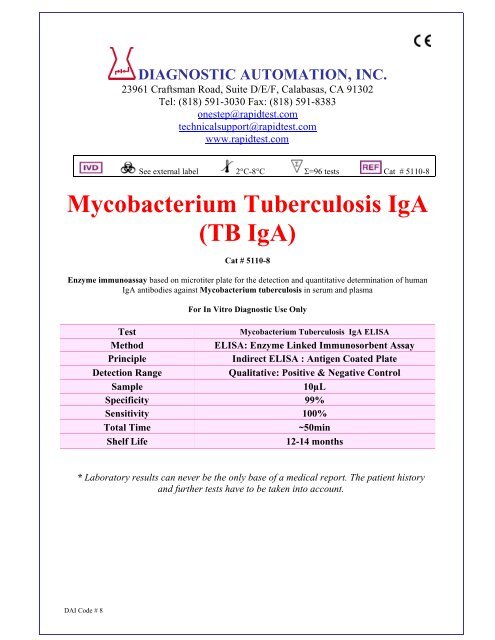
DIAGNOSTIC AUTOMATION, INC.<br />
23961 Craftsman Road, Suite D/E/F, Calabasas, CA 91302<br />
Tel: (818) 591-3030 Fax: (818) 591-8383<br />
onestep@rapidtest.com<br />
technicalsupport@rapidtest.com<br />
www.rapidtest.com<br />
See external label 2°C-8°C Σ=96 tests Cat # 5110-8<br />
Mycobacterium Tuberculosis <strong>IgA</strong><br />
(<strong>TB</strong> <strong>IgA</strong>)<br />
Cat # 5110-8<br />
Enzyme immunoassay based on microtiter plate for the detection and quantitative determination of human<br />
<strong>IgA</strong> antibodies against Mycobacterium tuberculosis in serum and plasma<br />
For In Vitro <strong>Diagnostic</strong> Use Only<br />
Test<br />
Mycobacterium Tuberculosis <strong>IgA</strong> ELISA<br />
Method<br />
ELISA: Enzyme Linked Immunosorbent Assay<br />
Principle<br />
Indirect ELISA : Antigen Coated Plate<br />
Detection Range<br />
Qualitative: Positive & Negative Control<br />
Sample 10µL<br />
Specificity 99%<br />
Sensitivity 100%<br />
Total Time<br />
~50min<br />
Shelf Life<br />
12-14 months<br />
* Laboratory results can never be the only base of a medical report. The patient history<br />
and further tests have to be taken into account.<br />
DAI Code # 8
Intended Use<br />
The <strong>Diagnostic</strong> <strong>Automation</strong> Mycobacterium tuberculosis <strong>IgA</strong> antibody ELISA kit has been<br />
designed for the detection and the quantitative determination of specific <strong>IgA</strong> antibodies against<br />
Mycobacterium tuberculosis in serum and plasma. Further applications in other body fluids are<br />
possible and can be requested from the Technical Service of <strong>Diagnostic</strong> <strong>Automation</strong>. This assay is<br />
intended for in-vitro diagnostic use only. Laboratory results can never be the only base of a medical<br />
report. The patient history and further tests have additionally to be taken into account.<br />
General Information<br />
Mycobacterioses (tuberculosis, leprosy, atypical mycobacterioses, paratuberculosis, and perhaps<br />
Crohn’s Disease) are the infectious diseases of men and animals with the largest diffusion on earth.<br />
The infectious agents of tuberculosis are acid-resistant rod-like formed bacteria of the family<br />
Mycobacteriaceae, genus Mycobacterium. The germ was detected by Robert Koch in 1882. Owing<br />
to the very high infectious power of pathogenic mycobacteria, early diagnosis is essential to prevent<br />
spreading of the disease. Convergence of various approaches are necessary to control the<br />
mycobacterioses, immune reactions and bacterial shedding being variable during the diseases.<br />
However, usual diagnostic procedures were up to now unsatisfactory and did not allow to<br />
distinguish among different mycobacterial species. The illness is normally transferred by droplets<br />
of saliva from infected persons. The target of the infection are mostly the lungs, but also other<br />
organs like the brain, intestinal tract, bones, lymph nodes and kidneys can be afflicted. Tuberculosis<br />
is not only found in developing countries with 8 million of new infections yearly, but also in<br />
industrialized civilizations, as an actual disease with some thousands of cases yearly. Without<br />
treatment, the disease leads in 50% of the cases to death within less than two years. Clinical<br />
symptoms are fatigue, loss of weight, lack of appetite, light fever, nocturnal sweat and pain in the<br />
chest. Especially patients with HIV are threatened by tuberculosis due to their impaired immune<br />
system. A vaccination with living attenuated bacteria is possible (BCG = Bacille Calmette Guérin).<br />
This is mostly done with newborn or young children. With older patients, before the vaccination<br />
there is normally performed the tuberculin test (Pirquet or Mantoux), where a small amount of<br />
tuberculin is injected under the skin. In a positive case, there exist antibodies against Mycobacteria,<br />
and a vaccination is not necessary. Up to recently, there have not existed any serological methods to<br />
detect tuberculosis antibodies in serum. The only available procedure was besides the skin<br />
tuberculin test the direct microscopical identification of the dyed bacteria in sputum. Meanwhile<br />
specific antigens have been prepared either by purification of natural material or by recombinant<br />
methods. This ELISA test kit for the determination of IgG antibodies uses a cocktail of highly pure<br />
proteins in order to determine an immune response against the bacteria in human serum. A fresh or<br />
chronically active infection can be diagnosed by <strong>IgA</strong> and IgM tests, which are also available.<br />
Principle of the Test<br />
The <strong>Diagnostic</strong> <strong>Automation</strong> Mycobacterium tuberculosis <strong>IgA</strong> antibody test kit is based on the<br />
principle of the enzyme immunoassay (EIA). Mycobacterium tuberculosis antigen is bound on the<br />
surface of the microtiter strips. Diluted patient serum or ready-to-use standards are pipetted into the<br />
wells of the microtiter plate. A binding between the <strong>IgA</strong> antibodies of the serum and the<br />
immobilized Mycobacterium tuberculosis antigen takes place. After a one hour incubation at room<br />
temperature, the plate is rinsed with diluted wash solution, in order to remove unbound material.<br />
Then ready-to-use anti-human-<strong>IgA</strong> peroxidase conjugate is added and incubated for 30 minutes.<br />
After a further washing step, the substrate (TMB) solution is pipetted and incubated for 20 minutes,<br />
inducing the development of a blue dye in the wells. The color development is terminated by the<br />
addition of a stop solution, which changes the color from blue to yellow. The resulting dye is<br />
measured spectrophotometrically at the wavelength of 450 nm. The concentration of <strong>IgA</strong> antibodies<br />
is directly proportional to the intensity of the color.<br />
DAI Code # 8<br />
2
Limitations, Precautions and General Comments<br />
• Only for in-vitro use! Do not ingest or swallow! The usual laboratory safety precautions as well<br />
as the prohibition of eating, drinking and smoking in the lab have to be followed.<br />
• All sera and plasma or buffers based upon, have been tested respective to HBsAg, HIV and HCV<br />
with recognized methods and were found negative. Nevertheless precautions like the use of latex<br />
gloves have to be taken.<br />
• Serum and reagent spills have to be wiped off with a disinfecting solution<br />
(e.g. sodium hypochlorite, 5%) and have to be disposed of properly.<br />
• All reagents have to be brought to room temperature (18 to 25 °C) before performing the test.<br />
• Before pipetting all reagents should be mixed thoroughly by gentle tilting or swinging. Vigorous<br />
shaking with formation of foam should be avoided.<br />
• It is important to pipet with constant intervals, so that all the wells of the microtiter plate have<br />
the same conditions.<br />
• When removing reagents out of the bottles, care has to be taken that the stoppers are not<br />
contaminated. Further a possible mix-up has to be avoided. The content of the bottles is usually<br />
sensitive to oxidation, so that they should be opened only for a short time.<br />
• In order to avoid a carry-over or a cross-contamination, separate disposable pipet tips have to be<br />
used.<br />
• No reagents from different kit lots have to be used, they should not be mixed among one another.<br />
• All reagents have to be used within the expiry period.<br />
• In accordance with a Good Laboratory Practice (GLP) or following ISO9001 all laboratory<br />
devices employed should be regularly checked regarding the accuracy and precision. This refers<br />
amongst others to microliter pipets and washing or reading (ELISA-Reader) instrumentation.<br />
• The contact of certain reagents, above all the stopping solution and the substrate with skin, eye<br />
and mucosa has to be avoided, because possible irritations and acid burns could arise, and there<br />
exists a danger of intoxication.<br />
Reagents Provided<br />
Store kit components at 2-8 o C and do not use after the expiry date on the box outer label. Before<br />
use, all components should be allowed to warm up to ambient temperature (18-25 o C). After use, the<br />
plate should be resealed, the bottle caps replaced and tightened and the kit stored at 2-8 o C. The<br />
opened kit should be used within three months.<br />
Components<br />
Volume / Qty.<br />
Mycobacterium tuberculosis antigen coated microtiter strips 12<br />
Calibrator A (Negative Control)<br />
2 mL<br />
Calibrator B (Cut-Off Standard)<br />
2 mL<br />
Calibrator C (Weak Positive Control)<br />
2 mL<br />
Calibrator D (Positive Control)<br />
2 mL<br />
Enzyme Conjugate<br />
15 mL<br />
Substrate<br />
15 mL<br />
Stop Solution<br />
15 mL<br />
Sample Diluent<br />
60 mL<br />
Washing Buffer (10×)<br />
60 mL<br />
Plastic foils 2<br />
Plastic bag 1<br />
DAI Code # 8<br />
3
1. Microtiter Strips<br />
12 strips with 8 breakable wells each, coated with a M. tuberculosis antigen mixture (recombinant<br />
Mycobacterium tuberculosis antigens, with 18, 36 and 40 kDa). Ready-to-use.<br />
2. Calibrator A (Negative Control)<br />
2 mL, protein solution diluted with PBS, contains no <strong>IgA</strong> antibodies against Mycobacterium<br />
tuberculosis. Addition of 0.01 % methylisothiazolone and 0.01 % bromonitrodioxane. Ready-touse.<br />
3. Calibrator B (Cut-Off Standard)<br />
2 mL human serum diluted with PBS, contains a low concentration of <strong>IgA</strong> antibodies against<br />
Mycobacterium tuberculosis. Addition of 0.01 % methylisothiazolone and 0.01 %<br />
bromonitrodioxane. Ready-to-use.<br />
4. Calibrator C (Weak Positive Control)<br />
2 mL, human serum diluted with PBS, contains a medium concentration of <strong>IgA</strong> antibodies against<br />
Mycobacterium tuberculosis. Addition of 0.01 % methylisothiazolone and 0.01 %<br />
bromonitrodioxane. Ready-to-use.<br />
5. Calibrator D (Positive Control)<br />
2 mL, human serum diluted with PBS, contains a high concentration of <strong>IgA</strong> antibodies against<br />
Mycobacterium tuberculosis. Addition of 0.01 % methylisothiazolone and 0.01 %<br />
bromonitrodioxane. Ready-to-use.<br />
6. Enzyme Conjugate<br />
15 mL, anti-human-<strong>IgA</strong>-HRP (rabbit), in protein-containing buffer solution. Addition of 0.01 %<br />
methylisothiazolone and 0.01 % bromonitrodioxane and 5 mg/L Proclin Ready-to-use.<br />
7. Substrate<br />
15 mL, TMB (tetramethylbenzidine). Ready-to-use.<br />
8. Stop Solution<br />
15 mL, 0.5 M sulfuric acid. Ready-to-use.<br />
9. Sample Diluent<br />
60 mL, PBS/BSA buffer. Addition of 0.095 % sodium azide. Ready-to-use.<br />
10. Washing Buffer<br />
60 mL, PBS + Tween 20, 10x concentrate. Final concentration: dilute 1+9 with distilled water. If<br />
during the cold storage crystals precipitate, the concentrate should be warmed up at 37°C for<br />
15 minutes.<br />
11. Plastic Foils<br />
2 pieces to cover the microtiter strips during the incubation.<br />
12. Plastic Bag<br />
Resealable, for the dry storage of non-used strips.<br />
Materials Required but not Provided<br />
• 5 µL-, 100 µL- and 500 µL micro- and multichannel pipets<br />
• Microtiter Plate Reader (450 nm)<br />
• Microtiter Plate Washer<br />
• Reagent tubes for the serum dilution<br />
• Bidistilled water<br />
DAI Code # 8<br />
4
Specimen Collection and Handling<br />
Principally serum or plasma (EDTA, heparin) can be used for the determination. Serum is separated<br />
from the blood, which is aseptically drawn by venipuncture, after clotting and centrifugation. The<br />
serum or plasma samples can be stored refrigerated (2-8°C) for up to 48 hours, for a longer storage<br />
they should be kept at -20 °C. The samples should not be frozen and thawed repeatedly. Lipemic,<br />
hemolytic or bacterially contaminated samples can cause false positive or false negative results.<br />
For the performance of the test the samples (not the standards) have to be diluted 1:101 with readyto-use<br />
sample diluent (e.g. 5 µL serum + 500 µL sample diluent).<br />
Assay Procedure<br />
1. Preparation of Reagents<br />
Washing Solution: dilute before use 1+9 with distilled water. If during the cold storage crystals<br />
precipitate, the concentrate should be warmed up at 37°C for 15 minutes.<br />
• Strict adherence to the protocol is advised for reliable performance. Any changes or<br />
modifications are the responsibility of the user.<br />
• All reagents and samples must be brought to room temperature before use, but should not be left<br />
at this temperature longer than necessary.<br />
• Standards and samples should be assayed in duplicates.<br />
• A standard curve should be established with each assay.<br />
• Return the unused microtiter strips to the plastic bag and store them dry at 2-8°C.<br />
2. Assay Steps<br />
1. Prepare a sufficient amount of microtiter wells for the standards, controls and samples in<br />
duplicate as well as for a substrate blank.<br />
2. Pipet 100 µL each of the diluted (1:101) samples and the ready-to-use standards and controls<br />
respectively into the wells. Leave one well empty for the substrate blank.<br />
3. Cover plate with the enclosed foil and incubate at room temperature for 60 minutes.<br />
4. Empty the wells of the plate (dump or aspirate) and add 300 µL of diluted washing solution.<br />
This procedure is repeated totally three times. Rests of the washing buffer are afterwards<br />
removed by gentle tapping of the microtiter plate on a tissue cloth.<br />
5. Pipet 100 µL each of ready-to-use conjugate into the wells. Leave one well empty for the<br />
substrate blank.<br />
6. Cover plate with the enclosed foil and incubate at room temperature for 30 minutes.<br />
7. Empty the wells of the plate (dump or aspirate) and add 300 µL of diluted washing solution.<br />
This procedure is repeated totally three times. Rests of the washing buffer are afterwards<br />
removed by gentle tapping of the microtiter plate on a tissue cloth.<br />
8. Pipet 100 µL each of the ready-to-use substrate into the wells. This time also the substrate blank<br />
is pipetted.<br />
9. Cover plate with the enclosed foil and incubate at room temperature for 20 minutes in the dark<br />
(e.g. drawer).<br />
10. To terminate the substrate reaction, pipet 100 µL each of the ready-to-use stop solution into the<br />
wells. Pipet also the substrate blank.<br />
11. After thorough mixing and wiping the bottom of the plate, perform the reading of the absorption<br />
at 450 nm (optionally reference wavelength of 620 nm). The color is stable for at least<br />
60 minutes.<br />
DAI Code # 8<br />
5
Evaluation<br />
The mean values for the measured absorptions are calculated after subtraction of the substrate blank<br />
value. The difference between the single values should not exceed 10 %.<br />
Example<br />
OD Value corrected OD Mean OD Value<br />
Substrate Blank 0.013<br />
Negative Control 0.022 / 0.023 0.009 / 0.010 0.010<br />
Cut-Off Standard 0.599 / 0.613 0.586 / 0.600 0.593<br />
Weak Positive Control 1.099 / 1.046 1.086 / 1.033 1.060<br />
Positive Control 2.021 / 1.893 2.008 / 1.880 1.944<br />
The above table contains only an example, which was achieved under arbitrary temperature and<br />
environmental conditions. The described data constitute consequently no reference values which<br />
have to be found in other laboratories in the same way.<br />
1. Qualitative Evaluation<br />
The calculated absorptions for the patient sera, as mentioned above, are compared with the value<br />
for the cut-off standard. If the value of the sample is higher, there is a positive result.<br />
For a value below the cut-off standard, there is a negative result. It seems reasonable to define a<br />
range of +/-20 % around the value of the cut-off as a grey zone. In such a case the repetition of the<br />
test with the same serum or with a new sample of the same patient, taken after 2-4 weeks, is<br />
recommended. Both samples should be measured in parallel in the same run.<br />
The positive control must show at least the double absorption compared with the cut-off standard.<br />
2. Quantitative Evaluation<br />
The ready-to-use standards and controls of the Mycobacterium antibody kit are defined and<br />
expressed in arbitrary units (U/mL). This results in an exact and reproducible quantitative<br />
evaluation. Consequently for a given patient follow-up controls become possible. The values for<br />
controls and standards in units are printed on the labels of the vials.<br />
For a quantitative evaluation the absorptions of the standards and controls are graphically drawn<br />
against their concentrations. From the resulting reference curve the concentration values for each<br />
patient sample can then be extracted in relation to their absorptions. It is also possible to use<br />
automatic computer programs.<br />
3. Interpretation<br />
Although antibody may occur in healthy persons in rare cases, normally they indicate the colonisation with<br />
Mycobacterium tuberculosis. Positive IgM results are related to the early stage of an infection. During its<br />
course a seroconversion towards IgG antibodies take place. The simultaneous presence of IgG and IgM<br />
antibodies denote an infection at its early stage or a reactivation in chronic infections. The sole occurrence of<br />
IgG is a sign for a completed immunological response. <strong>IgA</strong> antibodies occur after the initial activation of<br />
immunoreaction as indicated by the presence of IgM and are associated to a high inflammatory potential.<br />
Since they are not affected by energy effects as Igg antibodies, they are useful marker for patients which<br />
show a reduced IgG response due to pre existing immune depression. Briefly the humoral response can be<br />
summarized as follows:<br />
- IgM (-) IgG (-) <strong>IgA</strong> (-) no infection<br />
- IgM (+) IgG (-) <strong>IgA</strong> (-) infection at a very early stage<br />
- IgM (-) IgG (+) <strong>IgA</strong> (+/-) completed infection<br />
- IgM (+) IgG (+) <strong>IgA</strong> (+/-) infection at an early stage or re-infection<br />
- IgM (-) IgG (-) <strong>IgA</strong> (+) completed infection in patients suffering from IgG reducing effects.<br />
DAI Code # 8<br />
6
Assay Characteristics<br />
Mycobacterium<br />
IgG <strong>IgA</strong> IgM<br />
ELISA<br />
Intra-Assay-<br />
7.6 % 7.9 % 7.9 %<br />
Precision<br />
Inter-Assay-<br />
9.4 % 7.4 % 7.4 %<br />
Precision<br />
Inter-Lot-Precision 3.1 – 9.9 % 5.7 – 8.9 % 5.7 – 8.9 %<br />
Analytical<br />
1.09 U/mL 1.34 U/ml 1.22 U/mL<br />
Sensitivity<br />
Recovery 86 – 95 % 87 – 96 % 87 – 91 %<br />
Linearity 82 – 113 % 78 – 111 % 78 – 118 %<br />
Cross-Reactivity No cross-reactivity to Helicobacter pylori and Bordetella pertussis.<br />
Interferences<br />
No interferences to bilirubin up to 0.3 mg/mL, hemoglobin up to 8.0 mg/mL<br />
and triglycerides up to 5.0 mg/mL<br />
Clinical Specificity 99 % 99 % 100 %<br />
Clinical Sensitivity 100 % 100 % 100 %<br />
References<br />
1. Bloom BR, Murray CJL. Tuberculosis: commentary on a reemergent killer. Science, 1992,<br />
257:1055-64.<br />
2. Kochi A. Global tuberculosis situation and the control strategy of the WHO. Tubercle, 1991,<br />
72:1-6.<br />
3. Marks LG. Genetics of tuberculosis. Medical clinics of North America, 1993, 77(6):1219-33.<br />
4. Aziz A, Siddiqui SH, Ishaq M. Drug resistance of Mycobacterium tuberculosis from treated<br />
patients in Pakistan. Tubercle, 1989, 70:45-51.<br />
5. Outbreak of multidrug-resistant tuberculosis Texas, California and Pennsylvania. Morbidity and<br />
mortality weekly report, 1990, 39:369-72.<br />
6. <strong>TB</strong> morbidity United States, 1995. Morbidity and mortality weekly report, 1996, 45:365-70.<br />
7. Barnes PF, Lee HQ, Davidson PT. Tuberculosis in patients with HIV infection. Medical clinics<br />
of North America, 1993, 77(6):1369-89.<br />
8. Directorate-General for Chest Diseases. Tuberculosis control guide. Egypt, National<br />
Tuberculosis Control Programme, Ministry of Health, November 1994.<br />
9. Snider DE Jr, La Montagne JR. The neglected global tuberculosis problem: a report of the 1992<br />
World Congress on tuberculosis. Journal of infectious diseases, 1994, 169:1189-96.<br />
10. Tuberculosis control as an integral part of primary health care. Geneva, World Health<br />
Organization, 1988.<br />
11. Toman K. Tuberculosis case-finding and chemotherapy. Questions and answers. Geneva, World<br />
Health Organization, 1979:3-74.<br />
DAI Code # 8<br />
7
Date Adopted Reference No.<br />
2011-03-01 DA-<strong>TB</strong> <strong>IgA</strong>-2011<br />
DIAGNOSTIC AUTOMATION, INC.<br />
23961 Craftsman Road, Suite D/E/F, Calabasas, CA 91302<br />
Tel: (818) 591-3030 Fax: (818) 591-8383<br />
ISO 13485-2003<br />
Revision Date: 03-16-2011<br />
DAI Code # 8<br />
8