MEMORANDUM FOR: RADIOLOGISTS DEPLOYING ... - Radiology
MEMORANDUM FOR: RADIOLOGISTS DEPLOYING ... - Radiology
MEMORANDUM FOR: RADIOLOGISTS DEPLOYING ... - Radiology
Create successful ePaper yourself
Turn your PDF publications into a flip-book with our unique Google optimized e-Paper software.
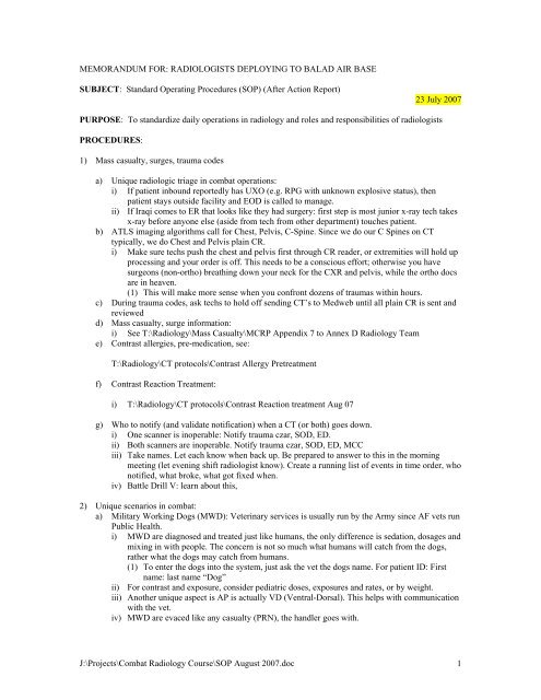
<strong>MEMORANDUM</strong> <strong>FOR</strong>: <strong>RADIOLOGISTS</strong> <strong>DEPLOYING</strong> TO BALAD AIR BASE<br />
SUBJECT: Standard Operating Procedures (SOP) (After Action Report)<br />
23 July 2007<br />
PURPOSE: To standardize daily operations in radiology and roles and responsibilities of radiologists<br />
PROCEDURES:<br />
1) Mass casualty, surges, trauma codes<br />
a) Unique radiologic triage in combat operations:<br />
i) If patient inbound reportedly has UXO (e.g. RPG with unknown explosive status), then<br />
patient stays outside facility and EOD is called to manage.<br />
ii) If Iraqi comes to ER that looks like they had surgery: first step is most junior x-ray tech takes<br />
x-ray before anyone else (aside from tech from other department) touches patient.<br />
b) ATLS imaging algorithms call for Chest, Pelvis, C-Spine. Since we do our C Spines on CT<br />
typically, we do Chest and Pelvis plain CR.<br />
i) Make sure techs push the chest and pelvis first through CR reader, or extremities will hold up<br />
processing and your order is off. This needs to be a conscious effort; otherwise you have<br />
surgeons (non-ortho) breathing down your neck for the CXR and pelvis, while the ortho docs<br />
are in heaven.<br />
(1) This will make more sense when you confront dozens of traumas within hours.<br />
c) During trauma codes, ask techs to hold off sending CT’s to Medweb until all plain CR is sent and<br />
reviewed<br />
d) Mass casualty, surge information:<br />
i) See T:\<strong>Radiology</strong>\Mass Casualty\MCRP Appendix 7 to Annex D <strong>Radiology</strong> Team<br />
e) Contrast allergies, pre-medication, see:<br />
T:\<strong>Radiology</strong>\CT protocols\Contrast Allergy Pretreatment<br />
f) Contrast Reaction Treatment:<br />
i) T:\<strong>Radiology</strong>\CT protocols\Contrast Reaction treatment Aug 07<br />
g) Who to notify (and validate notification) when a CT (or both) goes down.<br />
i) One scanner is inoperable: Notify trauma czar, SOD, ED.<br />
ii) Both scanners are inoperable. Notify trauma czar, SOD, ED, MCC<br />
iii) Take names. Let each know when back up. Be prepared to answer to this in the morning<br />
meeting (let evening shift radiologist know). Create a running list of events in time order, who<br />
notified, what broke, what got fixed when.<br />
iv) Battle Drill V: learn about this,<br />
2) Unique scenarios in combat:<br />
a) Military Working Dogs (MWD): Veterinary services is usually run by the Army since AF vets run<br />
Public Health.<br />
i) MWD are diagnosed and treated just like humans, the only difference is sedation, dosages and<br />
mixing in with people. The concern is not so much what humans will catch from the dogs,<br />
rather what the dogs may catch from humans.<br />
(1) To enter the dogs into the system, just ask the vet the dogs name. For patient ID: First<br />
name: last name “Dog”<br />
ii) For contrast and exposure, consider pediatric doses, exposures and rates, or by weight.<br />
iii) Another unique aspect is AP is actually VD (Ventral-Dorsal). This helps with communication<br />
with the vet.<br />
iv) MWD are evaced like any casualty (PRN), the handler goes with.<br />
J:\Projects\Combat <strong>Radiology</strong> Course\SOP August 2007.doc 1
3) Daily activities, shift-unique radiologist roles/ responsibilities:<br />
a) Day (7—1500):<br />
i) If patient has a CD and wants their images, you can save as .jpg images.<br />
(1) For CT data, easier to make a CD off the Phillips (not officially).<br />
(2) During this rotation, it was decided to not use patient thumb drives since there are some<br />
real concerns with viruses (Medweb does not get regular virus updates), files patients<br />
(some contractors, Army, may have different rules) may have inappropriate files on their<br />
drive that should not be connected to our PC’s (this happened during this rotation). Also,<br />
we should not take responsibility for patient’s personal drives as they may not know how<br />
to manage files and have the only copy of something they have worked hours or days on.<br />
If we take it from them, we have agreed to be responsible for it; so just don’t accept it.<br />
Let them know we used to, but now only do CD’s (preferably sealed, with HIPAA<br />
stickers, compliance, etc.).<br />
(3) Finish all JPTA notes: this takes priority over outpatients in that surgeons and ICU docs and<br />
other staff need to know the latest on all their patients at a glance. Outpatients can wait.<br />
(4) It is also recommended that techs and radiologists drink enough water during surges and take<br />
short food breaks to avoid exhaustion/ mistakes, etc. for the trauma codes to follow. You<br />
never know when it may go on for 12 or more hours. It does not do anyone any good to pass<br />
out or make mistakes or get kidney stones and be out of commission.<br />
(5) Provide snapshot reviews to the night shift radiologist of cases where imaging significantly<br />
guided life, limb or eyesight guiding therapy, AE or surgical triage.<br />
(6) Provide any equipment issues to forward to the night shift person to report (or answer to) in<br />
morning meeting. For example if a CT or PACS goes down for any length of time, the night<br />
shift person needs to answer to that if asked.<br />
(7) If busy during transition to next shift, stay until workload manageable by the incoming 1500-<br />
1100 radiologist.<br />
ii) Evening (1500-2300):<br />
(1) Provide snapshot reviews to the night shift radiologist of cases where imaging<br />
significantly guided life, limb or eyesight guiding therapy, AE or surgical triage.<br />
(2) Provide any equipment issues to forward to the night shift person to report (or answer to)<br />
in morning meeting. For example if a CT or PACS goes down for any length of time, the<br />
night shift person needs to answer to that if asked.<br />
(3) Read all exams, which may include providing a protocol for exams, and hand-deliver all<br />
reports to the appropriate location/person.<br />
(4) Enter a note into JPTA on all admitted patients.<br />
(5) Load patient CDs if one is available.<br />
(6) Be available to all staff as needed.<br />
(7) Make CDs of exams as needed<br />
(8) Pushing exams to LRMC as needed.<br />
iii) Night (2300-0830): ICU, ward reads, trauma PRN<br />
(1) The CASF runs start coming in around 2130, with other runs at 0200, 0400.<br />
(a) Basically a radiologist and tech respond to ER for every helicopter inbound, trauma code<br />
or not. This will save you time in the long run by making sure printed JPTA or CHCS<br />
notes on prior CT’s (about 8 studies per night) and or CD’s help prevent further imaging.<br />
About 10% of CD’s do not work or are incomplete, about 5% are sent prior to the<br />
patients showing up by telerad/ Medweb from sites pushing (don’t get your hopes up on<br />
this one, we have tried).<br />
(2) Inpatient census (no longer required as of June 2007). If you would like a census:<br />
(a) Early AM (0100), print hospital inpatient census from JPTA https://jpta.fhp.osd.mil<br />
(b) Reports, then 24 hr reports, export to Excel, then trim excess, then sort by ward.<br />
(c) To sort, need to delete all merged cells<br />
(d) Print for techs around 0200 (before portables).<br />
J:\Projects\Combat <strong>Radiology</strong> Course\SOP August 2007.doc 2
(e) The prior radiologists exported /printed this JPTA for ICU portables for techs<br />
(3) There is a meal served starting 0130, recommend eating early (if you can) as the ICU images<br />
start rolling in around 0200. I keep some cereal or other food aside since you often do not get<br />
time to get to the DFAC with ER, traumas, follow-up head CTs, ICU, etc.<br />
(4) Recommend reading ICU 1 first, then delivering those reports, then working your way down<br />
the hall. Hand delivering allows direct feedback to the caring nurse, getting more history,<br />
interaction (plus an opportunity to get up from sitting). Don’t just leave the reports without<br />
handing off to someone and explaining what is going on, especially with positive findings or<br />
tube recommendations.<br />
(5) Before the morning CT follow-ups the flight docs will likely want to go over CT findings on<br />
anything involving closed spaces (sinuses, all head injuries, lung contusions, BLI, etc.). I<br />
found it easier to do a mini-rounds after their first round with JPMRC (Joint Patient<br />
Movement Requirements Center, the ones that tell the flight docs to repeat C-Spines) and<br />
PAD. JPMRC will kickback any spine controversy (why we put “C, T and L spine negative”<br />
in JPTA).<br />
(6) The follow-up head CTs start around 0500.<br />
(7) Morning report (clinical openers): I bring an SOD list of all ED cases/ admits, especially<br />
where imaging effected intervention. Occasionally we help get the imaging results strait, but<br />
the SOD and trauma czar have reviewed fairly completely.<br />
(a) Clinical openers is where you can get a good feel for what other night duties are<br />
important to get the next day started.<br />
(b) This is also where you report any positive TB CXR or CT chest. We had quite a few<br />
during our rotation.<br />
(8) ICU rounds: 0800 or right after morning report in radiologist reading station. I list the ICU<br />
CXR’s I read that night on the SOD sheets in the order I read them. I started by<br />
(9) Nights are a good time to work on LOE and award bullets, SOP, AAR and other<br />
administrative duties.<br />
(10) The quietest time seems to be between 0430 and 0500, I was able to get two 40 minute naps<br />
during the first month (we were relatively slow our first month before the Arrowhead Ripper<br />
Offensive).<br />
(11) The flight commander goes to 2/3’s of the flight commanders meeting (chaired by the<br />
squadron commander) during the entire rotation. During our rotation they were Monday<br />
mornings 9-10. When the flight commander works nights, it was easy to stay an extra hour or<br />
two since they were staying for the clinical openers and ICU rounds.<br />
(a) When the flight commander works days, the night radiologist stays to cover the dept.<br />
(outpatients, ER and trauma codes) while the flight commander goes to the meeting.<br />
(b) Occasionally there are other morning meetings (to provide for those on shifts).<br />
(c) When the flight commander works 3-11, then the night person can go to the meetings<br />
while the day radiologist covers the department. The flight commander can come in early<br />
for late afternoon meetings on that shift.<br />
b) Reporting, outpatients:<br />
i) Keep it simple, I minimize to findings that matter. I discourage reporting on appropriate age<br />
related changes (DJD). I make it known to the ordering providers that I do not report on these<br />
and discourage ordering of LS spine plain CR to R/O DJD, or “scoliosis,” etc.<br />
c) Administrative:<br />
i) Counting: (this is done for monthly exam tracking)<br />
(1) The counting is done by the night shift radiologist for CT and the day R.T. shift leader for<br />
plain film, ER, routine, US. This is recorded in a small green binder by pen/ tickmark.<br />
(2) Documents for entering into XL (from the book) are on<br />
T:\<strong>Radiology</strong>\Rad Numbers\2007\Imaging_Numbers_May-Sept_07<br />
There is also an informative email in the Rad Numbers folder as to the history and<br />
reasoning behind the current counting system.<br />
J:\Projects\Combat <strong>Radiology</strong> Course\SOP August 2007.doc 3
Need to also update the Ancillary slide monthly, first week of each month.<br />
(3) Head =1, C spine = 1, CAP = 3, Stone protocol = 2 (abd, pelvis), face and brain = 2<br />
(4) Panscan (Head, cspine, CAP) = 5. Pan with CTA (circle of Willis and neck vessels).<br />
(5) If spending time doing reconstructions, count as an additional exam.<br />
(6) Example, if doing a CTA, add 2 to Pan scan<br />
(7) Count outside CT’s sent by telerad or CD same way. Put in CD reads in book.<br />
(8) Pelvic, transvag counts as an additional<br />
ii) Clinical openers presentations:<br />
(1) You need to check T:/EMDSS/Saturday Clinical Openers regularly to see<br />
when radiology is presenting. It will come without warning, so you need<br />
to check periodically.<br />
iii) Identification:<br />
(1) All Iraqi locals have a trauma number assigned to them when they arrive<br />
in Balad. Iraqi non-locals also have a number assigned, however it is a 9<br />
digit number. The first 3 digits indicate where they are from.<br />
(2) ALL Americans/Coalition Force use the full name and SSN. The last 4<br />
numbers are not acceptable for any identification in <strong>Radiology</strong>, except<br />
Iraqi locals.<br />
(3) Note: it is not acceptable to use “bay #’s” in that it only leads to confusion,<br />
more time consumption for other patients to follow in that several patients<br />
may end up in the same bay during surges. If anyone (like an ER doc or<br />
staff) asks for a CXR (or other), let them know you need PAD to give<br />
them a number first, or it will not do the patient (and those to follow) any<br />
good.<br />
iv) If you want to look up patients after discharge, look up last 4 and/ or name in<br />
Medweb, get the date they arrived, put\ that in JPTA then find their records in<br />
retrospect under patient search.<br />
d) <strong>Radiology</strong> trauma protocols (see 1.a, above)<br />
i) Protocolization (Radiologist and RT respond to trauma codes; radiologic triage)<br />
(1) Doctor orders imaging studies, with guidance from radiologist.<br />
(a) Does this by filling out imaging request (519), or verbal order to RN who fills it out.<br />
(i) 519 has what patient needs completed (CT, CXR), and a brief history as to what<br />
the radiologist is to look for. If this is not followed to the letter, confusion could<br />
arise as to what patient needs what study and for what reason. An analogy with<br />
pharmacy would be a doctor ordering morphine for a patient.<br />
ii) Inject in arm opposite majority of injury (minimize beam-hardening/ great vessels).<br />
iii) Utilization: Panscan/ blast protocol. Consider pain<br />
distraction, and contrast, in that, someone from a blast may<br />
not feel his lumbar vertebral fractures. Saves everyone time<br />
if we just go for the pancan.<br />
e) Oral contrast for CT: take cold water bottle, add 1/3 bottle oral contrast with flavor of choice.<br />
i) Drink half bottle in first half hour, then save lower 1/5 for just prior to scanning.<br />
ii) Note: there is no barium for GI studies. For intussuception, we would likely use air enema<br />
reduction.<br />
f) CT Myelogram:<br />
i) Lumbar Technique:<br />
(1) Stop medications that lower seizure threshold 24-48 hours prior<br />
(2) Prone or oblique bolstered position<br />
(a) Sterile prep of L1 to L3 level<br />
J:\Projects\Combat <strong>Radiology</strong> Course\SOP August 2007.doc 4
(b) In prone position insert styleted spinal needle 2-3 cm off midline at L2-3 level<br />
angled to course under the ipsilateral lamina under fluoroscopy.<br />
(c) In oblique bolstered position insert needle straight down in a bull’s eye fashion into<br />
the spinal canal through the boneless region under the lamina under fluoroscopy.<br />
(d) Remove stylet to confirm free flow of CSF out of hub after tactile sensation of<br />
passing the posterior longitudinal ligaments and thecal sac is felt.<br />
(3) Inject myelographic contrast under fluoroscopy to ensure free flow in thecal space.<br />
ii) Cervical Technique:<br />
(1) Stop medications that lower seizure threshold 24-48 hours prior<br />
(2) Place patient in prone position and ensure true lateral view on fluoroscopy.<br />
(3) Sterile prep of C1 – C4 on lateral neck<br />
(4) Place styleted spinal needle under fluoroscopy using bull’s eye technique anterior to the<br />
spinal lamina line at C1-2, staying in the posterior third of the spinal canal.<br />
(5) When needle on AP fluoroscopy is medial to lateral margin of dens, remove needle to<br />
check for free flow of CSF<br />
(6) Inject myelographic contrast under fluoroscopy to ensure free flow in thecal space.<br />
(a) DO NOT INJECT UNLESS FREE FLOW OF CSF IS SEEN<br />
(b) INJECTION IN TO THE CORD CAN CAUSE PERMANENT PARALYSIS<br />
iii) Myelographic Contrast<br />
(1) Dose: 3.06 g limit of intrathecal iondinated contrast medium in adults, 2.94g limit in<br />
children<br />
(2) Concentration: 300 mg / ml (10cc) limit in adults, 210 mg /ml limit in children.<br />
(a) Fluoroscopy should be used to determine correct amount of contrast administered<br />
(b) Patient with small thecal sacs require less than those with capacious ones<br />
(c) 200 cc / ml concentration for - pediatric patients, cervical , thoracic and lumbar<br />
myelograms<br />
(d) 300 cc / ml concentration for- total columnar myelography or cervical myelogram<br />
from lumbar injection.<br />
(3) Manufactures:<br />
(a) Use only contrast approved for intrathecal injection<br />
(b) Risk of arachnoiditis and nerve injury with non-approved contrast that is not free of<br />
preservatives or is not ultrafiltered<br />
(c) Bracco<br />
(i) Isovue-M 200 or Isoview-M 300<br />
(d) GE Healthcare<br />
(i) Omnipaque 180 or 240 or 300. (Do Not Use 140 or 350)<br />
iv) CT Protocol<br />
(1) Select slice thickness depending on region on interest and dose requirements depending<br />
on body habitus<br />
(a) Slice Thickness - 1mm , Increment - 1mm, Collimation -16 x 0.75, Pitch 0.69<br />
(b) Slice Thickness - 2mm, Increment - 1mm, Collimation -16 x 1.5, Pitch 0.95<br />
(2) Coronal and Sagittal Reconstructions<br />
v) Postprocedure Orders<br />
(1) Restrict activity for 24 hours with no heavy lifting or bending<br />
(2) Vigorous rehydration.<br />
(3) Elevation of head to limit contrast flowing intracranially. (increased risk of seizure)<br />
(4) Resumption of restricted medications after 24 hours.<br />
(5) Epidural blood patches for CSF leaks very effect at relieving spinal headaches.<br />
g) PACS Admin:<br />
i) Medweb Windows login: When you see DoD prompt, just hit return.<br />
(1) There is no logon or password. If you are asked one, hard reboot.<br />
(2) then, Medweb login: username: raddoc, Raddoc0!<br />
(3) For PC’s around the facility, providers can use the following indirect link to Medweb:<br />
(a) http://153.29.184.10/cobalt/<br />
(i) Login the same as above: username: raddoc, Raddoc0!<br />
J:\Projects\Combat <strong>Radiology</strong> Course\SOP August 2007.doc 5
ii) To read studies on Medweb, search by usual criteria on top bar. Click on ball by patient name<br />
to open. Play with tools to become familiar; fairly intuitive really.<br />
(1) Tools: example, magnifying glass, click magnifying glass, then crop what you want<br />
magnified, then push down on scrollbar to move magnified image.<br />
(2) Preset windows (lung, bone, abd, ect.) are on a small dropdown bar above the tools.<br />
iii) When patient is transferred and has CD, import into Medweb then give CD back to patient<br />
(1) Insert CD, open acrdir.exe folder on desktop, select “My Computer” click on name, if<br />
asked do you want DICOM, select yes, send, then send again, don’t pay attention to<br />
status bar when it stays on. Remove disk and give back to patient.<br />
(2) When asked to burn a CD for a patient being transferred: let them know we can push to<br />
LARMC, but go ahead and make one. Let them know the department or function asking<br />
for a CD needs to provide the CD (per previous rotations). <strong>Radiology</strong> does not have<br />
funding for CD’s. To burn a CD:<br />
(a) Click the CD or envelope on the Medweb icon on the patient line.<br />
(b) Save DICOM to PC, to desktop, then drag the folder to the CD ICON on taskbar.<br />
(3) If study has not been sent across (tech got busy with something else), to send to get read:<br />
(a) Validate study icon, then send study icon, the click Medweb on the window<br />
iv) Phillips workstation: from cold boot, or just turning on, Username md (should default), pass:<br />
leave blank, just hit return.<br />
(1) License agreement window, just hit yes.<br />
(2) Saving studies on Philips:<br />
(a) Batch tab, degrees, # of images (or essentially frame rate)<br />
(b) Select X vs. Y axis; save batch as…click save, label.<br />
(c) To select multiple images (rather than whole study)<br />
(d) Batch, slelect first image, click “From” then stop at last image, click “To”<br />
(e) Save batch with floppy icons below batch window.<br />
v) Telerad: Pushing exams to LARMC from Medwed: 2 options:<br />
1) Get a manifest for the shifts from the FCC located in the same office as the PAD. They<br />
print a finalized list after the flight leaves which may be mid morning.<br />
a) For each patient, find the last 4 of the SSN from JPTA<br />
b) Enter the last 4 into Medweb’s Find and search at least week<br />
c) If anything comes up, push to LRMC:<br />
i) For each exam, click on the folder icon (4 th from left)<br />
ii) Select “DICOM storage device” (4 th icon down) and then “Next”<br />
iii) Select “LRMC” from the drop down menu and then “Finish”<br />
iv) Select “Finish”<br />
2) Check in JPTA for aerovac’d patients.<br />
a) Under Reports, select “Evac and Transfer Reports”<br />
b) Find all patients going to LRMC<br />
c) Find films in Medweb and push as in 1b & c above<br />
d) In addition, read all inpatient JPTA notes to find if they will be aerovac’d and send<br />
images as above.<br />
J:\Projects\Combat <strong>Radiology</strong> Course\SOP August 2007.doc 6
vi) When done reading plain CR and CT, check off “read” in Medweb to track what is<br />
completed.<br />
vii) To move images from wrong folder<br />
(1) Close image folder, delete, then resend from Orex, change, modify then send<br />
h) List of bases, station names, phone #’s: see above chart<br />
i) Ultrasound: how to use after hours<br />
i) Turn on Sonosite (power button with circle).<br />
ii) Patient button, enter name, SSN exam type<br />
iii) Select transducer<br />
iv) Perform the US<br />
v) At the end of exam, to save import to PACS:<br />
(1) Click “Patient” then “End exam” (tab under screen), then “Done”<br />
(a) You will then see blank entry for next patient<br />
j) Volume imaging with Philips workstation<br />
i) Bring up study, select volume, rt click, bone removal, remove residual<br />
ii) Sculpting, exclude or include field selection<br />
iii) Unclick “Show Couch”<br />
iv) For CTA of brain, can do MIP<br />
v) Vessel tracking:<br />
(1) Select AVA icon under analysis, click on the one sequence you want to analyze (CTA<br />
carotid), click solid arrowhead to right of “Bone Removal” for “Vessel Extraction.”<br />
(a) Then click the Add vessel icon, enter a name (helpful for multiple), swap axial on<br />
side windows (for example), click on “Seed” icon, plant seeds in center of vessel<br />
along the desired track. Can select auto or manual track (if not clearly identified).<br />
vi) Virtual bronchoscopy:<br />
(1) Analyze (select one series), then groups, then thorax, then three folders Icon,<br />
k) How to generate “Rich-man’s 3-D x-ray” for inlet, outlet views.<br />
i) On the directory (before opening CT), select analysis, pick coronal (on icons of spinal cord<br />
graphic), pick # of images<br />
ii) Click on MPR (stick figure), select rotate, anatomy icon to pivot.<br />
iii) Pull trans scout line to center of ROI, select thickness (1cm), select number of images (4),<br />
magnify, select curved arrow to oblique (takes a moment)<br />
iv) To save file, save series to Medweb… label<br />
v) AVA is volume rendering<br />
vi) To change thresholds, select icon with 3 folders (who knows why)<br />
4) Educational<br />
a) Each Saturday at 0715, EMDSS presents a 5-10 minute topic at the morning stand-up.<br />
i) Normally the first briefing for each flight is a general overview providing a general awareness<br />
of capabilities and procedures.<br />
ii) The schedule is located at:<br />
(1) T:/EMDSS/Saturday Clinical Operners<br />
(2) Please access the file, and populate accordingly.<br />
b) About three times during the rotation, one of the radiologists presents an academic topic (in<br />
addition to above), again, 5-10 minutes. Example briefings are at the following location:<br />
T:\Clinical Openers\Clinical Openers Specialty Presentations<br />
This is also where you will be asked to save your presentation the day before at the latest. Be sure<br />
to associate any videos (i.e. 3D, CT scrolling) with your presentation, PRN.<br />
5) Unique terms to combat radiology:<br />
a) Fatally injured:<br />
i) KIA = Killed in Action (don’t even make it to hospital, i.e. decapitiated)<br />
ii) DOA = Dead on Arrival (no such thing in combat)<br />
J:\Projects\Combat <strong>Radiology</strong> Course\SOP August 2007.doc 7
iii) DOW = Died of Wounds (if make it to hospital): don’t want these to count up<br />
iv) This matters for JTTS Joint Theater Trauma System<br />
b) “Make a hole” need to know this since not knowing could put you in the way of trauma progress.<br />
i) Basically means move out of the way, a patient litter needs to move through (for example).<br />
6) Process, utilization review:<br />
a) Left over plain films and CD's: No matter how hard we try and give these to the nurses,<br />
the PAD, etc. they still make their way back to us. MSgt Candelario can help us develop<br />
a solid SOP statement on disposition for those that do not make it (validated by PAD).<br />
b) <strong>Radiology</strong> exams done routinely just because. Okay, did what I could to mitigate doing a<br />
CXR every time a patient is transferred from ER to OR, to ICU to ward. I lost that one, we<br />
will continue to do them on intubated patients. On the C spine and head CT on all blast<br />
patients, nobody has produced documentation or audit trail on that, so jury is still out. If<br />
you hear of that, go ahead and do, but take names (politely) ask for policy (JPMRC AFI,<br />
etc.) or any documentation.<br />
7) Accountability<br />
a) After alarm red, 100% accountability is required.<br />
b) Find phone and call radiology at 443-8527<br />
c) MCC at 443-8508, or go to DFAC to sign in<br />
d) When paged, shift NCO will contact down the roster (not recall); send runners PRN<br />
e) Pages will have number to call (likely radiology), then followed by one of the following<br />
i) 100 = need 100% accountability of all personnel<br />
ii) 411= just looking for info, call when able<br />
iii) 911 = report ASAP (or call to find out, if able)<br />
8) REFERENCES:<br />
a) After Action Report, AEF cycles 6/7, 7/8 (great references)<br />
b) Protocols, other items on shared drive: T:\<strong>Radiology</strong>\xxx<br />
c) We could not find a prior SOP.<br />
d) Balad phone book<br />
e) Mirvis Stu, Shan, “Imaging in Trauma and Critical Care”<br />
Colonel Les Folio, USAF, MC, SFS<br />
<strong>Radiology</strong> Flight Commander<br />
J:\Projects\Combat <strong>Radiology</strong> Course\SOP August 2007.doc 8


