HigH-Content AnAlysis
HigH-Content AnAlysis
HigH-Content AnAlysis

You also want an ePaper? Increase the reach of your titles
YUMPU automatically turns print PDFs into web optimized ePapers that Google loves.
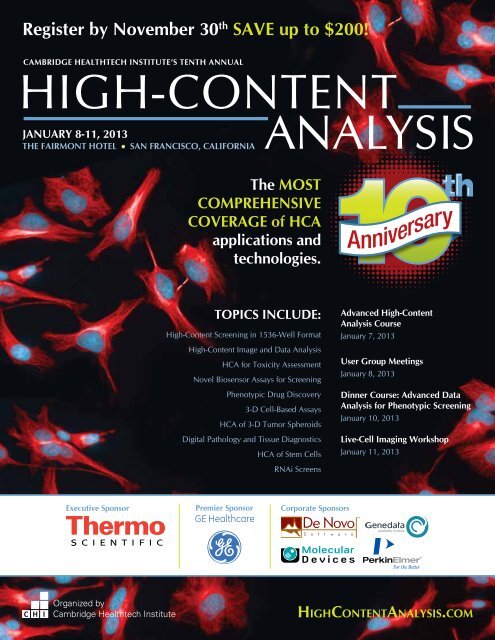
Register by November 30 th SAVE up to $200!<br />
Cambridge Healthtech Institute’s TENTH Annual<br />
High-<strong>Content</strong><br />
January 8-11, 2013<br />
The Fairmont Hotel l San Francisco, California<br />
Analysis<br />
The most<br />
COMPREHENSIVE<br />
COVERAGE of HCA<br />
applications and<br />
technologies.<br />
Anniversary<br />
TOPICS INCLUDE:<br />
High-<strong>Content</strong> Screening in 1536-Well Format<br />
High-<strong>Content</strong> Image and Data Analysis<br />
HCA for Toxicity Assessment<br />
Novel Biosensor Assays for Screening<br />
Phenotypic Drug Discovery<br />
3-D Cell-Based Assays<br />
HCA of 3-D Tumor Spheroids<br />
Digital Pathology and Tissue Diagnostics<br />
HCA of Stem Cells<br />
RNAi Screens<br />
Advanced High-<strong>Content</strong><br />
Analysis Course<br />
January 7, 2013<br />
User Group Meetings<br />
January 8, 2013<br />
Dinner Course: Advanced Data<br />
Analysis for Phenotypic Screening<br />
January 10, 2013<br />
Live-Cell Imaging Workshop<br />
January 11, 2013<br />
Executive Sponsor<br />
Premier Sponsor<br />
Corporate Sponsors<br />
Organized by<br />
Cambridge Healthtech Institute<br />
High<strong>Content</strong>Analysis.com
Cambridge Healthtech<br />
Institute’s TENTH Annual<br />
January 8-11, 2013<br />
CHI offers comprehensive sponsorship<br />
packages which include presentation<br />
opportunities, exhibit space and branding,<br />
as well as the use of the pre- and post-show<br />
delegate lists. Customizable sponsorship<br />
packages allow you to achieve your objectives<br />
before, during, and long after the event. Signing<br />
on early will allow you to maximize exposure to<br />
hard-to-reach decision makers!<br />
Agenda Presentations<br />
Showcase your solutions to a guaranteed,<br />
highly-targeted audience. Package includes a<br />
15- or 30-minute podium presentation within<br />
the scientific agenda, exhibit space, on-site<br />
branding and access to cooperative marketing<br />
efforts by CHI.<br />
User Group Meetings - Sold Out!<br />
Co-locate your user group meeting with High-<br />
<strong>Content</strong> Analysis 2013. CHI will help market the<br />
event, manage logistical operations, develop the<br />
agenda, and more. CHI can handle the entirety<br />
of the meeting, or aspects of your choosing.<br />
Pre-Conference Workshops/Short Courses<br />
Includes a 15-minute or 30-minute podium<br />
presentation during pre-conference workshop,<br />
as well as your company logo displayed on<br />
pre-conference workshop materials and<br />
on-site signage.<br />
Focus Groups<br />
CHI will gladly provide you the opportunity of<br />
running a focus group on-site. This exclusive<br />
gathering can be useful to conduct market<br />
research, gather feedback on a new product<br />
idea and gather marketing intelligence from<br />
industry experts on a specific topic.<br />
Invitation-Only VIP Dinner/Hospitality Suite<br />
Sponsor will select invitees from the<br />
conference pre-registration list for an evening<br />
of networking at the hotel or a top local venue.<br />
CHI will extend invitations, conduct follow-up<br />
and monitor responses. Reminder cards will<br />
be placed in the badges of delegates who will<br />
be attending.<br />
Exhibit Information<br />
Exhibitors will enjoy facilitated networking<br />
opportunities with qualified decision makers at<br />
High-<strong>Content</strong> Analysis, making it the perfect<br />
platform to launch a new product, collect<br />
feedback and generate new leads. Exhibit space<br />
sells out quickly, so reserve yours today!<br />
High-<strong>Content</strong><br />
Analysis<br />
Sponsorship & Exhibit Information<br />
Anniversary<br />
Looking for additional ways to drive leads<br />
to your sales team Cambridge Healthtech<br />
Institute can help with custom lead<br />
generation programs!<br />
We offer clients numerous options for custom<br />
lead generation programs to address their<br />
marketing and sales needs. Some of our<br />
programs include:<br />
• Live Webinars • White Papers<br />
• Market Surveys • Podcasts and More!<br />
Benefits of working with Cambridge<br />
Healthtech Institute for your lead generation<br />
needs:<br />
• Your campaign will receive targeted<br />
promotion to Cambridge Healthtech<br />
Institute’s unparalleled database of over<br />
800,000 individuals, all of which are involved<br />
in all sectors of the life sciences – lists can<br />
be segmented based on geography, research<br />
area, title and industry.<br />
• All custom lead generation programs are<br />
promoted through our experienced marketing<br />
team that will develop and drive targeted<br />
campaigns to drive awareness and leads to<br />
your lead generation program.<br />
• For our webinar programs, we offer<br />
assistance in procuring speakers for your<br />
web symposia through our extensive roster<br />
of industry-recognized speakers across<br />
multiple disciplines within life sciences, as<br />
well as provide an experienced moderator<br />
and dedicated operations team that will<br />
coordinate all efforts.<br />
• If choosing a white paper program, we can<br />
offer editorial experience and provide an<br />
industry-recognized author to write your<br />
white paper.<br />
To customize your participation as a sponsor<br />
or exhibitor at this event, please contact:<br />
Katelin Fitzgerald<br />
Manager, Business Development<br />
781-972-5458 | kfitzgerald@healthtech.com<br />
Hotel & Travel Information<br />
Conference Hotel:<br />
The Fairmont San Francisco<br />
950 Mason Street<br />
San Francisco, CA 94108<br />
Phone: 415-772-5000<br />
Discounted Room Rate: $219 s/d<br />
Discounted Room Rate Cut-off Date: December<br />
10, 2012<br />
Please call the hotel directly to reserve your<br />
sleeping accommodations. You will need to<br />
identify yourself as a Cambridge Healthtech<br />
Institute conference attendee to receive the<br />
discounted room rate with the host hotel.<br />
Reservations made after the cut-off date or after<br />
the group room block has been filled (whichever<br />
comes first) will be accepted on a space and rateavailability<br />
basis. Rooms are limited, so please<br />
book early.<br />
Flight Discounts:<br />
Special discount rentals have been established<br />
with American Airlines for this conference.<br />
• Call American Airlines 1-800-433-1790 and use<br />
Conference code 9413BL.<br />
• Go online www.aa.com and enter Conference<br />
code 9413BL in promotion discount box.<br />
• Contact our dedicated travel agents at 1-877-<br />
559-5549 or chi@protravelinc.com.<br />
Car Rental Discounts:<br />
Special discount rentals have been established<br />
with Hertz for this conference. Please use one of<br />
the following methods:<br />
• Call Hertz 1-800-654-3131 and use our Hertz<br />
Convention Number (CV): 04KL0003<br />
• Go online www.hertz.com and use our Hertz<br />
Convention Number (CV): 04KL0003<br />
Lead Sponsoring Publication<br />
Sponsoring Publication<br />
Sponsoring<br />
Organization<br />
Media<br />
Partners<br />
2 | High-<strong>Content</strong> Analysis High<strong>Content</strong>Analysis.com
Cambridge Healthtech<br />
Institute’s TENTH Annual<br />
January 8-11, 2013<br />
High-<strong>Content</strong><br />
Analysis<br />
Anniversary<br />
Monday, January 7, 2013 Pre-Conference Short course<br />
Advanced High-<strong>Content</strong> Analysis Course*<br />
Beyond the State-of-the-Art in HCA<br />
8:30-9:00 am Registration for Advanced High-<strong>Content</strong> Analysis Course<br />
9:00-10:30 Part 1: The HCS/A Platforms: What’s under the Hood<br />
Anthony M. Davies, Ph.D., Director, Irish National Center for High-<strong>Content</strong> Screening and Analysis (INCHA)<br />
10:30-11:00 Coffee Break<br />
11:00 am-12:30 pm Part 2: Data Analysis and Management<br />
Karol Kozak, Ph.D., Head, Data Handling Unit and High-<strong>Content</strong> Screening, ETH Zurich<br />
12:30-1:30 Enjoy Lunch on Your Own<br />
1:30-3:00 Part 3: HCA/HCS Assay Development<br />
Paul A. Johnston, Ph.D., Research Associate Professor, Pharmaceutical Sciences, School of Pharmacy, University of Pittsburgh<br />
3:00-3:30 Refreshment Break<br />
3:30-5:00 Part 4: Questions and Panel Discussion<br />
5:00 Close of Course<br />
h Please visit www.High<strong>Content</strong>Analysis.com for a detailed course description.<br />
*Separate registration required<br />
Tuesday, January 8, 2013 User Groups<br />
9:00 am-12:30 pm GE Healthcare User Group Meeting<br />
9:00 am-2:00 pm Molecular Devices User Group Meeting<br />
2:00-6:00 pm PerkinElmer User Group Meeting<br />
2:30-7:00 pm Thermo Scientific User Group Meeting<br />
5:00-6:00 pm Conference Pre-Registration<br />
Wednesday, January 9, 2013 Main Conference<br />
7:30-8:30 am Conference Registration<br />
8:30-8:40 Welcome Remarks from Conference Director<br />
Julia Boguslavsky, Executive Director, Conferences, Cambridge Healthtech Institute<br />
8:40-8:45 Welcome Remarks from Executive Sponsor<br />
Scott Keefer, MBA, Manager, Product Management, Thermo Scientific High <strong>Content</strong> Products<br />
Sponsored by<br />
8:45-9:45 Opening Panel Discussion<br />
Tenth Anniversary of HCA: Progress Report<br />
Hear an update from participants of the Inaugural HCA meeting 10 years ago, including recent developments in technology<br />
and applications, as well as remaining unmet needs and future directions.<br />
Chairperson: Anthony M. Davies, Ph.D., Director, Irish National Center for High-<strong>Content</strong> Screening and Analysis (INCHA)<br />
Panelists:<br />
Paul A. Johnston, Ph.D., Research Associate Professor, Pharmaceutical Sciences, School of Pharmacy, University<br />
of Pittsburgh<br />
Jonathan A. Lee, Ph.D., Senior Research Advisor, Quantitative Biology, Eli Lilly & Co.<br />
Joe Trask, Ph.D., Head, Cellular Imaging Core, The Hamner Institutes for Health Sciences<br />
9:45-10:45 Coffee Break in the Exhibit Hall with Poster Viewing<br />
Anniversary<br />
Sponsored by<br />
High<strong>Content</strong>Analysis.com High-<strong>Content</strong> Analysis | 3
Cambridge Healthtech<br />
Institute’s TENTH Annual<br />
January 8-11, 2013<br />
High-<strong>Content</strong><br />
Analysis<br />
Anniversary<br />
High-<strong>Content</strong> Screening in 1536-Well Format<br />
10:45-10:50 Chairperson’s Opening Remarks<br />
Richik N. Ghosh, Ph.D., Director, Research & Applications, Thermo<br />
Scientific High <strong>Content</strong> Products<br />
10:50-11:15 Developing a High-Throughput High-<strong>Content</strong><br />
Infrastructure and the Impact of 1536-Well HCS on BMS<br />
Drug Discovery<br />
Debra Nickischer, Research Scientist II, Cellular Systems, Molecular<br />
Sciences and Candidate Optimization, Bristol-Myers Squibb<br />
There is a growing interest within the drug discovery industry to utilize<br />
more physiologically relevant cellular models in early hit-identification<br />
efforts, with the goal of identifying higher-quality drug candidates and<br />
enabling better prediction of liabilities. For high-throughput screening with<br />
large compound libraries, this translates to a need to enable sophisticated<br />
assay platforms such as high-content screening in a miniaturized, 1536-<br />
well microplate format. With focus on specific BMS programs, we will<br />
discuss considerations and challenges for developing high-throughput HCS<br />
capabilities and will outline innovative approaches to analyze and interpret<br />
the large volumes of content-rich data this platform provides.<br />
11:15-11:40 A Miniaturized 1536-Well HCS Pipeline to Identify<br />
Small Chemical Compounds Improving Muscle Function<br />
Enrico Schmidt, Ph.D., Lab Head and Investigator III, Center for<br />
Proteomic Chemistry, Integrated Lead Finding 1, Novartis Pharma AG<br />
11:40-11:55 New CCD Camera Technology for Sponsored by<br />
High-<strong>Content</strong> Screening<br />
Audra Ziegenfuss, Technical Product Manager, Thermo<br />
Scientific High <strong>Content</strong> Products<br />
As camera technology advances, higher sensitivity and better resolution<br />
are the results. We will be discussing a new CCD camera used in highcontent<br />
analysis and comparing it to current technologies being used in<br />
the high-content space.<br />
Sponsored by<br />
11:55-12:10 pm Enabling High-Throughput HCS<br />
with the Cell Insight<br />
Debra Nickischer, Research Scientist II, Cellular Systems,<br />
Molecular Sciences and Candidate Optimization, Bristol-Myers Squibb<br />
We will discuss how BMS is utilizing the Thermo Scientific CellInsight HCS<br />
Platform in Lead Discovery. We will describe our automation configurations<br />
with the Thermo Scientific Orbitor plate handlers; how the CellInsights<br />
have enabled our high-throughput, high-content screens; and the impact<br />
on BMS drug discovery.<br />
12:10- 12:25 New Developments in High<br />
Sponsored by<br />
<strong>Content</strong> Imaging Systems for Faster and<br />
More Efficient Screening<br />
HaiGuang Zhang, Ph.D., Senior Application<br />
Consultant, GE Healthcare<br />
Rapid acquisition without compromising on image quality is essential<br />
for successful high content screening, particularly in high well-density<br />
formats. GE Healthcare is continuing to push the boundaries of<br />
HCS by incorporating the latest advances in hardware and software<br />
technologies for optical imaging. We will discuss new components and<br />
features of the IN Cell Analyzer systems that are enabling more rapid,<br />
robust and efficient hit identification and assessment.<br />
12:25-12:40 Sponsored Presentation<br />
(Opportunity available. Contact Katelin Fitzgerald at kfitzgerald@<br />
healthtech.com or 781-972-5458.)<br />
12:40-1:45 Enjoy Lunch on Your Own<br />
High-<strong>Content</strong> Image and Data Analysis<br />
10:45-10:50 Chairperson’s Opening Remarks<br />
10:50-11:15 Flat Field Correction and Multi-Parametric<br />
Regression Models for High-<strong>Content</strong> Analysis<br />
Peter Horvath, Ph.D., Data Analysis Specialist, Light Microscopy and<br />
Screening Center, ETH Zurich<br />
11:15-11:40 <strong>Content</strong>-Based Searching of Bioimage Databases<br />
Robert F. Murphy, Ph.D., Professor, Computational Biology and<br />
Biological Sciences, Biomedical Engineering and Machine Learning;<br />
Director, Ray and Stephanie Lane Center for Computational Biology,<br />
Carnegie Mellon University<br />
We have developed OMERO.searcher as the first of a series of open source<br />
tools that can augment the capabilities of a bioimage database system such<br />
as OMERO. OMERO.searcher Server can be installed on top of an OMERO<br />
database to allow both internal and external users to perform image content<br />
searches. These searches can be done to find images similar to a selected<br />
image (or images) already in the database, or using images on a user’s own<br />
computer. External users can also use OMERO.searcher Local Client to<br />
search one or more remote databases using similarity to local images, and<br />
searching of databases that use systems other than OMERO can also be<br />
easily enabled. I will discuss our experience with adapting other advanced<br />
image analysis tools for use with OMERO databases.<br />
11:40-12:05 pm Making an Individual Cell’s Voice Heard: The<br />
Beauty of High-<strong>Content</strong> Screening<br />
Tiao Xie, Ph.D., Leader, Image and Data Analysis Core (IDAC), Harvard<br />
Medical School<br />
A wide spectrum of image analysis tools/approaches have been utilized<br />
at IDAC to quantify the image datasets from the diverse image-based<br />
screening assays. The recent development of commercial image analysis<br />
packages has enabled us to robustly analyze data generated from the more<br />
traditional image-based assays such as cell viability, mitotic index and protein<br />
expression/translocation assays. However, some assays that target very<br />
specific morphological changes of whole cells or sub-cellular structures<br />
require more customized analysis solutions to extract specific information<br />
from the images. Moreover, the massive scale of image datasets generated<br />
from high-throughput screens also call for integrated data management<br />
approaches, including image storage, sharing, visualization, analysis and<br />
secondary data handling. Several successful stories will be presented to<br />
demonstrate our high-content screening capacity at Harvard, from image<br />
acquisition/storage, to high-content analysis and data visualization.<br />
12:05-12:20 Complex Challenges in the Field Sponsored by<br />
of Cellular Level Research Require NEW Paths<br />
in Your Road to Discovery<br />
Robert Graves, Ph.D., Senior Application Specialist, GE<br />
Healthcare<br />
For today’s highly automated systems for image acquisition requires insight<br />
into cells and intracellular components. The tools you need for everyday<br />
assays are complex, unique applications requires a comprehensive<br />
package of image analysis tools for a broad range of imaging experiments.<br />
Come and learn about our highly adaptive software configured for a range<br />
of skills and experience to meet your challenges in the modern lab.<br />
12:20-12:35 Sponsored Presentation<br />
(Opportunity available. Contact Katelin Fitzgerald at kfitzgerald@<br />
healthtech.com or 781-972-5458.)<br />
12:35-1:45 Enjoy Lunch on Your Own<br />
4 | High-<strong>Content</strong> Analysis High<strong>Content</strong>Analysis.com
Cambridge Healthtech<br />
Institute’s TENTH Annual<br />
January 8-11, 2013<br />
High-<strong>Content</strong><br />
Analysis<br />
Anniversary<br />
HCA for Toxicity Assessment<br />
1:45-2:10 High-<strong>Content</strong> Screening of Zebrafish Embryos for<br />
Drug Safety Assessment<br />
Jyotshna Kanungo, Ph.D., Principal Investigator, Lead Scientist, Zebrafish<br />
HTS Laboratory, National Center for Toxicological Research, FDA<br />
Zebrafish embryos are being routinely used in chemical toxicity<br />
assessments. Since these tests require minimal amounts of test<br />
compounds, test durations are less time-consuming than typical toxicity<br />
tests, and multi-parametric high-content assays based on a variety of<br />
sub-lethal endpoints can easily be performed including those useful<br />
for the assessment of a chemical’s teratogenicity. We have developed<br />
several endpoints for high-content analysis of drug effects on the zebrafish<br />
embryos. In one approach, using multi-well plates, a high-content<br />
automated imaging procedure is employed to measure axon length in<br />
zebrafish embryos in vivo to screen for drug effects and determine dose<br />
levels that are developmentally toxic. Automated fluorescent image<br />
acquisition of the transgenic embryos is performed in vivo with embryos<br />
arrayed in 384-well plates, and post-acquisition image analysis provides<br />
average axon lengths in the control and experimental groups.<br />
2:10-2:35 Removing the Edge Effect Limitation of<br />
Cytotoxicity Testing<br />
Peter J. O’Brien, D.V.M., Ph.D., Veterinary Clinical Pathologist, Pathology,<br />
University College Dublin<br />
The edge effect is one of the most limiting factors in performing<br />
cytotoxicity tests conducted over multiple days. Microenvironments of<br />
wells at the edges of microtiter plates can restrict cell growth and function<br />
and response to treatment. This effect is also found, although to a lesser<br />
extent, at the wells near to the edge and found to a greater extent with<br />
wells at plate corners. A recently-developed microtitre plate has largely<br />
eliminated this adverse effect, with a proprietary method for maintaining<br />
a constant microenvironment across all wells. The positive impact of<br />
this development on sample throughput, measurement precision, and<br />
accuracy of cytotoxicity assessment will be demonstrated.<br />
2:35-3:00 Contribution of Physicochemical Properties to Toxicity<br />
Shuyan Lu, Principal Scientist, Drug Safety Research & Development, Pfizer<br />
We examined the relationship between physicochemical properties,<br />
such as partition coefficient (clogP), topological polar surface area (TPSA),<br />
acid dissociation constant (pK(a)), and in vitro mechanistic endpoints<br />
generated using a high-content imaging approach. We demonstrate<br />
in our initial analysis that compounds with clogP>2 and pK(a)>5.5<br />
flagged more endpoints than compounds with clogP ≤ 2 and pK(a) ≤<br />
5.5. When this knowledge was applied to eight different mechanistic<br />
cytotoxicity endpoints (cell loss, apoptosis, ER stress, DNA fragmentation,<br />
mitochondrial potential, nuclear size, neutral lipids/steatosis and lysosomal<br />
mass), we found that compounds with such properties preferentially<br />
flagged in the lysosomal endpoint. We also saw a slight enrichment<br />
of such compounds in the endpoints cell loss, DNA fragmentation<br />
and nuclear size. In addition we demonstrated the contribution of<br />
physicochemical property to cardiotoxicity induced by imatinib.<br />
3:00-3:15 Cardiotoxicity of Oncology Drugs: Sponsored by<br />
High <strong>Content</strong> Analysis of Kinase Inhibitors in<br />
a Human Stem Cell Derived<br />
Cardiomyocyte Model<br />
Nick Thomas, Ph.D., Principal Scientist, Cell<br />
Technologies, GE Healthcare<br />
Industrial scale production of cardiomyocytes from human stem cells<br />
provides the potential to develop novel and improved physiologically<br />
relevant and human-predictive assays for cardiac drug liabilities. We<br />
have used a four color multi-parameter assay on IN Cell Analyzer to<br />
profile the phenotypic effects of a large panel of anticancer drugs<br />
currently in clinical use and in development. Multi-parameter profiling<br />
and clustering provides an efficient means to characterize cardiotoxic<br />
drug actions and to gain insights into mechanisms of cell injury.<br />
Image Analysis Technology Showcase<br />
1:45-2:15 New Developments in Accelerating Sponsored by<br />
the High-<strong>Content</strong> Screening Work Flow<br />
Grischa Chandy, Product Manager, Cellular Imaging,<br />
Molecular Devices, LLC<br />
Evan F. Cromwell, Ph.D., Director, Research, Molecular Devices, LLC<br />
Next-generation high-content tools for imaging offer automated<br />
techniques for modeling diseases and predictive toxicology. We<br />
will present examples of multi-parametric assays for testing the<br />
effects of compounds in a variety of assays and highlight the new<br />
innovative software that provides step-by-step assistance for designing<br />
customized, reusable, and distributable analysis algorithms.<br />
2:15-2:45 Get the Big Picture in Phenotypic Sponsored by<br />
Screening for Drug Discovery<br />
Oliver Leven, Head, Professional Services, Genedata<br />
High <strong>Content</strong> Screening is now routine in drug<br />
discovery, but integration of the HCS dataflow is still problematic.<br />
HCS images from phenotypic screening are reduced to numerical<br />
results and then are stored in isolated, instrument-specific image data<br />
management systems. Often there is no integration with central result<br />
data warehouses, and assay and compound results may be stored<br />
without any reference back to original image data. We will present<br />
a data management workflow that starts after initial image capture,<br />
supports data analysis and interpretation, and allows result mining using<br />
past experiments and maintains references to well images.<br />
2:45-3:15 Analysis and Reporting for High- Sponsored by<br />
<strong>Content</strong> Screening with FCS Express 4 Image<br />
Cytometry by De Novo Software<br />
David Novo, Ph.D., President & CEO, De Novo Software<br />
De Novo Software has developed premier analysis and reporting tools<br />
for research and clinical flow cytometry for a decade. Leveraging this<br />
experience, we developed an image cytometry package for HCA. A<br />
flow cytometry analysis environment for image cytometry data allows<br />
fully interactive data and image review at a single-cell level. Create<br />
sophisticated reports with 1-click.<br />
Sponsored by<br />
3:15-4:15 Refreshment Break in the<br />
Exhibit Hall with Poster Viewing<br />
PRESENT A POSTER AND SAVE $50!<br />
Cambridge Healthtech Institute encourages attendees to gain further<br />
exposure by presenting their work in the poster sessions. To secure a poster<br />
board and inclusion in the conference materials, your abstract must be<br />
submitted, approved and your registration paid in full by November 16, 2012.<br />
Register online, or by phone, fax or mail. Please indicate that you would<br />
like to present a poster. Once your registration has been fully processed,<br />
we will send an email with a unique link and instructions for submitting<br />
your abstract using our online abstract submission tool. Please see below<br />
for more information.<br />
Reasons you should present your research poster at this conference:<br />
• Your poster will be exposed to our international delegation<br />
• Receive $50 off your registration<br />
• Your poster abstract will be published in our conference materials<br />
• Your research will be seen by leaders from top pharmaceutical, biotech,<br />
academic and government institutes<br />
Note: Posters should be portrait orientation, with maximum<br />
dimensions of 36 inches wide (3 feet) x 48 inches high (4 feet).<br />
High<strong>Content</strong>Analysis.com High-<strong>Content</strong> Analysis | 5
Cambridge Healthtech<br />
Institute’s TENTH Annual<br />
January 8-11, 2013<br />
High-<strong>Content</strong><br />
Analysis<br />
Anniversary<br />
3:15-4:15 Refreshment Break in the Sponsored by<br />
Exhibit Hall with Poster Viewing<br />
Novel Biosensor Assays for Screening<br />
4:15-4:40 Development, Optimization and Validation of an<br />
HCS Biosensor Assay to Identify Compounds that Disrupt<br />
AR-TIF2 Protein-Protein Interactions<br />
Paul A. Johnston, Ph.D., Research Associate Professor, Pharmaceutical<br />
Sciences, School of Pharmacy, University of Pittsburgh<br />
High Transcriptional Initiation Factor 2 (TIF2) coactivator expression levels are<br />
associated with prostate cancer (CaP) recurrence after androgen ablation<br />
therapy (AAT). We will describe the development and optimization of a novel<br />
high-content image-based AR-TIF2 protein-protein interaction biosensor<br />
(PPIB) assay that exploits features of protein targeting to organelles, AR and<br />
TIF2 functional domains, and fluorescent reporters to generate positional<br />
biosensors to measure and quantify the interactions between AR and TIF2<br />
in cells. We will validate the performance of the AR-TIF2 PPI HCS assay by<br />
screening the LOPAC and NIH Clinical Collection compound libraries in two<br />
distinct formats; to identify compounds that can either block the formation<br />
or that can disrupt established AR-TIF2 PPI complexes.<br />
4:40-5:05 Novel Approaches to High-<strong>Content</strong> Imaging of<br />
Insulin Receptor Trafficking, Insulin Signaling and Endosomal<br />
Calcium Signaling<br />
James Johnson, Ph.D., Associate Professor, Cellular and Physiological<br />
Sciences, University of British Columbia<br />
We have generated insulin receptor reporters that do not impair<br />
signaling function in cells and will present data on insulin receptor<br />
trafficking and signaling in pancreatic beta-cells. We have developed the<br />
first calcium biosensor capable of measuring calcium in the lumen on<br />
the endosome and will present our analysis of the role for endosomes<br />
as dynamic calcium buffers. We present multiplexing approaches to<br />
generating rich data sets reporting on insulin signaling.<br />
5:05-5:30 High-<strong>Content</strong> Screening for Small Molecule<br />
Inhibitors of HIV Nef<br />
Andreas Vogt, Ph.D., Associate Professor, Drug Discovery Institute,<br />
University of Pittsburgh<br />
The HIV-1 accessory protein Nef is essential for high-titer viral replication<br />
and AIDS progression. The cellular activities of Nef are critically dependent<br />
on a variety of protein-protein interactions, including formation of Nef<br />
oligomers. Nef mutations that interfere with oligomerization prevent HIV<br />
replication in cell culture, suggesting that the Nef oligomerization interface<br />
is a rational target for Nef-directed anti-HIV therapy. In this talk, I will<br />
present the development and validation of a high-content, bimolecular<br />
fluorescence complementation assay for Nef dimerization inhibitors.<br />
HCS at NCATS<br />
5:30-5:55 Supporting Drug Discovery and Reposition in<br />
NCATS Using HCS Technology<br />
Zhuyin (Julie) Li, Ph.D., Biology Team Leader, Division of Pre-Clinical<br />
Innovation, National Center for Advancing Translational Sciences, NIH<br />
The mission of the newly created National Center for Advancing<br />
Translational Sciences (NCATS) in NIH is to catalyze the generation of<br />
innovation methods and technologies that will enhance the development,<br />
testing, and implementation of diagnostics and therapeutics across a wide<br />
range of human diseases and conditions. This presentation will highlight<br />
successful applications of HCS in target validation, compound screening,<br />
MOA study, toxicity investigation and drug repositions in NCATS.<br />
High-<strong>Content</strong> Image and Data Analysis (continued)<br />
4:15-4:40 Using New Cell Dyes and Automated Image<br />
Processing to Evaluate Cellular Responses to Small Molecules<br />
David W. Andrews, Ph.D., Director and Senior Scientist, Biological<br />
Sciences, Sunnybrook Research Institute, Toronto; Professor,<br />
Biochemistry, University of Toronto<br />
Here we describe a simple approach to quantify the responses in<br />
adherent cells to small molecules based on multivariate analysis of<br />
cells stained with new mix and read dyes. These dyes are non-toxic,<br />
non-fluorescent in water and available in several emission/excitation<br />
wavelengths compatible with existing HCA instruments. We compare<br />
multivariate analysis and clustering with more traditional measures<br />
of analysis and find that it provides high Z’ sensitivity and specificity<br />
resulting in improved classification in screening.<br />
4:40-5:05 A Label-Free Random Cell Motility Assay Based<br />
on Image Correlation Spectroscopy<br />
Michael Prummer, Ph.D., Scientist, High-<strong>Content</strong> Screening,<br />
F. Hoffmann-La Roche AG<br />
Cell migration is central to embryonic development, wound healing,<br />
inflammation, and cancer. Sparse metastatic cells or T-cells show<br />
isotropic and random motion, which is difficult to characterize<br />
with classical tools like scratch assays. We use image correlation<br />
spectroscopy (ICS) to quantify the speed and mode of random cell<br />
motility without labeling, identification or trajectory reconstruction. ICS<br />
offers a toolbox to analyze free, directed, hindered, or confined random<br />
walks. The random motility (RAMOT) assay is validated using THP1<br />
immune cells, cytoskeleton modulators and Monte-Carlo simulations.<br />
Combining ICS and HCS, the RAMOT assay opens up new routes in<br />
label-free image-based drug discovery.<br />
5:05-5:30 Image Analysis and Modeling of<br />
Cardiomyocyte Hypertrophy<br />
Jeffrey Saucerman, Ph.D., Assistant Professor, Biomedical<br />
Engineering, University of Virginia<br />
Cardiomyocyte hypertrophy plays a key role in the transition to<br />
heart failure. We are developing automated microscopy and image<br />
analyses to quantify the hypertrophy dynamics of individual live<br />
primary cardiomyocytes. I will present two applications. In the first,<br />
we characterized the kinetics of myocyte hypertrophy in response to<br />
transient receptor agonists. In the second example we used automated<br />
imaging to validate model predictions about the quantitative role of 11<br />
parallel hypertrophy pathways.<br />
5:30-5:55 An Evolving View of Cancer: High-<strong>Content</strong><br />
Analysis and Mathematical Modeling to Study Cancer Cell<br />
Heterogeneity and Resistance<br />
Arijit Chakravarty, Ph.D., Senior Scientist, Modeling and Simulation,<br />
DMPK, Millenium Pharmaceuticals<br />
The changing picture of the landscape of carcinogenesis and tumor<br />
response to therapy frames cancer as a disease of genomic instability<br />
and somatic Darwinian evolution. Developing realistic model systems<br />
and methodologies to study heterogeneity and evolution in populations<br />
of cancer cells would be the first step in leveraging the emerging<br />
picture of cancer in oncology drug development. In this presentation I<br />
will discuss the challenges posed by tumor heterogeneity and evolution,<br />
and the methods by which high-content analysis techniques, coupled<br />
with mathematical modeling, allow us to study this process.<br />
6:00-7:00 Networking Reception in the Exhibit Hall with Poster Viewing Sponsored by<br />
Phenotypic Drug Discovery<br />
6 | High-<strong>Content</strong> Analysis High<strong>Content</strong>Analysis.com
Cambridge Healthtech<br />
Institute’s TENTH Annual<br />
January 8-11, 2013<br />
High-<strong>Content</strong><br />
Analysis<br />
Anniversary<br />
Thursday, January 10, 2013<br />
7:30-8:15 am Breakfast Presentation Sponsored by<br />
Spots to Spheres: Parasites to 3-D Cancer Models for Drug Discovery<br />
Vicky M. Avery, Ph.D., Professor, Chief Investigator and Head, Discovery Biology (Avery Lab), Eskitis Institute for Cell &<br />
Molecular Therapies, Griffith University<br />
The focus of our laboratory is drug discovery and how we can improve the process, or obtain more information from the data we generate. This<br />
presentation will provide exemplars of the image-based assays utilized to identify new small molecule inhibitors and modulators of parasitic<br />
diseases and cancer, as well as those used to investigate the mechanisms involved.<br />
8:15-8:25 Award Presentations<br />
Cellume Award<br />
Poster Competition<br />
GE Image Competition<br />
Sponsored by<br />
Sponsored by<br />
8:25-8:30 Chairperson’s Opening Remarks<br />
8:30-8:55 The Value of Phenotypic Assays to Drug Discovery<br />
David C. Swinney, Ph.D., CEO, Institute for Rare and Neglected<br />
Diseases Drug Discovery (iRND3)<br />
Drug discovery strategies include target-based molecular approaches and<br />
phenotypic-based empirical approaches. Our recent analysis revealed the<br />
phenotypic approach as the more successful strategy for first-in-class<br />
medicines. We rationalized that this success was influenced by the unbiased<br />
identification of a molecular mechanism of action (MMOA) that contributed to<br />
a useful therapeutic index. The value and success of phenotypic approaches<br />
will be further increased through efforts to bridge the gap between molecular<br />
approaches and phenotypic approaches such that once an effective MMOA<br />
has been confirmed with phenotypic assays, molecular approaches can be<br />
efficiently utilized to optimize leads to medicines.<br />
8:55-9:20 Modern Phenotypic Drug Discovery Is a Viable<br />
Pharma Strategy<br />
Jonathan A. Lee, Ph.D., Senior Research Advisor, Quantitative Biology,<br />
Eli Lilly & Co.<br />
The majority of first-in-class NMEs originate from phenotypic screening<br />
(PS); however, this approach is not commonly used by the pharmaceutical<br />
industry. We addressed perceived issues with PS by conducting an antiangiogenesis<br />
MTS. Identification of novel chemical scaffolds that are<br />
differentiated, structurally and mechanistically, from anti-angiogenic standards<br />
of care and which modulate molecular targets not previously associated with<br />
angiogenesis demonstrates that PS interrogates relevant biology regardless<br />
of target validation status. Modern PS combines the biological complexity<br />
of physiology-based systems with the high-throughput compound testing<br />
capacity and operational robustness of biochemical methodologies.<br />
9:20-9:45 High-<strong>Content</strong> Phenotypic Screening for Target<br />
Identification at Pfizer: High-Throughput Flow Cytometry<br />
and Live-Cell Imaging of Human Primary Cells<br />
Regis Doyonnas, Ph.D., Senior Principal Scientist, Stem Cell<br />
Engineering and High-<strong>Content</strong> Screening Lab, Hit Discovery and Lead<br />
Profiling, PDM-NCE, Worldwide Research & Development, Pfizer<br />
9:45-10:45 Coffee Break in the Exhibit Hall Sponsored by<br />
with Poster Viewing<br />
3-D Cell-Based Assays<br />
8:25-8:30 Chairperson’s Opening Remarks<br />
8:30-8:55 Overcoming the Challenges of Implementing 3-D Cell-<br />
Based Assays in Automated High-<strong>Content</strong> Screening Workflows<br />
Anthony M. Davies, Ph.D., Director, Irish National Center for High-<br />
<strong>Content</strong> Screening and Analysis (INCHA)<br />
To meet the needs of our in-house drug discovery and target identification<br />
programs, we have developed a novel 3-D cell culture system that has<br />
been specifically designed for use with HCS/A imaging and automated<br />
liquid handling. Unlike the 3-D assay systems currently used, our<br />
technology does not rely upon solid gel matrices, scaffolds, micropatterned<br />
surfaces or hanging drop assay systems to achieve reproducible<br />
cancer spheroid growth, hence offering advantages in flexibility and ease<br />
of use in an HCS/HTS environment. In this presentation we examine<br />
(i) Some of the most commonly used 3-D assay systems and their<br />
advantages and disadvantages when used in conjunction with HCA; (ii)<br />
In the same context we will also present our own 3-D cell suspension<br />
technology and the latest data derived from its use.<br />
8:55-9:20 3-D Hepatocyte Culture to Profile Compounds<br />
Marc Bickle, Ph.D., Head, HT Technology Development Studio, Max<br />
Planck Institute of Molecular Cell Biology and Genetics<br />
3-D cell culture increases the physiological relevance of cellular systems.<br />
In this context we have established a 3-D hepatocyte cell culture system,<br />
where polarized hepatocytes form bile canaliculi. In this system we<br />
have established assays to probe secretion, endocytosis, mitochondria,<br />
peroxisomes, autophagy and lipid droplets in order to profile compound<br />
action on these various cellular pathways. These assays will help predict<br />
the toxicity of compounds and establish their mode of action.<br />
9:20-9:50 Sponsored Presentations<br />
(Opportunities available. Contact Katelin Fitzgerald at kfitzgerald@<br />
healthtech.com or 781-972-5458.)<br />
9:50-10:45 Coffee Break in the Exhibit Hall Sponsored by<br />
with Poster Viewing<br />
High<strong>Content</strong>Analysis.com High-<strong>Content</strong> Analysis | 7
Cambridge Healthtech<br />
Institute’s TENTH Annual<br />
January 8-11, 2013<br />
High-<strong>Content</strong><br />
Analysis<br />
Anniversary<br />
Studying the Tumor Microenvironment:<br />
HCA of 3-D Tumor Spheroids<br />
10:45-11:10 3-D High-<strong>Content</strong> Screening for the Identification<br />
of Compounds that Target Chemo-Resistant Tumor Cells<br />
Carsten Wenzel, High-<strong>Content</strong> Analysis, Bayer Healthcare<br />
Limited predictability of cellular assays based on 2-D cell culture for the<br />
efficacy of drug candidates in vivo has fostered the search for novel culture<br />
techniques for drug discovery in oncology. Based on significant differences<br />
in gene expression between cells cultured in 2-D and 3-D, as well as<br />
more physiological growth conditions that resemble the situation in tumor<br />
tissue, spheroid-based 3-D cell culture is being recognized as a potential<br />
test platform to better predict drug efficacies in vivo. Here, we present our<br />
current work on evaluating and implementing different methods for the<br />
automated generation of 3-D spheroids in microtiterplates, visualization<br />
of selected markers, automated microscopy and image analysis. By this<br />
approach we identified several compounds that specifically target cells in<br />
quiescent tumor spheroid core regions. Additionally we could show that<br />
these hits, combined with cytostatics, show a significant increase in cell<br />
death in our spheroid model compared to single treatment conditions.<br />
11:10-11:35 Elucidation of Cell Signaling Using High-<strong>Content</strong><br />
Imaging of Patient-Derived Breast Tumor Spheroids with<br />
Cancer Stem Cell Characteristics<br />
Fredika M. Robertson, Ph.D., Professor, Experimental Therapeutics,<br />
Center for Targeted Therapy, Translational Therapeutics Laboratory,<br />
University of Texas MD Anderson Cancer Center<br />
Using tumor spheroids, we have identified specific gene signatures<br />
and genetic abnormalities that are involved in cancer metastasis which<br />
we find to be associated with changes in cell signaling pathways that<br />
can be targeted using small molecule inhibitors. This presentation will<br />
discuss the use of the combination of high-content live-cell imaging,<br />
confocal microscopy, flow cytometry, and histological approaches that<br />
allow imaging of these surrogates of metastasis and the evaluation of<br />
a patient’s tumor cells for drug responses in real time.<br />
11:35-12:00 pm 4-D MAME Models for Live-Cell Imaging<br />
of Interactions between Breast Tumor Cells and Their<br />
Microenvironment: Adapting for Compound Screening<br />
Bonnie Sloane, Ph.D., Distinguished Professor and Chair,<br />
Pharmacology and Karmanos Cancer Institute, Wayne State University<br />
To define druggable pathways involved in breast cancer progression, our<br />
laboratory has pioneered the development of techniques for functional<br />
imaging of protease activity associated with live human breast cells and<br />
of 3- and 4-D co-culture models that recapitulate breast tumor architecture<br />
as well as the cellular and non-cellular tumor microenvironment, i.e.,<br />
MAME (mammary architecture and microenvironment engineering)<br />
models. Use of these techniques and models in concert with various<br />
types of imaging probes has allowed us to image, quantify and follow<br />
the dynamics of proteolysis in the tumor microenvironment and to test<br />
interventions that impact directly or indirectly on proteolytic pathways.<br />
12:00-12:25 The Multicellular Tumor Spheroid: A Systems<br />
Biology Approach for High-<strong>Content</strong> Imaging and Cancer<br />
Drug Discovery<br />
Daniel V. LaBarbera, Ph.D., Assistant Professor, Pharmaceutical<br />
Sciences, Skaggs School of Pharmacy and Pharmaceutical Sciences,<br />
University of Colorado<br />
In vitro 3-D models, particularly multicellular tumor spheroids (MCTS), offer<br />
a systems biology approach to bridge information from in silico, molecular<br />
target, and 2- D cell-based screening, with in vivo models to increase the<br />
predictability of cancer drug discovery. Here we will discuss our current<br />
research targeting the metastatic phenotype in cancer using MCTS models<br />
suitable for high-content imaging.<br />
12:25-1:55 Enjoy Lunch on Your Own<br />
8 | High-<strong>Content</strong> Analysis High<strong>Content</strong>Analysis.com
Cambridge Healthtech<br />
Institute’s TENTH Annual<br />
January 8-11, 2013<br />
High-<strong>Content</strong><br />
Analysis<br />
Anniversary<br />
Phenotypic Drug Discovery (continued)<br />
10:45-11:10 A Chemical Approach to Controlling Cell Fate<br />
Sheng Ding, Ph.D., William K. Bowes, Jr. Distinguished Investigator<br />
and Professor, Pharmaceutical Chemistry, Gladstone Institute of<br />
Cardiovascular Disease, University of California San Francisco<br />
Recent advances in stem cell biology may make possible new<br />
approaches for the treatment of a number of diseases. A better<br />
understanding of molecular mechanisms that control stem cell fate as<br />
well as an improved ability to manipulate them are required. Toward<br />
these goals, we have developed and implemented high-throughput<br />
cell-based phenotypic screenings of arrayed chemical libraries to<br />
identify and further characterize small molecules that can control stem<br />
cell fate in various systems. This talk will provide the latest examples<br />
of discovery efforts in my lab that have advanced our ability and<br />
understanding toward controlling stem cell fate, including self-renewal,<br />
survival, differentiation and reprogramming of pluripotent stem cells.<br />
11:10-11:35 Screening of Protein Kinase Inhibitors to<br />
Identify Target and Liability Kinases in Axon Growth<br />
John L. Bixby, Ph.D., Professor, Pharmacology and Neurological<br />
Surgery; Vice Provost for Research, University of Miami<br />
We screened 995 protein kinase inhibitors (PKIs) in primary neurons to<br />
identify critical kinase targets and “hit” PKIs that promote growth of<br />
neuronal processes. We analyzed the activity profiles of hit and non-hit<br />
PKIs against a kinase panel, narrowing the set of informative kinases.<br />
A machine learning model was trained using these kinases and will be<br />
used to perform in silico searches for novel hits using experimentally<br />
acquired or predicted activity profiles. Our novel approach allows us<br />
to search for hits based on biochemical profiles rather than SAR, and<br />
addresses the problem of PKI polypharmacology.<br />
11:35-12:00 pm Zebrafish as a Tool for Rapid, in vivo<br />
Identification of Complex Behavioral Chemotypes<br />
Andrew J. Rennekamp, Ph.D., Postdoctoral Researcher,<br />
Cardiovascular Research Center and Division of Cardiology,<br />
Massachusetts General Hospital, Harvard Medical School<br />
Zebrafish larvae are well suited for in vivo, high-content screens and<br />
yet they are neurocompetent organisms exhibiting complex behavioral<br />
phenotypes with neurophysiology that is remarkably similar to humans.<br />
Using zebrafish, we have identified several reproducible behaviors<br />
amenable to high-content screening. By probing these behavioral<br />
phenotypes with compounds having known pharmacological targets<br />
in humans, we are able to identify neurological pathways relevant to<br />
human diseases. Subsequently, we are able use the zebrafish as a<br />
tool for the identification of novel drugs which perturb those important<br />
pathways via previously unknown mechanisms.<br />
12:00-12:25 Image-Based High-Dimensional Profiling of<br />
Cell Response to Perturbation<br />
Auguste Genovesio, Ph.D., Group Leader, Image Analysis, Broad<br />
Institute of MIT and Harvard<br />
From each image in a high-content experiment, hundreds of cells<br />
can be detected and analyzed leading to millions of high-dimensional<br />
cell profiles for an experiment. These profiles may be a rich source of<br />
information about the corresponding RNAi or small molecule treatments,<br />
revealing complex and subtle phenotypes and identifying families of<br />
treatment conditions. How to maximize the insights gained from these<br />
huge datasets is currently a challenging question for the community. In<br />
this talk, we discuss the data analysis of high dimensional datasets of<br />
this kind and show applications in drug discovery and basic research.<br />
HCA of Stem Cells<br />
1:55-2:00 Chairperson’s Opening Remarks<br />
2:00-2:25 Development of Screening Platforms for<br />
Modifiers of Cell Fate in Human Pluripotent Stem Cells<br />
April Pyle, Ph.D., Assistant Professor, Eli & Edythe Broad Center<br />
of Regenerative Medicine and Stem Cell Research, University of<br />
California, Los Angeles<br />
Differentiated cells from human pluripotent stem cells (hPSCs) provide an<br />
unlimited source of cells for use in regenerative medicine. However, prior<br />
to establishment of patient specific cells for cell therapy it is important<br />
to understand the basic regulation of cell fate decisions in hPSCs. One<br />
caveat is that hPSCs survive poorly upon dissociation, which limits<br />
genetic manipulation because of poor cloning efficiency of individual<br />
hPSC, and hampers production of large-scale culture of hPSCs. Improving<br />
our understanding of pathways that are important for hPSC growth<br />
could improve our ability to culture, differentiate and derive new hPSCs.<br />
Our large scale screening work provides a comprehensive evaluation of<br />
chemical space for modifiers of cell fate in hPSCs.<br />
2:25-2:50 Patient-Specific Cell-Based Disease Models for<br />
Drug Discovery<br />
Anne Bang, Ph.D., Director, Cell-Based Disease Modeling and<br />
Screening, Sanford-Burnham Medical Research Institute<br />
Patient-specific primary cells and induced pluripotent stem cells (iPSCs)<br />
complement traditional cell-based drug discovery assays, allowing testing<br />
on differentiated features not reflected by immortalized lines. We used<br />
patient cells to develop a phenotypic assay for muscular dystrophy<br />
that distinguishes between affected and unaffected siblings, faithfully<br />
recapitulating key molecular features of the disease. A high-content screen<br />
of patient cells was conducted with the goals of identifying early treatment<br />
candidates, and probes to gain a greater understanding of underlying<br />
cellular defects. We will discuss screening results and development of<br />
iPSC-based models for testing of drugs on disease relevant cell types.<br />
2:50-3:15 HCS to Discover Drugs for Heart Failure<br />
Mark Mercola, Ph.D., Professor and Director, Muscle Development and<br />
Regeneration Program, Sanford-Burnham Medical Research Institute<br />
There is an urgent need for therapies that reverse the course of<br />
ventricular dysfunction in heart failure, which remains a leading cause<br />
of morbidity and mortality in Western countries. Current therapies<br />
do not treat the underlying cause—death or damage of heart muscle<br />
cells combined with increased scarring. Our research is focused on<br />
developing high-content screening assays and instrumentation to<br />
discover targets and screen for molecules active in cardiac regeneration<br />
and cardiomyocyte contractility. The presentation will discuss<br />
modeling regeneration using stem cells in high-throughput analysis of<br />
cardiomyocyte physiology, and demonstration of small molecules that<br />
promote differentiation and RNA molecules that sustain contractility in<br />
the failing heart.<br />
3:15-4:15 Refreshment Break in the Sponsored by<br />
Exhibit Hall with Poster Viewing<br />
12:25-1:55 Enjoy Lunch on Your Own<br />
High<strong>Content</strong>Analysis.com High-<strong>Content</strong> Analysis | 9
Cambridge Healthtech<br />
Institute’s TENTH Annual<br />
January 8-11, 2013<br />
High-<strong>Content</strong><br />
Analysis<br />
Anniversary<br />
Digital Pathology and Tissue Diagnostics<br />
1:55-2:00 Chairperson’s Opening Remarks<br />
2:00-2:25 Digital Pathology: Development, Challenges and<br />
Strategies Using HCA to Measure in vivo Responses<br />
Joe Trask, Ph.D., Head, Cellular Imaging Core, The Hamner Institutes<br />
for Health Sciences<br />
In this talk I will discuss the similarities between digital pathology and<br />
high-content analysis and how these disciplines when taken together<br />
are used to extract in-depth multiparameteric data to better understand<br />
the biological outcome from in vivo response from tissue sections.<br />
In a test case scenario of a small animal study, rats were treated<br />
with a synthetic steroidal antiandrogen drug cyproterone acetate<br />
(CPA) or 2,3,7,8-Tetrachlorodibenzo-p-dioxin (2,3,7,8-TCDD); whole<br />
liver sections were isolated, processed, and labeled with multiple<br />
probes to identify cellular damage including apoptosis using activated<br />
caspase-3, TUNEL, and nuclei with either fluorescent or chromagen<br />
indicators. I will discuss the limitation of imaging technology to capture<br />
tissue sections, the challenges and the methods employed to address<br />
autofluorescence in liver tissue and development of a multiplexed<br />
bioprobe assay using automated image analysis techniques.<br />
2:25-2:50 High-<strong>Content</strong> Analysis of Peripheral Patient<br />
Tissues for Analyzing Biological Processes and Drug<br />
Response in Neurodegeneration<br />
Justin D. Boyd, Ph.D, Scientist, Laboratory of Drug Discovery in<br />
Neurodegeneration, Harvard NeuroDiscovery Center, Harvard<br />
Medical School, Brigham and Women’s Hospital<br />
Neurodegeneration is ranked among the top causes of death in the<br />
United States. The majority of neurodegenerative diseases have no<br />
effective therapy. A challenge to discover disease-modifying drugs for<br />
neurodegeneration is that the diseases manifest in the brain. But what<br />
if we could use blood to monitor a disease process Processes that<br />
contribute to the degeneration in the brain have been reported in blood.<br />
We are developing a platform to monitor a disease process in blood cells<br />
from neurodegenerative disease patients to 1) characterize different<br />
forms of the disease as they relate to these processes, 2) to identify<br />
drugs that elicit a modification of a disease-causing dysfunction and 3) to<br />
identify patients most likely to respond to specific treatments.<br />
2:50-3:20 Sponsored Presentations<br />
(Opportunities available. Contact Katelin Fitzgerald at kfitzgerald@<br />
healthtech.com or 781-972-5458.)<br />
Sponsored by<br />
3:20-4:15 Refreshment Break in the<br />
Exhibit Hall with Poster Viewing<br />
RNAi Screens<br />
4:15-4:40 SeedSeq—Off-Target Transcriptome Database<br />
Karol Kozak, Ph.D., Head, Data Handling Unit and High-<strong>Content</strong><br />
Screening, ETH Zurich<br />
Owing to a tolerance for mismatches and gaps in base-pairing with<br />
target transcripts, small RNAs could have up to hundreds of potential<br />
target sequences in a genome, and some small RNAs in mammalian<br />
systems have been shown to affect the levels of many messenger<br />
RNAs (off-targets) besides their intended target transcripts (ontargets).<br />
The Reference Sequence (RefSeq) collection aims to provide<br />
a comprehensive, integrated, non-redundant, well-annotated set of<br />
sequences, including mRNA transcripts. We performed a detailed<br />
analysis of off-targeted transcripts based on latest RefSeq version.<br />
We developed SeedSeq, an extended version of the RefSeq database<br />
including information about off-target transcripts.<br />
4:40-5:05 A Genome-Wide RNAi Screen Using C. elegans<br />
Identifies a Drug that Protects against Toxicity Associated<br />
with Protein Aggregation<br />
Stephen C. Pak, Ph.D., Assistant Professor, Pediatrics, University of<br />
Pittsburgh School of Medicine<br />
The accumulation of misfolded protein aggregates within cells is<br />
a common cause of tissue injury and degenerative disease (e.g.,<br />
Alzheimer’s, Huntington’s, ALS and alpha-1-antitrypsin (AT) deficiency).<br />
To better understand the genetic factors that influence pathogenesis<br />
of protein aggregation disorders, we modeled AT-deficiency in a<br />
microscopic nematode, C. elegans. Transgenic animals expressing the<br />
mutant AT protein accumulated large intracellular aggregates, similar to<br />
those detected in human hepatocytes. Using the ArrayScanVTi imaging<br />
platform, we developed a high-content assay for the quantification<br />
of misfolded protein accumulation. Using whole, live animals, we<br />
performed a genome-wide RNAi screen to identify genetic modifiers<br />
of AT-deficiency. Moreover, using computational methods, we found<br />
compounds with known inhibitory activity against the genetic modifiers.<br />
When tested in human cell models of AT-deficiency, one compound<br />
significantly reduced accumulation of mutant AT protein aggregates.<br />
5:05-5:30 High-<strong>Content</strong> Assays to Assess Cellular<br />
Stress Responses<br />
Hakim Djaballah, Ph.D., Director, HTS Core Facility, Molecular Pharmacology<br />
and Chemistry Program, Memorial Sloan-Kettering Cancer Center<br />
High-content assays provide the ultimate measure of cellular stress<br />
responses. We explored this feasibility by investigating responses upon<br />
exposure to chemicals, or during DNA/RNA transfection using an in-house<br />
developed panel of indicators/reporters. This has allowed for the first time the<br />
ability to classify both performance and cytotoxicity of these various agents.<br />
I will describe our established platform and discuss our results, especially<br />
regarding the use of transient transfection in chemical and RNAi screening.<br />
6:00-9:00 Dinner Course*<br />
Biting into Big Data: Advanced Data Analysis for Phenotypic Screening<br />
Instructor: Mark A. Collins, Ph.D., CEO, PurpleBio Consulting<br />
With the continued resurgence of phenotypic drug discovery, driven in part<br />
by high-content technologies (imaging, flow, etc.) there is now a demand<br />
for appropriate methods of analysis and mining of this rich multi-parameter<br />
“big data.” This dinner course aims to lay out in practical terms, the<br />
design of experiments for PDD campaigns and the biological relevance of<br />
phenotypic “fingerprints”; analysis and quality control of multi-parameter<br />
phenotypic data; data-mining of phenotypic fingerprints and large-scale<br />
meta-analyses of phenotypic data coupled with other information, e.g.<br />
genome and structure activity relationships (SAR). In addition tools,<br />
resources and strategies for implementing phenotypic screening data<br />
analysis will be discussed.<br />
Drawing on data sets from both published papers and case-studies<br />
from high-content analysis and flow, the course attendees will gain a<br />
clear understanding of the practical approaches to maximizing the value<br />
from multi-parameter data and how to implement them in their own<br />
organization. This course will be a lively mix of interactive discussion,<br />
presentations and networking. Attendees will receive a comprehensive<br />
course book and web resources.<br />
*Separate registration required<br />
10 | High-<strong>Content</strong> Analysis High<strong>Content</strong>Analysis.com
Cambridge Healthtech<br />
Institute’s TENTH Annual<br />
January 8-11, 2013<br />
High-<strong>Content</strong><br />
Analysis<br />
Anniversary<br />
Friday, January 11, 2013<br />
Live-Cell Imaging Workshop*<br />
8:30-8:35 am Chairperson’s Opening Remarks<br />
8:35-9:00 FLIM HCA from Automated Multiwell Plate Assays<br />
to Live Disease Models<br />
Paul French, Ph.D., Professor, Physics; Head, Photonics Group,<br />
Imperial College London<br />
This talk will outline advances in our multidimensional fluorescence imaging<br />
platform to assay biomolecular interactions across the scales from cuvette<br />
and cell-based assays to live organisms. In particular, I will report on a rapid<br />
automated FLIM-FRET multiwell plate reader based on time-gated imaging,<br />
including an assay of HIV Gag protein oligomerisation where we achieve Z’<br />
factors > 0.6, and on 3-D FLIM and FRET of live zebrafish and mice using<br />
time-gated imaging combined with optical tomography.<br />
9:00-9:25 Localization of Tryptophan and NADH Interactions<br />
Using Three-Photon FLIM-FRET Microscopy<br />
Ammasi Periasamy, Ph.D., Professor, Biology; Director, W.M. Keck<br />
Center for Cellular Imaging (KCCI), University of Virginia<br />
Recent reports link tryptophan (TRP) metabolic activity to cancer<br />
development and progression. Increased TRP degradation may also occur in<br />
early-stage breast and lung cancer. To quantitate the TRP and nicotinamide<br />
adenine dinucleotide (NADH) interaction, we developed a novel three-photon<br />
excitation (3PE) fluorescence lifetime imaging and Förster resonance energy<br />
transfer (FLIM-FRET) microscopy method, able to differentiate tumorigenic<br />
from non-tumorigenic human live cells. Based on perturbation studies, where<br />
the addition of glycolytic substrates significantly quenches TRP lifetimes in<br />
tumorigenic HeLa cells, our results demonstrate the potential use of 3PE-<br />
FLIM-FRET as a tool for screening in the early stages of cancer.<br />
9:25-9:50 Monitoring Dynamic Protein Interactions in the<br />
Living Cell Nucleus<br />
Richard Day, Ph.D., Professor, Cellular and Integrative Physiology,<br />
Indiana University School of Medicine<br />
A critical challenge for public health research is to identify the molecular<br />
mechanisms that function in the dynamic control of the epigenome. The<br />
heterochromatin protein 1 alpha (HP1α) coordinates a network of protein<br />
interactions critical for epigenetic regulation during cellular differentiation, but<br />
the underlying mechanisms are not well understood. We are using frequency<br />
domain fluorescence lifetime imaging microscopy (FLIM) and Förster<br />
resonance energy transfer (FRET) to detect steady-state protein interactions<br />
involving HP1α. Combined with fluorescence correlation spectroscopy (FCS) to<br />
characterize the sub-nuclear protein diffusion, these techniques provide novel<br />
insights into protein network interactions inside the living cell nucleus.<br />
9:50-10:20 Sponsored Presentations<br />
(Opportunities available. Contact Katelin Fitzgerald at kfitzgerald@<br />
healthtech.com or 781-972-5458.)<br />
10:20-11:10 Coffee Break<br />
11:10-11:35 High-<strong>Content</strong> Screening with Secreted Peptides<br />
Produced by an in vitro Co-Culture Strategy<br />
Peter Antinozzi, Ph.D., Assistant Professor, Biochemistry, Wake Forest<br />
School of Medicine<br />
The proposed talk features a unique screening strategy which utilizes inwell<br />
synthesis of secreted proteins. This method circumvents the often<br />
difficult process of producing bioactive proteins by utilizing co-cultured<br />
cells which enable efficient post-translational modifications of secreted<br />
proteins. Two examples are demonstrated with high-content live-cell<br />
imaging of proliferation and cell survival events.<br />
11:35-12:00 pm FUEL for Thought: A Novel Approach to<br />
Detect Spatial Proximity on Mesoscopic Scales in vitro and<br />
in vivo Using Luminescence Excitation<br />
Spencer L. Shorte, Ph.D., Director, Plateforme d’Imagerie Dynamique<br />
(PFID), Imagopole, Institut Pasteur<br />
Bioluminescence Resonance Energy Transfer (BRET) improves the<br />
sensitivity of bioluminescence by red-shifting blue photons, and<br />
provides a measure of molecular co-localization at distances of up to<br />
10nm. However, BRET detection methods may overlook long-distance,<br />
radiating energy excitation-emission effects that are significant in the<br />
bioluminescent detection regime. Fluorescence by Unbound Excitation<br />
from Luminescence (FUEL) describes this radiating luminescence effect<br />
that excites fluorophores by epifluorescence at distances far beyond<br />
10nm, many microns, or even millimeters away in a manner completely<br />
distinct from BRET. Further, we show that detection of FUEL per se is<br />
sufficient to provide a detection of long-distance proximity in, and beyond<br />
the microscopic range both in vitro and in vivo. By enabling detection of<br />
mesoscopic proximity between luminescent and fluorescent probes in the<br />
context of living biological tissues FUEL promises utility as a novel tool for<br />
high-content analysis in cell and animal models.<br />
12:00-12:25 Tracking “DNA Repair Centers” in Living<br />
Mammalian Cells<br />
Sylvain Costes, Ph.D., Principal Investigator, Cancer and DNA Damage<br />
Response, Lawrence Berkeley National Laboratory<br />
Upon DNA damage, nuclear sub-domains are formed in the nucleus. These<br />
radiation-induced foci (RIF) are characterized by the local recruitment of<br />
DNA damage sensing proteins such as p53 binding protein (53BP1). We<br />
recently hypothesized that protein recruitment occurs in specific nuclear<br />
regions called “repair centers.” By integrating a small X-ray device with a<br />
microfluidics system on a fluorescent light microscope, we can monitor<br />
various mammalian cells expressing 53BP1-GFP while being exposed to<br />
ionizing radiation. Using novel RIF counting algorithms and cell tracking<br />
algorithms, the formation of DNA repair factories will be discussed as a<br />
function of cell cycle, cell lineage and cell type.<br />
12:25-12:50 Dynamic Imaging for High-<strong>Content</strong> Analysis of<br />
Three-Dimensional Tissue<br />
David Nolte, Ph.D., Professor, Physics, Purdue University<br />
An innovative label-free non-invasive imaging technology called Tissue<br />
Dynamic Imaging (TDI) extracts high content from live three-dimensional<br />
tissue culture responding to pharmaceuticals. Coherence-gated dynamic light<br />
scattering captures cellular dynamics through ultra-low-frequency speckle<br />
fluctuations that encode a broad range of cellular and subcellular motions as a<br />
new form of functional imaging contrast. The motions are altered by different<br />
mechanisms of action and generate drug-response spectrograms that act<br />
as fingerprints for phenotypic profiling. Applications span from early drug<br />
candidate screening to point-of-care and personalized medicine.<br />
12:50-1:15 Time-Lapse Imaging of Promoter Reporters in<br />
Stem Cell Colonies<br />
Anne L. Plant, Ph.D., Leader, Cell Systems Science Group, Biochemical<br />
Science Division, National Institute of Standards and Technology<br />
Much of our knowledge of stem cell lineage progression comes from studies<br />
involving fixed time points. We are developing image collection and analysis<br />
protocols for tracking pluripotent and differentiated cells by time lapse<br />
microscopy. Because the same individual cells and colonies can be observed<br />
over long times (>4 days), we can examine gene expression dynamics<br />
and heterogeneity and relate these features with changes in morphology,<br />
proliferation, location, and differentiation status in individual cells and colonies.<br />
Temporal features of promoter reporters can provide information that can<br />
lead to mathematical prediction of the kinetics of cell fate.<br />
1:15 Close of Conference *Separate registration required<br />
High<strong>Content</strong>Analysis.com High-<strong>Content</strong> Analysis | 11